
Case Report
Austin J Radiol. 2022; 9(5): 1203.
Rare Cerebrum Metastasis of Intraventricular Colorectal Cancer
Berrada K*, Adil H, Bakkari AE, Allioui S, Jerguigue H and Latib R
Department of Radiology, National Institute of Oncology, Mohammed V University in Rabat, Morocco
*Corresponding author: Berrada Kenza, Radiology Department, National Institute of Oncology, Mohammed V University in Rabat, Rabat, Morocco
Received: July 27, 2022; Accepted: August 19, 2022; Published: August 26, 2022
Abstract
Intraventricular metastases are very rare. Only a few intracerebral tumors, and other extra-cerebral tumors can be at the origin of this particular localization. Clear cell renal carcinoma, melanoma and pulmonary adenocarcinoma are the most frequent etiologies. Colorectal carcinoma is a very rare cause, only a few cases of intraventricular metastases are described in the literature. We describe the case of a patient followed for colorectal adenocarcinoma, who had a sudden alteration of the neurological state. A cerebral CT scan was performed, showing multiple intraventricular lesions.
Keywords: Vestibular schwannoma; MRI: Ice cream cone sign
Introduction
Colorectal cancer is classified among the most common cancers in the world (after breast cancer and prostate cancer) and it is the second digestive tract cancer in Morocco after stomach cancer [1]. The common sites of metastasis in colorectal cancer include the lung, liver, and the draining lymph nodes. Brain metastases are relatively rare in colorectal cancers, with a reported incidence ranging from less than 1% to 4% [2]. However, there is some evidence that the incidence of metastases to the central nervous system is increasing, possibly because of better treatment of the primary tumor with longer survival and better neuroimaging techniques.
Case Presentation
We report the case of a 66-year-old patient, operated a month ago for sigmoidal adenocarcinoma, who went to the emergency room for a confusional syndrome with anisocoria dating back a week. A cerebral CT scans without and after injection of contrast was carried out in our training. It has demonstrated multiple intra ventricular masse with irregular shapes and contours with necrotic centers rising in the periphery after injection of contrast product (Figures 1 and 2).
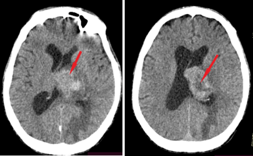
Figure 1: Axial CT sections with contrast enhancement of left intraventricular mass.
Discussion
Colorectal Cancer (CRC) is one of the most common malignant tumors in the world [3]. Common sites of metastasis in colorectal cancer include the lung, liver, and draining lymph nodes [4]. The most common site of CRC metastases is the liver; 15-25% of patients develop liver metastases synchronously, while 30-50% of patients develop liver metastases later [5]. In contrast, the incidence of brain metastases in CRC is very low (1.2-3.2%) [6] and usually occurs at a later stage of disease progression.
Solitary metastases of the choroid plexus are very rare. Intraventricular tumors make up only about 10% of Central Nervous System (CNS) tumors, and intraventricular metastases make up about 6 percent [7].
The rate of brain metastases discovered during the diagnosis of colorectal cancer is 0.1% with a 1.2% risk of developing brain metastases [8] during a follow-up period of nearly 4 years Intraventricular metastases secondary to colorectal cancer are rare. Only four cases have been described in the literature [9-11].
Metastases to the choroid plexus are rare, the vast majority of intracranial cancer metastases occur in the cerebral parenchyma. The most frequent pure intraventricular tumors are meningiomas and papillomas of the choroid plexus, and metastases are not ranked at the top of the list of differential diagnoses.
Despite its rich vascularization and lack of a blood-brain barrier, the choroid plexus is even less frequently affected by metastases than other areas of the brain [12]. Some hypotheses suggest that the low incidence of solitary metastases of the choroid plexus may be attributable to multiple factors [13]. First, the choroid plexus “soil“ may not be hospitable for metastases, because it easily accumulates immune complexes and is targeted by anti-basement membrane antibodies that can discourage tumor cell implantation [14]. Second, blood-borne tumors cannot be trapped and extravased due to high blood flow [15]. Third, the rarity of intraventricular metastases in the choroid plexus may be due to the absence of cell receptors [16].
Although histological analysis remains the examination of choice to confirm the metastatic origin of colorectal cancers, imaging also helps in diagnosis.
Enhanced contrast computed tomography is often the first line of imaging. On pre-contrast imaging, the mass can be isodense, hypodense or hyperdense (classical melanoma) compared to the normal cerebral parenchyma with varying amounts of surrounding vasogenic edema. After contrast administration, the improvement is also variable and can be intense, punctate, nodular or ring-enhancing if the tumor has exceeded its blood supply [17].
However, currentMRI technology has proven to be more sensitive than CT scans and is the preferred imaging of choice. Contrastenhanced MR is the current standard for small metastases detection, therefore, delayed sequences should be used to look for additional lesions. Spectroscopy is very specific, it shows intratumoral choline peak with no choline elevation in the peritumoral edema, a lipid peak is associated in any tumor necrosis results.
In our case, the MRI could not be performed because the patient was agitated and he was admitted to resuscitation for better management [18].
When considering intraventricular tumours, choroid plexus metastases must be included in the list of differential diagnoses even in a patient without a previous systemic cancer. Post-contrast enhancement, intraventricular haemorrhage, and peritumoural oedema on imaging, may indicate metastases from extracranial neoplasms.
References
- Mohamed Said Belhamidi, Mohamed Sinaa, Abdessamad Kaoukabi, Hicham Krimou, Mohamed Menfaa, et al. Service de Chirurgie Viscérale, Hôpital Militaire Moulay Ismail, Meknès, Maroc, 2 Laboratoire d’Anatomopathologie, Hôpital Miltaire Moulay Ismail, Meknès, Maroc.
- Schouten LJ, Rutten J, Huveneers HAM, Twijnstra A. Incidence of brain metastases in a cohort of patients with carcinoma of the breast, colon, kidney, and lung and melanoma. Cancer. 2002; 94: 2698-2705.
- Schouten LJ, Rutten J, Huveneers HAM, Twijnstra A. Incidence of brain metastases in a cohort of patients with carcinoma of the breast, colon, kidney, and lung and melanoma. Cancer. 2002; 94: 2698-2705.
- Schouten LJ, Rutten J, Huveneers HAM, Twijnstra A. Incidence of brain metastases in a cohort of patients with carcinoma of the breast, colon, kidney, and lung and melanoma. Cancer. 2002; 94: 2698-2705.
- Hiroaki Nozawa, Soichiro Ishihara, Kazushige Kawai, Kazuhito Sasaki, Koji Murono, et al. Brain Metastasis from Colorectal Cancer: Predictors and Treatment Outcomes. Oncology. 2017; 93: 309-314.
- Hiroaki Nozawa, Soichiro Ishihara, Kazushige Kawai, Kazuhito Sasaki, Koji Murono, et al. Brain Metastasis from Colorectal Cancer: Predictors and Treatment Outcomes. Oncology. 2017; 93: 309-314.
- Kohno M, Matsutani M, Sasaki T, et al Solitary metastasis to the choroid plexus of the lateral ventricle. Report of three cases and a review of the literature. J Neuro-oncol. 1996; 27: 47–52.
- Nozawa H, Ishihara S, Kawai K, Sasaki K, Murono K, Otani K, et al. Brain Metastasis from Colorectal Cancer: Predictors and Treatment Outcomes. Oncology. 2017; 93: 309-314.
- Kohno M, Matsutani M, Sasaki T, et al Solitary metastasis to the choroid plexus of the lateral ventricle. Report of three cases and a review of the literature. J Neuro-oncol. 1996; 27: 47–52.
- Hassaneen W, Suki D, Salaskar AL, Wildrick DM, Lang FF, Fuller GN, et al. Surgical management of lateral-ventricle metastases: report of 29 cases in a single-institution experience. Journal of neurosurgery. 2010; 112: 1046-1055.
- Kitajima K, Morita M, Morikawa M, Sugimura K. Choroid plexus metastasis of colon cancer. Magnetic resonance in medical sciences : MRMS : an official journal of Japan Society of Magnetic Resonance in Medicine. 2003; 2: 155- 158.
- Al-Anazi A, Shannon P, Guha A. Solitary metastasis to the choroid plexus. Case illustration. Journal of neurosurgery. 2000; 92: 506.
- Qasho R, Tommaso V, Rocchi G, Simi U, Delfini R. Choroid Plexus Metastasis from Carcinoma of the Bladder: Case Report and Review of the Literature. Journal of Neuro-Oncology. 2004; 45: 237-240.
- Leach JCD, Garrott H, King JAJ, Kaye AH. Solitary metastasis to the choroid plexus of the third ventricle mimicking a colloid cyst: a report of two cases. Journal of Clinical Neuroscience. 2004; 11: 521-523.
- Al-Anazi A, Shannon P, Guha A. Solitary metastasis to the choroid plexus. Case illustration. Journal of neurosurgery. 2000; 92: 506.
- Qasho R, Tommaso V, Rocchi G, Simi U, Delfini R. Choroid Plexus Metastasis from Carcinoma of the Bladder: Case Report and Review of the Literature. Journal of Neuro-Oncology. 2004; 45: 237-240.
- Kumar V, Abbas AK, Fausto N et-al. Robbins and Cotran pathologic basis of disease. W B Saunders Co. (2005) ISBN:0721601871.
- Fink KR, Fink JR. Imaging of brain metastases. Surgical Neurology International. 2013; 4: 209.