
Clinical Image
Austin J Radiol. 2022; 9(5): 1204.
Sigmoid Diverticulis
Berrada K*, Mribat M, Fenni JE and Lahkim M
Department of Radiology, Mohammed V Military Teaching Hospital, Faculty of Medicine and Pharmacy, Mohammed V University, Rabat, Morocco
*Corresponding author: Kenza Berrada, Department of Radiology, Mohammed V Military Teaching Hospital, Faculty of Medicine and Pharmacy, Mohammed V University, Rabat, Morocco
Received: July 26, 2022; Accepted: August 19, 2022; Published: August 26, 2022
Clinical Image
Sigmoid diverticulitis is a complication of colonic diverticulosis, and one of the presentations of diverticular disease. It is a very common pathology in Western countries due to the lack of fiber content in diets, leading sigmoid colon to make strong contractions and end up but formation of diverticula, with a very high risk of inflammation and infection. It mainly interests people over 45 years old, without any gender preference. The reflex is to always think of a sigmoid diverticulitis in front of an acute pain of the left iliac fossa with fever, leukocytosis, and supplement with an emergency scanner and failing that an abdomino-pelvic ultrasound. Imaging plays a vital role in both positive diagnosis and diagnosis of complications .Main complication are fistula, peritonitis, small bowl obstruction, abcess, and rarely portal venous pylephlebitis. Although CT is the modality of choice for the diagnosis and staging of colonic diverticulitis, it commonly shows sigmoid wall thinckening, fat stranding, and diverticulos appearing like aeric addition image (Figure A and B). Main differential diagnosis is colorectal carcinoma and acute appenditics. Acute non complicated sigmois is treated with antibiotherapy with a colonoscopy after recovery, when it’is complicated, elective and emergency surgery is a priority.
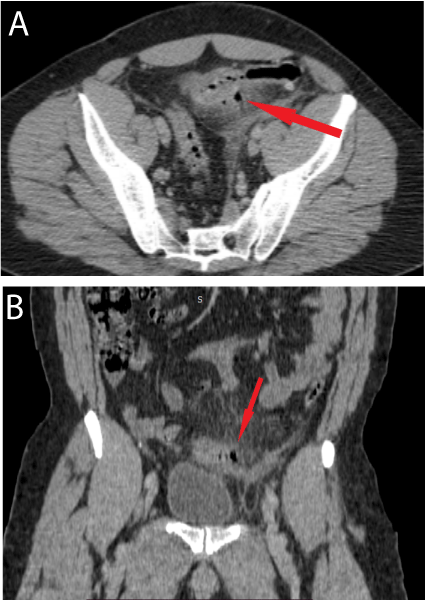
    Figure 1: Axial CT section (Figure A) and saggital section (Figure B)

with contrast enhancement sigmoid wall thinckening, fat stranding, and

diverticulos appearing like aeric addition image. Figure A: Axial CT section

with contrast enhancement sigmoid wall thinckening, fat stranding, and

diverticulos appearing like aeric addition image. Figure B: Saggital section

with contrast enhancement sigmoid wall thinckening, fat stranding, and

diverticulos appearing like aeric addition image.