Case Report
Austin J Radiol. 2022; 9(5): 1205.
Intraorbital Colobomatous Cyst with Microphthalmia in a Child Presented with Orbital Mass
Rawat A* and Yadav P
Assistant Professor (Radiodiagnosis), KGMU, Lucknow - 226003, India
*Corresponding author: Anil Rawat, Assistant Professor, Department of Radio-Diagnosis, KGMU, Lucknow, UP-226003, India
Received: September 02, 2022; Accepted: October 06, 2022; Published: October 13, 2022
Abstract
A colobomatous cyst is an extremely uncommon form of congenital abnormality that is frequently linked with a microophthalmic globe. It is caused by a problem with the closure of the optic vesicle as well as invagination.. Here we are presenting a case of orbital cyst associated with microophthalmos in an infant presented with small eye ball and orbital lesion, child was evaluated clinically and radiologically by USG and MRI. To the best of our knowledge very few cases have been reported in previous literature
Keywords: Colobomatous cyst; Microphthalmia; B-Scan; Orbital cyst; Congenital ocular anomaly
Introduction
After congenital cataract, microphthalmia is among the most frequent birth defects affecting the eye. However, intraorbital cysts are rarely linked to this condition. Due to a general lack of evidence documented in literature, most lesions continue to be incorrectly labelled as orbital tumours or teratomas.
Case Presentation
The parents of an eight-month-old female newborn brought her to an outpatient ophthalmology clinic due to gradually growing swelling under the right lower lid since birth and intermittent wetness of the right eye. The size of the cyst increased over time. (Figure 1) depicts an ocular examination of the right eye, which revealed a microphthalmic globe with bluish swelling throughout the right lower lid. There were no developmental abnormalities in the left eye at that time.

Figure 1: Bluish swelling present throughout the extent of right lower lid.
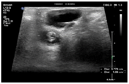
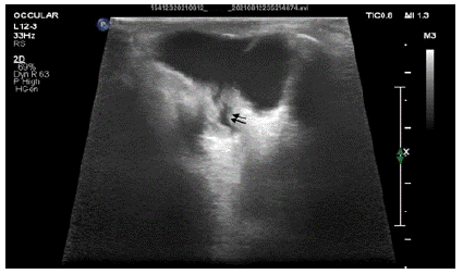
B-scan high resolution ultrasoundand MRI was performed which showed a well defined extraconal cyst in the inferior aspect of right eye with a microphthalmic globe (Figure 2). Thin streak of communication is seen with posteroinferior aspect of microphthamic globe (Figure 3).

    Figure 2: B mode ultrasound image showing well defined, oval anechoic

lesion with deformed microophthalmic globe displaced posteriorly.

    Figure 3: B mode ultrasound image showing thin streak of communication

between the orbital cyst and deformed microophthalmicglobe.
B-scan ultrasonography revealed a cystic lesion in the right orbit’s inferior compartment, with the right orbit’s tiny globe being shifted posterosuperiorly. The cysts’ interior contents were also homogeneous and their walls were thin. The cysts averaged about 3 mm in height and 2 mm in width. The antero-posterior diameter of the globe reduced, making it smaller in size.
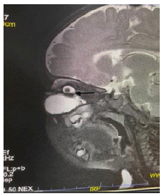
The 1.5 T GE MR scanner was used for the multiplanar MR imaging. In the axial, coronal, and sagittal planes, conventional T1- and T2-weighted, Fluid Attenuation Inversion Recovery (FLAIR), and diffusion weighted sequences were employed. (Figure 4) shows a scan of the right orbit, which shows a homogenous cystic lesion associated with a small sized globe in the lower part of the orbit. Its anteroposterior, transverse, and craniocaudal measurements were roughly 3.2x2.2x1.8cm. The cyst contained no solid component. There was no post-contrast enhancement or signs of haemorrhage inside the cyst. Communication of the cysts through a defect in the posterior half of the globes was suspected.

    Figure 4: T2W sagittal image showing well defined cyst arising in the

inferior half of the orbital cavity in association with microophthalmic

globe. Arrow showing thin streak of communication between cyst and the

microophthalmicglobe.
Treatment
Most of the time, functional improvement can’t be made, but surgery should only be considered as a last resort for looks. In the most severe and unsightly cases, doctors recommend enucleation, cyst removal, and then fitting a prosthesis. In our case, the cyst aspiration was done only.
Outcome & Follow-up
The child was undergone cyst aspiration. On follow up imaging the cyst size was significantly reduced, further management was explained to the mother for cosmetic resort.
Discussion
Microphthalmia is a condition in which the eye is small but still has parts like the lens, choroid, and retina. Anophthalmia is a condition in which there is no eye at all. This is caused by the optic vesicles not developing or their development being stopped at an early stage [2]. The rate of microphthalmia is between 1.4 and 3.5 out of every 10,000 births [3]. Microphthalmia with orbital cyst is very rare, and there is no agreed-upon rate of how often it happens [1]. Congenital microphthalmia with intraorbital cyst happens when the embryonic fissure doesn’t close properly and the inner layer of the optic cup grows too much [4]. An extremely rare neuroectodermallined cyst, the colobomatous cyst was first described by Thomas Bartholin in 1673. A microphthalmic globe and a protruding cyst in the infero-nasal quadrant of the eye are the most common clinical manifestations of this condition, though a non-existent cyst is possible [5,6].
Most of the time, a diagnosis needs to be made with the help of a clinical exam and imaging. Imaging methods like B scan ultrasonography, computed tomography, and magnetic resonance imaging can help confirm the diagnosis and tell it apart from a congenital cystic eye, meningocele, primary optic nerve sheath cysts, and teratomas of the orbit.
Our ability to distinguish this entity from other benign or malignant orbital masses in children is facilitated by imaging techniques such A and B scan ultrasonography, computed tomography, and magnetic resonance imaging.
In this case also, the combination of an MRI and an orbital ultrasound A+B scan proved to be invaluable in making a correct diagnosis. The isointensity of the two cavities on T1- and T2-weighted spin echo may aid in the diagnosis, making MRI the preferred imaging modality for proving that the vitreous cavity communicates with the colobomatous cyst.
These imaging techniques not only help with diagnosis, but can also be important in finding other anomalies, especially in the brain. Bilateral cases are more likely to be accompanied with systemic anomalies including cleft lip, basal encephalocele, mid brain malformation, microcephaly, agenesis of corpus callosum, and saddle nose. Preoperative imaging may be extremely useful in such situations [7]. There were no systemic issues associated with our deformity.
Colobomatous cysts have a wide variety of possible etiologies, including deep dermoids, teratomas, mucoceles, meningoceles, primary optic nerve sheath cysts, and congenital cystic eye. CT scans of dermoids should reveal a fatty core, but we observed that this was the case in only 41% of cases [8]. In contrast to our situation, deep dermoids often cluster higher up in the orbit, not lower down. Meningoceles have a pedicle leading into the skull cavity, while mucoceles have a direct connection to the ethmoid or frontal sinus.
Surgery should be viewed as a last choice for aesthetic purposes alone given that it offers no functional benefits. Even though it may be impossible to completely remove posteriorly positioned cysts, enucleation and cyst excision followed by prosthesis fitting is suggested in the most severe, unsightly cases.
Learning Points/Conclusion
• Colobomatous cyst with microphthalmia is an extremely rare congenital anomaly.
• Misdiagnosis of a neoplastic lesion is possible if the radiologist is unfamiliar with the condition’s complex and varied characteristics.
• Proper knowledge and early diagnosis of the condition is a must for early diagnosis and timely definitive management.
References
- K. Khurana, I. Khurana, A. K. Khurana, and B. Khurana, Anatomy and Physiology of Eye, CBS Publishers & Distributors Pvt Limited, Chennai, 2017.
- Stahnke T, Erbersdobler A, Knappe S, Guthoff RF, Kilangalanga NJ. Management of Congenital Clinical Anophthalmos with Orbital Cyst: A Kinshasa Case Report. Case Reports in Ophthalmological Medicine. 2018; 2018: 1-6.
- McLean CJ, Ragge NK, Jones RB, Collin JRO. The management of orbital cysts associated with congenital microphthalmos and anophthalmos. British Journal of Ophthalmology. 2003; 87: 860-863.
- Cui Y, Zhang Y, Chang Q, Xian J, Hou Z, Li D. Digital Evaluation of Orbital Cyst Associated with Microphthalmos: Characteristics and Their Relationship with Orbital Volume. PLoS ONE. 2016; 11: e0157819.
- Shields JA, Shields CL. Orbital cysts of childhood--classification, clinical features, and management. Survey of ophthalmology. 2004; 49: 281-299.
- Onwochei BC, Simon JW, Bateman JB, Couture KC, Mir E. Ocular colobomata. Survey of ophthalmology. 2000; 45: 175-194.
- Stahnke T, Erbersdobler A, Knappe S, Guthoff RF, Kilangalanga NJ. Management of Congenital Clinical Anophthalmos with Orbital Cyst: A Kinshasa Case Report. Case Reports in Ophthalmological Medicine. 2018; 2018: 1-6.
- Salvolini U, Menichelli F, Pasquini U. Computed assisted tomography in 90 cases of exophthalmos. J Comput Assist Tomogr 1978; 1: 81-100.
- Shields JA. Diagnosis and Management of Orbital Tumors. Philadelphia: Saunders, 1989; 89-122.
- Helveston EM, Malone E, Lashmet MH. Congenital cystic eye. Archives of ophthalmology. 1970; 84: 622-624.