
Case Report
Sarcoma Res Int. 2020; 5(1): 1044.
Multiple Giant Retroperitoneal Myelolipoma: A Case Report and Review of the Literature
Colarossi C, Picardo C, Di Mattia P, Aiello E, Colarossi L, De Zuanni M and Memeo L*
Department of Experimental Oncology, Mediterranean Institute of Oncology, Italy
*Corresponding author: Memeo L, Department of Experimental Oncology, Mediterranean Institute of Oncology, Via Penninazzo 7, 95029 Viagrande, (CT), Italy
Received: May 08, 2020; Accepted: October 09, 2020; Published: October 16, 2020
Abstract
Myelolipoma is an uncommon benign mesenchymal tumor consisting of mature adipocytes and hemopoietic elements mostly found in adrenal glands. Extra-adrenal myelolipoma occurs rarely and requires differential diagnosis with other soft tissue tumors. Here the authors present a case of a 55 years old man who underwent laparotomic surgery for three peri-renal masses. The aim of this report is to present the clinical, radiological and pathological features of an unusual retroperitoneal lesion that still represents a challenging diagnosis.
Keywords: Myelolipoma; Retroperitoneal mass; Liposarcoma
Abbreviations
AM: Adrenal Myelolipoma; ML: Myelolipoma
Introduction
Myelolipomas are rare benign tumor, composed of mature adipose and myeloid tissue, described for the first time by Gierke in 1905 [1] and classified by Oberling in 1929 [2]. They are usually asymptomatic, unilateral, single and located in the adrenal gland [3]. Despite the more frequent adrenal presentation, approximately 15% of myelolipomas can be found in an extra-adrenal location, more frequently in retroperitoneum [4], pelvis [5] and mediastinum [6]. Extra-adrenal myelolipoma may present a diagnostic challenge, especially when origins in retro peritoneum. Fat-containing retroperitoneal lesions require a differential diagnosis with a large number of neoplastic and no neoplastic conditions [7]. Commonly, they may be misidentified as well-differentiated liposarcoma on radiographic imaging given their similar gross morphological composition. We report a rare case of a 55 old man who presented three retroperitoneal nodules, a giant soft tissue tumor with two minor satellite tumors. The presence of three discontinuous nodules was suggestive of liposarcoma. The postoperative histological analysis allowed the diagnosis of myelolipoma.
Case Presentation
A 55 years old white male was admitted in emergency room complaining non-specific left abdominal pain. He was affected by obesity and chronic hypertension. No previous surgery was reported in anamnesis. On physical examination, abdominal palpation demonstrated a left abdominal, painful mass. Therefore, he was admitted in our oncologic surgery and abdominal CT scan was performed, which revealed the presence of three expansive solid masses occupying the left abdomen, the largest with a diameter of 19x13x12cm, and two minor satellite masses, which displaced adjacent organs, compressed the left ureter and encased the left renal artery. All the three masses appeared capsulated, with minimal contrast enhancement and mixed density containing soft tissue enhancement and fatty component (Figure 1). No abdominal lymphadenopathy was described. Laboratory tests at admission gave the following results: blood leukocytes 5,900/mm3 with normal differential cell count; red blood cells 5,470,000/mm3; blood hemoglobin 14.9 g/dl with a Mean Corpuscular Volume (MCV) of 83 fl; blood platelet count 175,000/ mm3; HTC 45.3%. The patient underwent exploratory laparotomy, and the three masses were removed en bloc. The post-operative course was regular and the patient was discharged on the seventh postoperative day. Laboratory tests at discharge: blood leukocytes 4,080/ mm3 with normal differential cell count; red blood cells 4,180,000/ mm3; blood hemoglobin 11.4 g/dl with a Mean Corpuscular Volume (MCV) of 80 fl; and blood platelet count 152,000/mm3; HTC 33,6%.
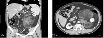
    Figure 1: Abdomen CT shows a retroperitoneal well circumscribed soft
tissue density mass with fat-containing containing components with low postcontrast
enhancement. A) coronal scan shows lateral dislocation of the left
kidney; B) axial scan shows the three components with different density.
The macroscopic examination of surgical sample showed three masses, with the largest mass measuring 19x15x6.5, and the adjacent nodules were 8 and 9 cm in diameter, respectively. They showed an oval shape with distinct margins and large hemorrhagic areas. The color was reddish brown (Figure 2).

Figure 2: Macroscopic image of the retroperitoneal three nodules.
The three lesions showed the same histological features. They were highly cellular and circumscribed by a thick fibrous capsule (Figure 3). The tumors showed a variable mixture of mature adipocytes islands with intermixed hematopoietic elements at numerous stages of differentiation. No blasts CD34+ were observed. Numerous megakaryocytes were present though the tumor, ranging from 10 to 20/HPF. There were wide hemorrhagic areas and an increase of reticulin fibers, stained with silver impregnation, but no signs of malignancy. All the tumors were diagnosed as myelolipoma.
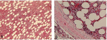
    Figure 3: Microscopic images of Myelolipoma; A) Trilinear population
intermixed with mature adipocytes (H&E 5x); B) the thick fibrous capsule
delimitationg the mass (H&E 20x).
Discussion
Myelolipoma is a benign tumor who usually origins in adrenal gland. Rarely it can be found in extra-adrenal sites. The pathogenesis is still unclear. Some theories suggest that myelolipomas arise from metaplastic transformation of adrenal stromal cells in response to stimuli such as necrosis, infection, and stress. Anyway, some authors showed karyotype abnormalities suggesting a clonal origin of adrenal myelolipomas. In one case a balanced translocation t (3,21) (q25, p21) was found [8]. Later, Bishop et al showed nonrandom X-chromosome inactivation in hematopoietic elements and adipocytes of 8 out of 11 cases of adrenal and extra-adrenal myelolipomas [9]. According to the literature, around 50 extra-adrenal myelolipomas have been described so far. Most of them were localized in perirenal [10] and presacral region [11], while 1/3 of the cases were localized in mediastinal and thoracic region [12]. Few cases have been described in spleen [13]. Some of them were associated with adrenal myelolipoma [14] and others with various other pathologies [15,16]. Interestingly some myelolipomas were reported to develop hematological malignancies [17,18]. Our case presented with three contiguous nodules, and this made the pre-operatory diagnosis more challenging. Furthermore, one of the three tumors measured 19 cm in diameter. Tumors of this size have been classified as giant myelolipomas [19]. Due to the voluminous size of the lesion, we decided to treat the lesion with laparoscopic surgery.
Although most AMs are small and asymptomatic, larger tumors may present with symptoms ranging from nonspecific abdominal pain to abscesses and spontaneous retroperitoneal hemorrhage [20]. Surgical intervention is required in symptomatic tumors and in tumors bigger than 7 cm, because of the potential risk of rupture. MLs can cause spontaneous retroperitoneal hemorrhage with high risk of deaths [21]. The main problem when a lipomatous tumor is localized in retroperitoneum is the differential diagnosis with liposarcomas which represent up to 45% of retroperitoneal soft tissue tumors [22]. The presence of two satellites tumor of the same macroscopic features in our patient, was suggestive of liposarcoma. Moreover, the synchronous presentation of myelolipomas is quite rare [23,24]. In conclusion, we recommend considering this entity in the preoperative stage, in case of retroperitoneal masses, even multiple.
Acknowledgment
Authors would like to thank Giovanni Ferlito and Mariarita Pulvirenti for technical assistance.
References
- Gierke E. Uber Knochenmarksgewebe in der Nebenniere. Z Path Anat. 1905; 37: 311.
- Oberling C. Les formation myelo-lipomateuses. Bull Cancer. 1929; 18: 234- 246.
- Decmann A, Perge P, Tóth M, Igaz P. Adrenal myelolipoma: a comprehensive review. Endocrine. 2018; 59: 7-15.
- Vigutto G, Lauro A, Vaccari S, Pirini MG, Diegoli M, D’Andrea V, et al. Giant Retroperitoneal Myelolipoma: An Unusual Diagnostic GI Challenge-Case Report and Review of the Literature. Dig Dis Sci. 2019; 64: 3431-3435.
- Sethi S, Thakur S, Jacques S, Aoun HD, Tranchida P. Myelolipoma of the Pelvis: A Case Report and Review of Literature. Front Oncol. 2018; 8: 251.
- Shen C, Zhou K, Lai Y, Fan J, Liu L, Che G. Review of primary extra-adrenal myelolipoma of the thorax. J Surg Res. 2017; 207:131-137.
- Shaaban AM, Rezvani M, Tubay M, Elsayes KM, Woodward PJ, Menias CO. Fat-containing Retroperitoneal Lesions: Imaging Characteristics, Localization, and Differential Diagnosis. Radiographics. 2016; 36: 710-734.
- Bishop E, Eble JN, Cheng L, Wang M, Chase DR, Orazi A, et al. Adrenal myelolipomas show nonrandom X-chromosome inactivation in hematopoietic elements and fat: support for a clonal origin of myelolipomas. Am J Surg Pathol. 2006; 30: 838-843.
- Chang K, Chen P, Huang Z, Lin YM, Kuo PL. Adrenal myelolipoma with translocation (3;21)(q25;p11). Cancer Genet Cytogenet. 2002; 134: 77-80.
- Dan D, Bahadursingh S, Hariharan S, Ramjit C, Naraynsingh V, Maharaj R. Extra-adrenal perirenal myelolipoma. A case report and review of literature. G Chir. 2012; 33: 62-65.
- Singla AK, Kechejian G, Lopez MJ. Giant presacral myelolipoma. Am Surg. 200; 69: 334-338.
- Shen C, Zhou K, Lai Y, Fan J, Liu L, Che G. Review of primary extra-adrenal myelolipoma of the thorax. J Surg Res. 2017; 207: 131-137.
- Zeng Y, Ma Q, Lin L, Fu P, Shen Y, Luo QY, et al. Giant Myelolipoma in the Spleen: A Rare Case Report and Literature Review. Int J Surg Pathol. 2016; 24: 177-180.
- Zieker D, Königsrainer I, Miller S, Vogel U, Sotlar K, Steurer W, et al. Simultaneous adrenal and extra-adrenal myelolipoma - an uncommon incident: case report and review of the literature. World J Surg Oncol. 2008; 6: 72.
- Giuliani A, Tocchi A, Caporale A, Demoro M, Miccini M, Di Bari M, et al. Presacral myelolipoma in a patient with colon carcinoma. J Exp Clin Cancer Res. 2001; 20: 451-454.
- Yildiz BD. Giant Extra-Adrenal Retroperitoneal Myelolipoma with Incidental Gastric Mesenchymal Neoplasias. Int Surg. 2015; 100: 1018-1012.
- Arora K, Sidhu J. Extra-Adrenal Myelolipoma Containing Small Lymphocytic Lymphoma/Chronic Lymphocytic Leukemia: A Case Report and Review of the Literature. Case Rep Hematol. 2016; 2016: 7364951.
- Gheith S, Boulay R, Cornfield D. Small lymphocytic lymphoma/chronic lymphocytic leukemia in a pelvic myelolipoma. Int J Clin Exp Pathol. 2009; 2: 95-98.
- Suárez-Peñaranda JM, Bermúdez Naveira A, Fraga M, Aliste-Santos C, Cordeiro C, Muñoz-Barús JI. Unusual Forms of Adrenal and Extra-Adrenal Myelolipomas. Int J Surg Pathol. 2014; 22: 473-477.
- Kumar S, Jayant K, Prasad S, Agrawal S, Parma KM, Roat R, et al. Rare adrenal gland emergencies: a case series of giant myelolipoma presenting with massive hemorrhage and abscess. Nephrourol Mon. 2015; 7: e22671.
- Liu HP, Chang WY, Chien ST, Hsu CW, Wu YC, Kung WC, et al. Intraabdominal bleeding with hemorrhagic shock: a case of adrenal myelolipoma and review of literature. BMC Surg. 2017; 17: 74.
- Vijay A, Ram L. Retroperitoneal liposarcoma: a comprehensive review. Am J Clin Oncol. 2015; 38:213-219.
- Lazarides AL, Scott EJ, Cardona DM, Blazer DG, Brigman BE, Eward WC. Simultaneous Primary Presacral Myelolipomas: Case Report and Review of the Literature. J Gastrointest Cancer. 2016; 47: 331-335.
- Yugandhar S, Sureka SK, Yadav P, Lal H. A rare case of extra-adrenal bilateral perirenal and periureteric myelolipoma. BMJ Case Rep. 2017.