
Case Report
Sarcoma Res Int. 2021; 6(1): 1045.
Prosthetic Reconstruction for Proximal Tibial Osteosarcoma with an Anterolateral Surgical Approach: Case Report
Khezami K¹*, Nouri H², Jenzri M³, Gharbi A¹ and Bennour MA¹
¹Department of Orthopedic Surgery, University Tunis El Manar, Tunisia
²Department of Adult Surgery, University El Manar II, Tunisia
³Department of Children Orthopedic, Institute of Kassab Manouba, Tunisia
*Corresponding author: Karim Khezami, Department of Orthopedic Surgery, University Tunis El Manar, Habib Bougatfa Hospital, Bizerte, Tunisia
Received: March 09, 2021; Accepted: March 30, 2021; Published: April 06, 2021
Abstract
We report the case of a 13-year-old boy with a primary malignant bone tumor of the proximal tibia. We had approached the bone tumor by the anterolateral route, which allowed resection and reconstruction by a massive prosthesis. The patient presented at the last follow-up of 30 months good knee joint mobility and good muscular strength with active knee flexion 90 degrees, 0 degrees active extension and muscle strength at 5.
Keywords: Osteosarcoma; Surgery; Reconstruction; Proximal Tibia
Introduction
Osteosarcoma is a more common primary malignant bone tumor accounting for 15% of the primary bone tumors, and the incidence rate is about 0.3 per million. It often occurs in adolescents [1] and is characterized by high morbidity and mortality [1,2]. The upper tibia is the osteosarcoma predilection site [1]. The location and technique of the biopsy are major determinants of the outcome of a limb-sparing resection. To minimize contamination of the anterolateral muscles, peroneal nerve, popliteal space, and knee joint authors recommend an anteromedial surgical approach for a proximal tibia biopsy and resection with reconstruction. The surgical and technical problems include intimate anatomic relationships, a difficult surgical approach, inadequate soft tissue coverage, and vascular complications. Our case report describes the technique that we used to perform with anterolateral approach the reconstruction, which permitted a safe resection and reconstruction of a large segment of proximal tibia.
Case Presentation
Our case report subject is a 13-year-old boy referred to our Hospital. He complained of spontaneous pain in the proximal third of his right tibia. The Radiography of knee right show a lytic lesion located at the proximal part of the tibia. We decided to further investigate the case through extensive imaging studies in order to elucidate the nature of the lesion and to establish its staging. CT and MRI show an upper metaphyso-epiphyseal lytic lesion of the tibia right, measuring 45*38 mm in the transverse plane extended by approximately 52 mm in height. It is responsible for a rupture of the metaphyseal posteroexternal cortex as well as epiphyseal with extension to deep muscular compartment, arriving in contact with the cortex of the head of the fibula without sign of invasion. This exam does not show a skip lesions, a extension in tibio-fibular joint, an intra-articular knee involvement and an involvement of the posterior vascular-nervous package. The surgical stage of patient is Stage IIB according to ennecking staging (Figure 1-5).
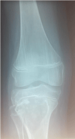
    Figure 1: Radiography of knee right show a lytic lesion located at the proximal
part of the tibia.
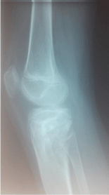
    Figure 2: Radiography of knee right show a lytic lesion located at the proximal
part of the tibia.
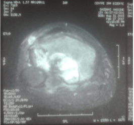
    Figure 3: Magnetic Resonance Imaging (MRI) show an upper metaphysoepiphyseal
lytic lesion of the tibia right, measuring 45*38 mm in the transverse
plane extended by approximately 52 mm in height.
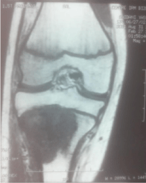
    Figure 4: Magnetic Resonance Imaging (MRI) show an upper metaphysoepiphyseal
lytic lesion of the tibia right, measuring 45*38 mm in the transverse
plane extended by approximately 52 mm in height.
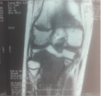
    Figure 5: Magnetic Resonance Imaging (MRI) show an upper metaphysoepiphyseal
lytic lesion of the tibia right, measuring 45*38 mm in the transverse
plane extended by approximately 52 mm in height.
The biopsy by an anterolateral approach is performed and extreme caution is taken to minimize contamination of the anterior muscles, peroneal nerve, patellar tendon, and knee joint. The biopsy site must be placed along the line of the definitive incision. A tourniquet is used to decrease the local contamination. The result of the anatomopathological examination confirms the diagnosis of common osteosarcoma.
Surgical Technique
After administration of neoadjuvant chemotherapy, the limb salvage surgery for proximal tibial osteosarcoma with involvement of the Proximal Tibio-Femoral Joint (PTFJ) included three main procedures:
Bloc resection of the primary tumor: With the patient on supine position, a single incision is used. The anterolateral incision encircling the biopsy scar was used. The anterior tibialis muscle is exposed. Its perimysium is opened and the muscle is retracted laterally, leaving the inner layer of the perimysium attached to the tibial periosteum, in order to preserve the wide margin of tumor resection. The neck of the fibula is identified and the common peroneal nerve is dissected. fibular collateral ligament and the femoral biceps tendon are released from the fibular head. Next, the anterior tibial artery was ligated and disconnected. The placement of the osteotomy plane was established at 3 cm distal from the primary tumor, based on the results of T1- weighed imaging. This resection is performed through the same incision, a lateral flap is developed to permit the exposure of the proximal fibula. The peroneal nerve must be exposed and retracted prior to resection. The capsule is transected circumferentially approximately 1 cm away from the tibia and the patellar tendon, to avoid contamination. The cruciate ligaments are visually explored (Figure 6-11).
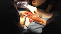
Figure 6: The anterolateral incision encircling the biopsy scar was used.
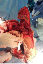
    Figure 7: The primary tumor was en bloc resected, which included the
proximal tibiofibular joint and the upper end of the fibula.
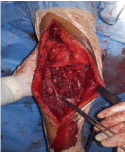
    Figure 8: The primary tumor was en bloc resected, which included the
proximal tibiofibular joint and the upper end of the fibula.

    Figure 9: The primary tumor was en bloc resected, which included the
proximal tibiofibular joint and the upper end of the fibula.
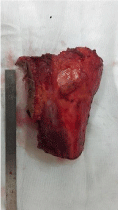
Figure 10: Proximal tibia resection piece.
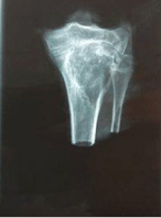
Figure 11: Proximal tibia resection piece.
Defect of bone and joint: After bloc resection including the PTFJ and the upper end of fibula (Figure 4), the custom prosthesis was secured using methyl methacrylate cement into both the tibia and the femur.
Reconstruction for the extensor mechanism and the defect of soft tissue: The extensor mechanism was repaired by reattachment of the patellar tendon to the slot on the tibial component, and suturing of the patellar tendon to a medial gastrocnemius rotation flap (Figure 5A). Since no deep fascia remained anteriorly, the metal endoprosthesis was left inadequately covered by only fat and skin after resection. A medial gastrocnemius rotation flap was used as a routine solution to cover the implant (Figure 5B). In addition, care was taken to repair the lateral collateral ligament and the biceps femoris tendon (Figure 12 and 13).
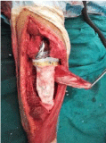
    Figure 12: A medial gastrocnemius flap is transposed to repair the extensor
mechanism by direct suturing of the patellar tendon and to cover the custom
prosthesis.
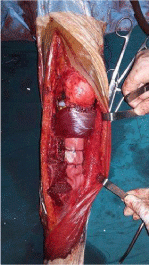
    Figure 13: A medial gastrocnemius flap is transposed to repair the extensor
mechanism by direct suturing of the patellar tendon and to cover the custom
prosthesis.
Postoperative Management
Large suction drains are used to prevent hematoma. Our patient was immobilized for six weeks in a long leg cast to permit the healing of the extensor mechanism to the gastrocnemius transfer. The patient received postoperative physiotherapy with progressive weight bearing. At 3 months, post-operatively we authorized walking without support. Evaluation showed knee mobility: passive flexion 90 degrees, 5 degrees passive extension. Active flexion at 3 months was of 40 degrees and -15 degrees active extension. Muscle strength in the quadriceps was 4 (the patient did knee extension against a force applied to the front of the leg, but didn’t perform full extension). 2 years postoperatively, the patient presented a fracture of the femur on a prosthesis during a leisure accident. As the fracture is not displaced much and the prosthesis is stable, we opted for orthopedic treatment with immobilization for three months. The evolution is marked by the consolidation of the fracture. Actually we consider the patient fully recovered locally with active knee flexion 90 degrees, 0 degrees active extension, muscle strength at 5 (normal) (Figure 14-17).
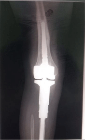
    Figure 14: Fracture on prosthesis on femoral side consolidated after three
months of orthopedic treatment with good knee mobility.
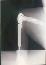
    Figure 15: Fracture on prosthesis on femoral side consolidated after three
months of orthopedic treatment with good knee mobility.

    Figure 16: The patient fully recovered locally with active knee flexion 90
degrees, 0 degrees active extension.

    Figure 17: The patient fully recovered locally with active knee flexion 90
degrees, 0 degrees active extension.
Discussion
The last two decades have witnessed a dramatic change in the approach to the treatment of osteosarcoma. The development of more effective chemotherapeutic agents, combined with advances in imaging technology, has permitted the surgeon to use limb salvage surgery to preserve limb function in patients with osteosarcoma of the extremities. The management of proximal tibial osteosarcoma still continues to be one of the most challenging areas in orthopedic oncology. Proximal tibial megaprosthetic reconstructions have been associated with less favorable outcome and function compared to other joints [3-8]. According to the location of the tumor involvement, the surgical approach was selected as either anteromedial or anterolateral of the knee joint. Proximal tibia resection was done using an anteromedial approach extending from the distal femur to the anteromedial tibia depending on tumor’s extension [3]. Many surgeons use the anteromedial approach to the proximal tibia because it allows for an excellent exposure of the vessels, fibula, and medial gastrocnemius muscle. The surgical and technical problems include intimate anatomic relationships, a difficult surgical approach, inadequate soft-tissue coverage, and vascular complications. In addition, unique to an arthroplasty of the proximal tibia is the need to reconstruct the patellar tendon (extensor mechanism). Extreme caution must be taken to minimize contamination of the anterior muscles, peroneal nerve, patella tendon, and knee joint when the biopsy is performed. The biopsy site must be placed along the line of the definitive incision. Though it is not routine practice to excise the head of the fibula when excising proximal tibial tumors, in case of disease extension into the proximal tibia fibular joint the head of the fibula is excised en bloc with the proximal tibia [9-11].
Prior to this the lateral popliteal nerve needs to be carefully dissected free to prevent injury to it. The upper end of the tibia poses unique problems. The complex vascular anatomy in the popliteal fossa adds to the challenges. A large posterior soft tissue component often causes tethering of the vessels and can necessitate delicate dissection to ensure that the posterior tibial vessels are dissected free of the tumor. Frequently, the anterior tibial vessels may need to be ligated as they pass from the posterior compartment anteriorly. The surgeon must ensure that the continuity of the posterior tibial vessels is maintained and he is not inadvertently ligating the tethered popliteal/posterior tibial vessels at the point where the anterior tibial branches out to pass over the interosseous membrane.
Conclusion
In conclusion, an anterolateral approach for a malignant tumor of the upper extremity of the tibia can be used especially when its location is posteroexternal without invasion of the vasculonervous pedicles and has devellopement especially intra osseous.
References
- Mirabello L, Troisi RJ, Savage SA. Osteosarcoma incidence and survival rates from 1973 to 2004. Cancer. 2009; 115: 1531-1543.
- Koh CH, Tai BC, Ang LW, Heng D, Yuan JM, Koh WP. All-cause and cause-specific mortality after hip fracture among Chinese women and men. Osteoporos Int. 2013; 24: 1981-1989.
- Grimer RJ. Endoprosthetic replacement of the proximal tibia. Bone Joint J. 1999; 81: 488-494.
- Malawer MM, Mchale KA. Limb-sparing surgery for high- grade malignant tumors of the proximal tibia: Surgical technique and a method of extensor mechanism reconstruction. Clin Orthop Relat Res. 1989; 239: 231-248.
- Bickels J, Wittig JC, Kollender Y, Neff RS, Kellar-Graney K, Meller I, et al. Reconstruction of the extensor mechanism after proximal tibia endoprosthetic replacement. J Arthroplasty. 2001; 16: 856-862.
- Unwin PS, Cannon SR, Grimer RJ, Kemp HB, Sneath RS, Walker PS. Aseptic loosening in cemented custom-made prosthetic replacements for bone tumours of the lower limb. J Bone Joint Surg Br. 1996; 78: 5-13.
- Horowitz SM, Lane JM, Otis JC, Healey JH. Prosthetic arthroplasty of the knee after resection of a sarcoma in the proximal end of the tibia. J Bone Joint Surg Am. 1991; 73A: 286-293.
- Eckardt JJ, Matthews JG, Eilber FR. Endoprosthetic reconstruction after bone tumor resections of the proximal tibia. Orthop Clin North Am. 1991; 22: 149-160.
- Horowitz SM, Glasser DB, Lane JM, Healey JH. Prosthetic and extremity survivorship after limb salvage for sarcoma. How long do the reconstructions last? Clin Orthop Relat Res. 1993; 293: 280-286.
- Mavrogenis AF, Pala E, Angelini A, Ferraro A, Ruggieri P. Proximal tibial resections and reconstructions: Clinical outcome of 225 patients. J Surg Oncol. 2013; 107: 335-342.
- Zhang Y, Yang Z, Li X, Chen Y, Zhang S, Du M, et al. Custom prosthetic reconstruction for proximal tibial osteosarcoma with proximal tibiofibular joint involved. Surg Oncol. 2008; 17: 87-95.