Research Article
Austin Spine. 2025; 2(1): 1004.
Relationship with the Severity of Spinal Cord Injury and the Location of Acute Intervertebral Disc Extrusion Occurred in Long-Haired Dachshunds and French Bulldogs in Taiwan
Jenshawn Wang1, Yi-Chun Lin1, Pei-Yu Kao1 and Ming-Hsien Chiang2,3
1Wangs Vet Clinic, Taipei, Taiwan
2Department of Anatomy and Cell Biology, College of Medicine, National Taiwan University, Taipei, Taiwan
3Zhonghe Duma Animal Hospital, New Taipei City, Taiwan
*Corresponding author: Ming-Hsien Chiang, Department of Anatomy and Cell Biology, College of Medicine, National Taiwan University, No. 1, Sec 1, Ren-Ai Road, Taipei, Taiwan Email: D01446001@ntu.edu.tw
Received: May 05, 2025 Accepted: May 28, 2025 Published: May 30, 2025
Abstract
Objective: The prevalence of intervertebral disc extrusion (IVDE) in longhaired Dachshunds (LHDs) is much higher than in French Bulldogs (FBs) in Taiwan. This study aimed to explore the relationship between the severity of spinal cord injury and the location of IVDE that occurred in these two breeds.
Study Design: Retrospective study.
Animals: 264 LHDs and 41 FBs with neurologic grades ranging from 1–5 caused by intervertebral disc disease (IVDD), 25 LHDs, and 10 FBs without clinical signs of IVDD.
Methods: The location of IVDE was detected by non-contract CT, and the height (H), width (D), and cross-sectional area (A) of compression materials and intervertebral foramen in the same slice were measured. All animals underwent decompressive surgery which confirmed the situation of the spinal cord.
Results: The neurologic grade of LHDs that IVDE happened in the thoracic spines was significantly more serious than IVDE occurred in the lumbar region, and the values of H, D, and A of the thoracic spine were significantly narrower than those in the lumbar spine (p<0.05). However, IVDE more frequently happened in the lumbar spine in FBs, and the values were not significantly different in patients of each grade (p>0.05).
Conclusions: In LHDs, the severity of paralysis caused by IVDE may be due to a narrow intervertebral foramen. However, this association was not significantly different among FBs.
Clinical Significance: The results of this study should help diagnose the location of acute IVDE in LDHs and FBs.
Introduction
Paralysis due to spinal cord injury (SCI) is the main reason owners consider euthanizing pets in animal hospitals in Taiwan. The most common cause of paralysis in dogs is SCI caused by intervertebral disc disease (IVDD) [1]. IVDD can be roughly divided into Hansen type I or II [2]. Hansen type II IVDD mostly involves chronic spinal cord compression that causes long-term chronic back pain and lameness. In contrast, Hansen type I IVDD is a situation in which the intervertebral discs rupture suddenly in a process called intervertebral disc extrusion (IVDE), which often causes acute paralysis in dogs [3]. Some studies have reported that IVDE often occurs in chondrodystrophic dog breeds including Dachshunds, French Bulldogs (FBs), Corgis, and Poodles. Hansen type II IVDD is more likely to occur in large non-chondrodystrophic dog breeds such as Golden Retrievers or Labrador Retrievers, etc [2]. In the United States, it has been reported that the prevalence of IVDD in Dachshunds is approximately 10– 12 times higher than that in other breeds. Both Dachshunds and French bulldogs are classified as chondrodystrophic breeds, but the prevalence of IVDE in French Bulldogs is not as high as that in Dachshunds [4]. Previous studies have investigated the relationship between congenital thoracolumbar vertebral abnormalities and IVDE in Dachshunds and French Bulldogs, but no significant differences have been found [4-6]. We hypothesized that the severity of paralysis in dogs may be related to the location of IVDE and aimed to identify differences in the characteristics of the spine associated with IVDE in Long-haired Dachshunds (LHDs) and French bulldogs (FBs).
Materials and Methods
Between January 2020 and December 2021, 264 LHDs and 41 FBs were treated at Wang’s Veterinary Hospital (Taipei, Taiwan) for acute paralysis of the hind limbs. All patients had their weight, age, and alteration status recorded, complete blood count, serum biochemical profile, and electrocardiography tests performed before general anesthesia. The location of IVDD and spinal cord compression was confirmed by non-contract computed tomography (CT) (TSX- 035A Aquilion Lightning, Toshiba, Tokyo, Japan). For each group, a neurological examination was performed, and the modified Frankel Score was used to assess the degree of paralysis of each patient from grades 1 to 5. Grade I indicates back pain with no neurological deficits; Grade 2 indicates ambulatory paraparesis, but the patient is still able to stand and walk independently; Grade 3 indicates non-ambulatory paraparesis in which the patients can move their limbs but cannot stand and walk independently; Grade 4 indicates an inability move the limbs but present deep pain perception (DPP); and Grade 5 indicates paraplegia with the loss of DPP (Table 1).
![]()
Neurological Grade
Grade 1
Back pain with no neurological deficits
Grade 2
Ambulatory paraparesis; but still able to stand and walk independently
Grade 3
Non-ambulatory paraparesis; and cannot stand and walk independently
Grade 4
Grade 5
Patients were paraplegic with the loss of DPP
Table 1: Different neurological grades in dogs. Grade 1 means patients
had back pain with no neurological deficits and as the grade increases, the
neurological deficit becomes more severe. Patients with grade 5 mean they
were paraplegic with loss of deep pain perception (DPP).
Data regarding IVDE and the spine of each patient were acquired with a helical Toshiba Aquilion Lightning TSX-035A (Toshiba Medical Systems Corporation, Tokyo, Japan) CT scanner. The imaging parameters in dogs weighing 2–10 kg were 200 mA and 120 VK, and those in dogs weighing 10–20 kg were 350 mA and 120 VK. The slice thickness was 0.5 mm. All animals were under general anesthesia and maintained with 1.5-3.0% isoflurane in oxygen during the CT scans. All CT images were evaluated by two board-certified veterinarians, and images were processed with version 2021.2 RadiAnt Dicom viewer (Medixant, Poznán, Poland) with a window length of 40 and width of 230. The obtained indices in this study were as follows: height of ruptured materials of the IVDE (IVDEH), height of the intervertebral foramen in the same slice (H), ratio of IVDEH to H x 100% (H%), width of the ruptured materials of the IVDE (IVDED), width of the intervertebral foramen in the same slice (D), ratio of IVDED to D x 100% (D%), area of the ruptured materials of the IVDE (IVDEA), area of the intervertebral foramen in the same slice (A), and ratio of IVDEA to A x 100% (A %) [7,8] (Figure 1). Other intervertebral foramina of patients were considered normal, and their data of H, D, and A were also measured as a control group. Following the diagnosis on CT, all patients in this study underwent hemilaminectomy surgery to confirm the condition of each intervertebral disc and spinal cord of patients, and after surgery, the recovery situations of the animals were recorded.
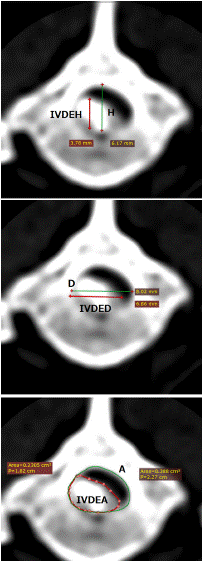
    Figure 1: Cross-sectional Computer tomography view. The height (H) of
the intervertebral foramen, IVDEH indicates the height of the intervertebral
foramen occupied by the ruptured disc materials and H% indicates the ratio
of IVDEH and H. D indicates the width of the intervertebral foramen, IVDED
indicates the width of the ruptured materials and D% indicates the ratio
of IVDED and D. A indicates the cross-sectional area of the intervertebral
foramen, IVDEA indicates the cross-sectional area of the intervertebral
foramen occupied by the ruptured material, and the proportion of compression
material occupying the intervertebral foramen is indicated by A%.
Statistical Analysis
Data in this study were collated and recorded using a Microsoft Excel 2013 (Microsoft Corporation, Redmond, WA, USA) spreadsheet. One-way ANOVA was performed using the software Prism X9 (Graph Pad Software, San Diego, USA), P < 0.05 was considered statistically significant, * means P<0.05, *** means P<0.001, and **** means P<0.0001.
Results
In the LHD group, grade 1 disease was present in 17 cases, grade 2 was present in 76 cases, grade 3 was present in 54 cases, grade 4 was present in 89 cases, and 28 cases were in grade 5; the total number of cases was 264. The average age, body weight, and Highest HU, H%, D%, and A% of compression materials of IVDE in the intervertebral foramina and their recovery periods after spinal cord surgeries in the LHD group of each neurological grade were shown in Table 2. The average age of those with grade 1 patients was 8.4±3.0 years, grade 2 was 8.2±2.8 years, grade 3 was 8.7±2.2 years, grade 4 was 8.0±2.9 years, and that of those with grade 5 patients was 6.6±3.0 years. Patients with grade 5 disease were younger than other grades, and a significant difference in the age of occurrence was found when comparing grade 3 (p<0.05).
![]()
    LHD
    Grade 1
    Grade 2
    Grade 3
    Grade 4
    Grade 5


    Numbers
    17
    76
    54
    89
    28


    Age (Years)
    8.4±3.0
    8.2±2.8
    8.7±2.2
    8.0±2.9
    6.6±3.0


    Weight (kg)
    7.25±1.8
    6.81±1.7
    6.82±1.5
    7.0±1.7
    7.01±1.4


    Highest HU
    293±300.1
    297±270.3
    209±182.5
    303±292.3
    425±234.1


    H%
    0.69±0.12
    0.71±0.15
    0.70±0.14
    0.758±0.14
    0.76±0.14


    D%
    0.47±0.18
    0.50±0.20
    0.43±0.17
    0.52±0.19
    0.64±0.18


    A%
    0.36±0.16
    0.46±0.39
    0.41±0.30
    0.44±0.18
    0.53±0.16


    Recovery days
    5.65±4.78
    7.57±7.32
    11.25±10.06
    15.34±12.42
    85.63±115.68
Table 2: Neurological grade by age, weight, Hounsfield unit (HU) value of
intervertebral disc material, and the proportion of intervertebral disc compression
to the height(H%), width (D%), and area (A%) of the intervertebral foramen,
and recovery period after spinal surgeries in LHDs. Patients of grade 5 were
significantly younger and had a longer recovery period than the patients of grade
3. The higher the HU value of the compression, the greater the compression ratio
was, but no significant difference was found in each grade.
The average body weight and Highest HU of IVDE of grade 1 patients in LHDs were 7.25±1.8 years and 293±300.1, grade 2 were 6.81±1.7 years and 297±270.3, grade 3 was 6.82±1.5 years and 209±182.5, grade 4 were 7.0±1.7 years and 303±292.3, and that of those with grade 5 patients were 7.01±1.4 years and 425±234.1, no significant differences found in each grade of patients.
![]()
    FB
    Grade 1
    Grade 2
    Grade 3
    Grade 4
    Grade 5


    Numbers
    7
    7
    4
    14
    9


    Age (Years)
    4.86±1.86
    5.29±3.68
    3.5±1.29
    4.71±1.77
    4.88±1.36


    Weight (kg)
    12.34±1.82
    11.74±1.68
    12.28±1.59
    12.33±2.67
    11.74±2.06


    Highest HU
    297±147.1
    223±142.0
    310±166.9
    271±150.0
    291±150.7


    H%
    0.68±0.24
    0.66±0.19
    0.64±0.14
    0.75±0.13
    0.68±0.16


    D%
    0.53±0.14
    0.46±0.15
    0.63±0.06
    0.57±0.18
    0.49±0.16


    A%
    0.31±0.12
    0.34±0.13
    0.38±0.07
    0.40±0.11
    0.38±0.09


    Recovery days
    3.83±1.47
    5.5±1.67
    5.25±5.85
    13±5.62
    44.75±43.28
Table 3: Neurological grade by age, weight, Hounsfield unit (HU) value of
intervertebral disc material, and the proportion of intervertebral disc compression
to the height(H%), width (D%), and area (A%) of the intervertebral foramen,
and recovery period after spinal surgeries in FBs. Patients of grade 5 were
significantly had a longer recovery period than the patients (P<0.05), but no
significant difference found in other indices.
![]()
    LHD
    FB


    Numbers (%)
    Grade
    Numbers (%)
    Grade


    T10–T11
    5 (1.9%)
    4.4±0.55


    T11–T12
    21 (8.0%)
    3.86±1.2
    1 (2.4%)
    2


    T12–T13
    43 (16.3)
    3.12±1.0
    2 (4.9%)
    4±0


    T13–L1
    67 (25.4%)
    3.39±1.1
    6 (14.6%)
    4.00±1.55


    L1–L2
    43 (16.3%)
    3.05±1.15
    9 (22.0%)
    2.89±1.45


    L2–L3
    38 (14.4%)
    3.05±0.96
    5 (12.2%)
    4.00±0.71


    L3–L4
    23 (8.7%)
    2.39±1.08
    10 (24.4%)
    3.50±1.43


    L4–L5
    6 (2. 3%)
    2.5±1.22
    6 (14.6%)
    2.33±1.51


    L5–L6
    2 (0.9%)
    2±0
    2 (4.8%)
    2.5.±2.12


    L7-S
    1 (0.4%)
    2


    Type II IVDD
    15 (5.7%)


    Gender


    Male
    143 (54.2%)
    34 (82.9%)


    Female
    121 (45.8%)
    7 (17.1%)


    Affected side


    Right side
    110 (41.7%)
    19 (46.3%)


    Left side
    120 (45.4%)
    21 (51.2%)


    Median
    34 (12.9%)
    1 (2.4%)


    Total
    264
    41
Table 4: Number of intervertebral disc disease (IVDD) cases in long-haired
dachshunds (LHDs) and French bulldogs (FBs) by male-to-female ratio and left
and right sides. LHDs were mostly affected in the thoracic spine, while FBs had
a higher incidence in the lumbar spine. The severity of thoracic disease in LHDs
was worse than that in the lumbar spine, but there was no such tendency in FBs.
There was no significant difference in the number between males and females
in LHDs, but IVDD was more affected by female FBs than male FBs (P<0.05).
There was no significant difference in the number of IVDD cases occurring on the
left and right sides in both LHDs and FBs.
H%, D%, and A% of compression material of IVDE in grade 1 patients of LHDs were 0.69±0.12, 0.47±0.18 and 0.36±0.16, those of grade 2 were 0.71±0.15, 0.50±0.20 and 0.46±0.39, grade 3 were 0.70±0.14, 0.43±0.17 and 0.41±0.3, grade 4 were 0.758±0.14, 0.52±0.19 and 0.44±0.18, and those with grade 5 patients were 0.76±0.14, 0.64±0.18 and 0.53±0.16 (Table 5). Patients with grade 5 disease had a larger ratio of D% and a significant difference was found when comparing with patients of grade 2 (**), grade 3 (****), and 4(*), no significant differences were found in H% and A% in each grade of patients (Figure 2).
![]()
    LHD

    FB


    H%
    D%
    A%


    H%
    D%
    A%


    Grade 1
    0.69±0.12
    0.47±0.18
    0.36±0.16

    Grade 1
    0.68±0.24
    0.53±0.14
    0.31±0.12


    Grade 2
    0.71±0.15
    0.50±0.20
    0.47±0.39

    Grade 2
    0.66±0.19
    0.46±0.15
    0.34±0.13


    Grade 3
    0.70±0.14
    0.43±0.17
    0.40±0.30

    Grade 3
    0.64±0.14
    0.63±0.06
    0.38±0.07


    Grade 4
    0.76±0.14
    0.52±0.19
    0.44±0.18

    Grade 4
    0.75±0.13
    0.57±0.18
    0.40±0.11


    Grade 5
    0.76±0.14
    0.64±0.18
    0.53±0.16

    Grade 5
    0.68±0.16
    0.49±0.16
    0.38±0.09
Table 5: The obtained indices in this study were as follows: H% was the ratio of the height of compression materials and the height of the intervertebral foramen in the same slice, D% was the ratio of the width of the materials and the width of the intervertebral foramen in the same slice, and A% was the ratio of the area of the ruptured materials and that of the intervertebral foramen in the same slice. Patients with grade 5 disease had a larger ratio of D% and a significant difference was found when comparing with patients of grade 2 (**), grade 3 (****), and 4(*) in long-haired dachshunds (LHDs), but no significant differences were found in H% and A% in each grade of patients in LHDs. No significant differences in indices were found in French bulldogs (FBs).
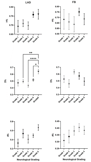
    Figure 2: Relationship between neurological grading and the ratio of the
height (H%), the width (D%), and the area (A%) of the ruptured materials
and the intervertebral foramen in the patients of long-haired dachshunds
(LHD) and French bulldogs (FB). The D% of grade 5 patients in LHDs was
significantly higher than that of grades 2(*), 3(***) and 4(*). However, there
was no significant difference found in the FBs group.
All patients in this study underwent non-contrast CT of the thoracolumbar spine, and spinal compression was confirmed by hemilaminectomy surgery. Among the cases, 249 of 264 LHDs (94.3%) had IVDE identified. Fifteen patients were finally confirmed to have type II disc herniation. The location of the ruptured materials in the spinal cord can be calculated and recorded if the HU value of the ruptured materials is above 50. When the HU value of the IVDE was less than 50, it was difficult to calculate the range of the compression area, and the data of IVDEH, IVDED, and IVDEA were excluded.
![]()
    Location
    H%
    H (mm)
    IVDEH
    D%
    D (mm)
    IVDED
    A (cm2)
    IVDEA
    A%


    T10–T11
    0.83±1.18
    4.66±0.75
    4.14±1.32
    0.63±0.20
    9.61±1.39
    4.96±1.62
    0.34±0.07
    0.19±0.10
    0.55±0.15


    T11–T12
    0.79±0.11
    4.83±0.73
    3.76±0.70
    0.68±0.17
    9.36±1.18
    5.94±1.78
    0.33±0.06
    0.2±0.08
    0.60±0.15


    T12–T13
    0.77±0.12
    4.75±0.71
    3.89±0.82
    0.59±0.18
    9.78±1.49
    5.52±1.81
    0.33±0.06
    0.18±0.09
    0.52±0.17


    T13–L1
    0.72±0.13
    4.47±0.67
    3.40±0.88
    0.51±0.19
    9.65±1.44
    4.58±1.99
    0.30±0.06
    0.14±0.08
    0.44±0.17


    L1–L2
    0.71±0.12
    4.55±0.59
    3.39±0.72
    0.45±0.18
    9.27±1.31
    4.04±1.79
    0.30±0.06
    0.11±0.06
    0.35±0.15


    L2–L3
    0.69±0.17
    4.76±0.66
    3.27±0.66
    0.41±0.16
    9.44±1.41
    3.89±1.29
    0.32±0.06
    0.10±0.05
    0.32±0.13


    L3–L4
    0.73±0.15
    4.91±0.60
    3.91±1.17
    0.43±0.13
    10.0±1.20
    4.30±1.65
    0.36±0.06
    0.17±0.16
    0.51±0.61


    L4–L5
    0.72±0.11
    5.32±0.63
    4.22±0.69
    0.45±0.15
    11.1±1.27
    5.51±2.15
    0.42±0.07
    0.16±0.08
    0.36±0.17


    L5–L6
    0.78±0.25
    4.87±0.74
    4.28±1.31
    0.58±0.20
    11.6±1.85
    7.33±4.62
    0.40±0.09
    0.30±0.26
    0.50±0.31
Table 6: The obtained indices in long-haired dachshunds(LHDs): height of ruptured materials of the ruptured intervertebral disc materials (IVDEH), the height of the vertebral foramen in the same slice (H), the ratio of IVDEH to H x 100% (H%), the width of the ruptured dick materials (IVDED), the width of the vertebral foramen in the same slice (D), ratio of IVDED to D x 100% (D%), area of the ruptured materials (IVDEA), area of the vertebral foramen in the same slice (A), and ratio of IVDEA to A x 100% (A %)of each intervertebral foramen. H, D, and A of thoracic spines were significantly narrower than those in lumbar spines in LHDs.
In those patients, additional imaging tests such as CT myelography or magnetic resonance imaging (MRI) may be needed. There were 32 cases in this group: 25 cases of IVDE were finally confirmed by surgery, 6 cases were type II disc herniation and in one case compression of the spinal cord was not found. The ability to regain independent walking after surgery is positively correlated with the level of paralysis before surgery. The recovery times in each grade of patients were 5.65±4.78 days in grade 1 patients, 7.57±8.02 days in grade 2, 11.25±10.06 days in grade 3, 15.34±12.42 days in grade 4, and 85.63±115.68 days in grade 5 patients. The recovery period is positively correlated to the body weight of patients and significantly longer for grade 5 paralysis before surgery, only 10 patients in total 28 dogs (35.7%) regained their ability to walk independently after surgery, and most of them underwent a spinal cord decompression surgery within 24 hours after paralysis. One patient regained walking without assistance after surgery but no DPP in hind limbs, and unable to control the urination was considered a spinal walk. One patient in grade 4 and 3 patients (13%) in grade 5 in this study followed by progressive myelomalacia and died a few days later. In LHDs, the most common location IVDE occurred in this study was T13–L1 in 67 cases (25.4%) and the average neurologic grading of patients was 3.39±1.1, those in T10–T11 were 5 (1.9%) cases and 4.4±0.55, in T11– T12 were 21 (8.0%) cases and 3.86±1.2, in T12–T13 were 43 cases (16.3%) and 3.12±1.0, in L1–L2 were 43 cases (16.3%) and 3.05±1.15, in L2–L3 were 38 (14.4%) cases and 3.05±0.96, in L3–L4 level was 23 (8.7%) cases and 2.39±1.0, in L4–L5 there were 6 (2.3%) cases and 2.5±1.22, in L5-L6 were 2 cases (0.9%) and 2 and only one patient occurred IVDE in L7-S (0.4%) with neurological grading of 2. In the LHD group, IVDE more frequently occurred in the thoracic spines and neurological grading was significantly more severe than IVDE occurred in the lumbar spine (Table 4).
![]()
    Location
    H%
    H (mm)
    IVDEH
    D%
    D (mm)
    IVDED
    A (cm2)
    A%
    IVDEA


    T12–T13
    0.84±0.16
    6.13±2.11
    4.99±0.81
    0.58±0.18
    6.13±2.11
    4.11±0.95
    0.37±0.08
    0.39±0.03
    0.14±0.03


    T13–L1
    0.71±0.17
    6.31±1.13
    4.52±1.48
    0.56±0.17
    6.31±1.13
    6.11±2.34
    0.53±0.18
    0.41±0.11
    0.22±0.12


    L1–L2
    0.74±0.08
    6.84±1.20
    5.11±1.16
    0.52±0.10
    6.84±1.20
    5.30±0.92
    0.51±0.11
    0.41±0.05
    0.21±0.07


    L2–L3
    0.64±0.18
    7.82±1.39
    4.85±0.92
    0.74±0.17
    7.82±1.39
    8.62±3.29
    0.64±0.16
    0.41±0.10
    0.26±0.07


    L3–L4
    0.72±0.17
    7.35±0.77
    5.34±1.58
    0.47±0.11
    7.35±0.77
    5.66±1.96
    0.59±0.11
    0.34±0.12
    0.20±0.09


    L4–L5
    0.66±0.25
    7.07±1.01
    4.62±1.81
    0.46±0.18
    7.07±1.01
    6.00±3.62
    0.7±0.23
    0.32±0.14
    0.22±0.16


    L5–L6
    0.52±0.01
    6.59±0.54
    3.42±0.31
    0.42±0.26
    6.59±0.54
    5.39±2.60
    0.56±0.07
    0.25±0.12
    0.13±0.04
Table 7: The obtained indices of French bulldogs (FBs): height of ruptured materials of the ruptured intervertebral disc materials (IVDEH), the height of the vertebral
foramen in the same slice (H), the ratio of IVDEH to H x 100% (H%), the width of the ruptured dick materials (IVDED), width of the vertebral foramen in the same
slice (D), ratio of IVDED to D x 100% (D%), area of the ruptured materials (IVDEA), area of the vertebral foramen in the same slice (A), and ratio of IVDEA to A x
100% (A %)of each intervertebral foramen. No significant differences in indices were found in FBs.
In the FB group, grade 1 disease was present in 7 cases, grade 2 in 7 cases, grade 3 in 4 cases, grade 4 in 14 cases, and 9 cases in grade 5 patients, the total number of cases was 41. The average age of grade 1 patients in the FB group was 4.86±1.86 years, grade 2 was 5.29±3.68 years, grade 3 was 3.5±1.29 years, grade 4 was 4.71±1.77 years, and that of those with grade 5 patients was 4.88±1.36 years, no significant difference was found between each grade. The average body weight and Highest HU of IVDE materials in grade 1 patients were 12.34±1.82 years and 297±147.1, grade 2 were 11.74±1.68 years and 223±142.0, grade 3 were 12.28±1.59 years and 310±166.9, grade 4 were 12.33±2.67 years and 271±150.0, and that of those with grade 5 patients were 11.74±2.06 years and 291±150.7. There were no significant differences found in each grade (Table 3). H%, D%, and A% of compression material of IVDE in grade 1 patients of the FB group were 0.68±0.24, 0.53±0.14 and 0.31±0.12, those of grade 2 were 0.66±0.19, 0.46±0.15 and 0.34±0.13, grade 3 were 0.64±0.14, 0.63±0.06 and 0.38±0.07, grade 4 were 0.75±0.13, 0.57±0.18 and 0.40±0.11, and those with grade 5 patients were 0.68±0.16, 0.49±0.16 and 0.38±0.09, no significant difference was found between each grade (Table 5). All patients of FBs in this study had IVDE identified during hemilaminectomy surgery.
The recovery times to regain independent walking ability after surgery in each grade of patients were 3.83±1.47 days in grade 1 patients, 5.5±1.67 days in grade 2, 5.25±5.85 days in grade 3, 13±5.62 days in grade 4, and 44.75±43.28 days in grade 5 patients (Table 3). The recovery period is significantly longer for grade 5 patients, 4 patients in total 9 dogs (44.4%) regained their ability to walk independently after surgery, but one patient followed progressive myelomalacia and died a few days later after the paralysis.
In FBs, the most common location IVDE occurred in this study was L3–L4 in 10 cases (24.4%) and the average neurologic grading of patients in this region were 3.5±1.43, no cases occurred in T10– T11, only one case (2.4%) in T11–T12 with a neurologic grading of 2, those in T12–T13 were 2 cases (4.9%) and 4±0, in L1–L2 were 9 cases (22.0%) and 2.89±1.45, in L2–L3 were 5 (12.2%) cases and 4±0.71, in L4–L5 there were 6 (14.6%) cases and 2.33±1.51, in L5-L6 were 2 cases (4.8%) and 2.5±1.51. In this study, IVDE more frequently occurred in the lumbar spines in the FB group, but there was no significant difference found in neurological grading between IVDE occurring in the lumbar spines or thoracic spines. Among all patients, 41.7% and 46.3% of IVDD affected the right side of the spinal cord in the LHD group and the FB group. 45.4% and 51.2% affected the left side of the intervertebral foramen. The results were similar in both groups, no significant difference was found. There were 143 male dogs (54.2%) and 121 female dogs (45.8%) in the LDH group, 34 male dogs (82.9%) and 7 females (17.1%) in the FB group, male patients were significantly more than females in the FBs group (Table 4).
Discussion
Paralysis often imposes a serious situation and cost for dogs and their owners, and was a reason for owners decided to euthanize their pets in Taiwan. According to past studies, the main causes of paralysis in animals include the compression of the spinal cord by IVDD, accidents, degenerative myelopathy, etc [9]. The main cause of acute paralysis in dogs is Hansen type I intervertebral disc herniation or IVDE, which particularly affects chondrodystrophic dog breeds2 including Dachshunds (miniature and standard), Fench Bulldogs, Shih-Tzus, Pekingese, Japanese Chins, Corgis, Bassett Hounds, Cocker Spaniels, and Beagles [10].
However, even in chondrodystrophic dog breeds, the prevalence of IVDE in LHDs is much higher than that in other dogs. It has been reported that the proportion of Dachshunds suffering from intervertebral disc herniation is approximately 15.7%, of which Standard Smooth-Haired Dachshunds account for approximately 24.4% of cases [11]. To reduce the occurrence of paralysis in Dachshunds, breeding candidates are determined by selecting dogs according to intervertebral disc calcification and by excluding highrisk dogs from breeding in some countries [12,13].
This study revealed that the location of IVDE may be significantly related to the severity of paralysis in LHDs, but in FBs this relationship was not clear. In LHDs, acute IVDE more frequently happened in thoracic spines than occurred in lumbar spines. T13–L1 was the most segment IVDE affected, and 136/264 (51.5%) cases happened in T10-L1 region. In FBs, IVDE more frequently happened in lumbar spines, with only 9/41(21.9%) cases in the T11-L1 region in this study. Neurological grading of IVDE happened in T10-T11 in LHDs was significantly higher than those at L3–L4 (**) and L4-L5 (*), those at T11–T12 was higher than that at L3–L4 (***) and L4–L5 (*), and that at T13–L1 was higher than that at L3–L4 (**) (Figure 3). However, this correlation was not significantly different in each spine of the FB group.
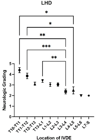
    Figure 3: In the long-haired dachshund (LHD) group, the average
neurological grade caused by intervertebral disc diseases in the thoracic
spine was higher than that in the lumbar spine. In particular, the neurological
grade caused by compression at T10–T11 was significantly higher than that
at L3–L4 (*) and L4-L5 (*), that at T11–T12 was higher than that at L3–L4
(**) and L4–L5 (*), and that at T13–L1 was higher than that at L3–L4 (*).
However, this correlation was not significantly different in the French bulldog
(FB) group
In a past study, the ratio of the spinal cord to the vertebral foramen in small breeds of dogs was higher than that in large dogs [8]. A higher ratio of the cross-sectional area of the ruptured materials in the vertebral foramen might associated with the severity of paralysis in dogs. However, there was no significant difference between the height of the ruptured materials in the intervertebral foramen and the neurologic grade of paralysis [7]. In this study, we measured the height of materials of the IVDE (IVDEH), the height of the vertebral foramen in the same slice (H), and their ratio them (H%). Width of the materials (IVDED), width of the vertebral foramen in the same slice (D), and ratio of IVDED to D x 100% (D%). Area of materials of the IVDE (IVDEA), area of the vertebral foramen in the same slice (A), and the ratio of IVDEA to A x 100% (A %) [7,8]. The results showed that only the proportion of the width (D%) of IVDE materials in intervertebral foramen in LHDs was significantly related to the clinical neurologic grading of paralysis (p<0.05) in this study, but in the FB group no significantly different was found (Figure 2). If the proportion of the compressed area in the intervertebral foramen was greater than 30%, the proportion of ruptured materials in the vertebral foramen was associated with the severity of paralysis (p<0.05).
We also measure the height (H), width (D), and cross-sectional area (A) of each intervertebral foramina of the thoracolumbar spines of LHDs and FBs. The values of H, D, and A of thoracic spines were significantly narrower than those in lumbar spines of LHDs, most narrow H and A were found in the T13-L1 segment, and most narrow D were found in L1-L2 in LHDs. However, no significant differences were found in the values of H, D, and A of the thoracic and lumbar spines of FBs (Figure 4).
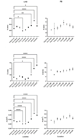
    Figure 4: Correlation of the location of intervertebral disc extrusion (IVDE)
affected and the characters of intervertebral discs in long-haired dachshunds
(LHDs) and French bulldogs (FBs). The values of H, D, and A indicated the
height (H), width (D), and area (A) of the intervertebral foramen of each spine,
H in T10-T11 was significantly different from T13-L1(*), L3-L4(**), L4-L5(***)
and L5-L6(*) in LHDs. D in T10-T11 was significantly different from L3-L4(*),
L4-L5(****) and L5-L6(****) and A in T10-T11 was significantly different from
T13-L1(****), L1-L2(****), L3-L4(*), L4-L5(****) and L5-L6(****)in LHDs. No
significant difference was found in H, D, and A of each spine in FBs
LDHs tended to have spinal cord compression in the thoracic spine, in contrast, fore-limbs paralysis in FBs tended to be concentrated with the lumbar spine in this study. IVDE occurred in T10-T11 causing the most severe damage to the spinal cord and causing higher grading of paralysis in LHD (Table 4). We compared the value of H, D, and A in T10-T11 intervertebral foramen with other spines of LHDs and FBs, H in T10-T11 was significantly different from T13-L1(*), L3-L4(**), L4-L5(***) and L5-L6(*) in LHDs. D in T10-T11 was significantly different from L3-L4(*), L4-L5(****) and L5-L6(****) and A in T10-T11 was significantly different from T13-L1(****), L1-L2(****), L3-L4(*), L4-L5(****) and L5-L6(****)in LHDs (Figure 4). Because in this study we found a higher D% might lead to a higher neurologic grading of lameness, a narrow D in thoracic spines might be a reason why IVDE happened in this region with more severe damage to the spinal cord when the intervertebral discs ruptured suddenly in LHDs. The dynamics of intervertebral discs ruptured at each segment and the degree to which spinal cord damage was affected require further study. No significant difference was found in H, D, and A of each spine in FBs might be the same reason that no significant difference in neurologic grading of IVDE happened in thoracic and lumbar spines for FBs.
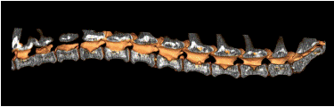
    Figure 5: The picture shows the cross-sectional view of the spine of a longhaired
dachshund after the hemilaminectomy surgery. The intervertebral
foramina of thoracic vertebrae was narrower than the lumbar vertebrae,
an intervertebral disc extrusion affected T11-T13, and a spinal cord
decompression surgery was performed.
Some papers have reported that the rate of myelomalacia after grade 5 paralysis was 33% in FBs [4]. However, in this study, there were 28 LHDs with grade 5 paralysis, and 3 (10.3%) of them developed myelomalacia and died within 1 week after paralysis. In addition, of 9 FBs with grade 5 paralysis, 1 (11.1%) died after myelomalacia occurred. The lower occurrence of myelomalacia in this study may be due to the performance of durotomy after spinal decompression surgery, which significantly reduces the occurrence of myelomalacia [14,15].
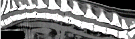
    Figure 6: In the study using computer tomography, the following anomalies
were found in a French bulldog: 1. Congenital hemivertebra 2. Calcification
of intervertebral discs 3. Protrusion and compression of the spinal cord due
to an intervertebral disc 4. A cavity in the center of an intervertebral disc 5.
Extrusion of an intervertebral disc and compression of the spinal cord.
The size of the body, body condition score, and ratio of the length and height of Dachshunds may increase the risk of IVDE [16,17]. The ratio of the cross-sectional area of the spinal cord to that of the vertebral foramen (CFAR) based on MRI has been reported, and the CFAR in the thoracic region is lower than that in the lumbar region in healthy dogs. However, in patients with spinal disease, the CFAR in the thoracic spine is similar to that in the lumbar region [18]. Vertical motions such as jumping or climbing may increase the stress on the intervertebral discs of the thoracic spine compared with those in the lumbar region. Instant acute compression of the spinal cord may be more serious in narrow areas such as the thoracic region.
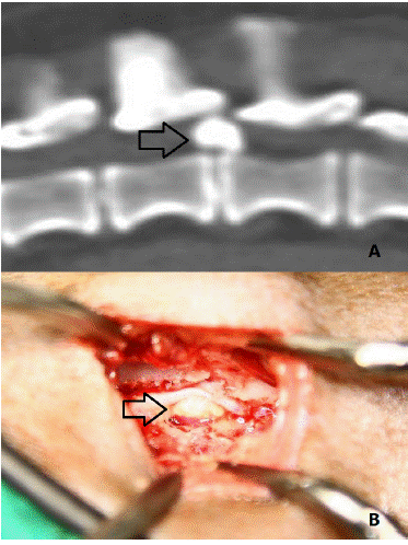
    Figure 7: Computed tomography showing the location of spinal cord
compression (arrow) in a patient with acute paralysis caused by
intervertebral disc extrusion (A) and what was observed during surgery (B)
In this study, T13–L1 was also the most prone to grade 5 disease in LHDs, accounting for 39.3% of cases (11/28), which is similar to the findings of previous reports [19]. In T12-T13 for 3 cases, in T11-T12 for 7 cases, and T10-T11 for 2 cases, the T11–L1 level was affected in 82.1% (23/28) of cases with grade 5 disease in this study, was significantly higher than the prevalence of IVDE occurred in the region, a narrow intervertebral foramen than those in the lumbar region might be the reason. Grade 5 disease may be secondary to myelomalacia, which is a more serious situation [20,21]. Early surgery in patients with grade 5 disease may provide a higher recovery rate and decrease the risk of myelomalacia [22]. Although many FBs have thoracic spinal malformations such as vertebral aplasia and hypoplasia or hemivertebra, these malformations are usually not the cause of acute paralysis [4,5]. Kyphosis of the thoracic spine may increase the risk of IVDE in FBs [6]. We found that the incidence of the lumbar spinal IVDE was higher than that of thoracic spinal IVDE in FBs, with 32 of 41 (78.0%) cases of acute spinal compression occurring at L1–L6. Notably, the lumbar spine is less likely to have congenital vertebral abnormalities than the thoracic spine [23,24]. Kyphosis of the spines might not increase the prevalence of acute IVDE and spinal cord damage in FBs.
Several methods can be used to detect acute paralysis caused by IVDE: myelography, CT, and MRI. Previous studies have shown that myelography has detection rates of 78.9%, 83.6%, and 53–74%, and CT has detection rates of 85.3%, 81.8%, 81–94%, and 88.6% [19,25,26], and MRI is 98.5% accurate for detecting IVDE [27]. Due to the risk of epilepsy with myelography [28-30] and the longer anesthesia time with MRI, CT has the advantage of being a faster and safer method to diagnose IVDE.
The HU value of the spinal cord on CT is approximately 4–55 HU (mean attenuation: 31.3±8.6 HU; range: 4–55 HU), and that of herniated disc material in previous studies was 219±95 HU (range: 104–407 HU) [31]. In this study, the ruptured materials in the vertebral foramen were diagnosed by CT using an HU value greater than 50, and 232 cases of 264 (87.8%) LHDs in this study were diagnosed using non-contrast CT, which is similar to other reports, but 40/41(97.6%) cases in FBs their HU value of IVDE materials were greater than 50. There were 32 (12.1%) of 264 patients in LHDs and one patient (2.4%) in FBs with an HU value less than 50 that could not be diagnosed with non-contrast CT. After decompression surgeries, 25 (78.1%) cases of them in LHDs were finally diagnosed with IVDE, and 7 cases were Hansen type II intervertebral disc herniation. In one case with grade 5 paralysis, no obvious abnormality was identified at surgery. Ultimately, this dog died because of respiratory failure after being paralyzed for several days due to myelomalacia. Only one patient in FBs with an HU value less than 50 was confirmed as acute IVDE during spinal surgery. After decompression surgeries in this study, 257/264 (97.3%) cases of LHDs and 41/41(100%) in this study IVDE materials were confirmed and removed during the surgeries.
In this study, most LHDs and FBs with acute paralysis had disease caused by IVDE, higher HU values are known to be significantly associated with clinical duration and may represent chronic cases [31,32]. But in this study, no significant difference was found between the HU value of materials of IVDE of each neurologic grading of paralysis patients both in LHDs and FBs. In this study, the HU value of the ruptured materials was higher than 765, the higher HU value corresponded to a lower neurologic grade (p<0.05), and higher HU values would not lead to more severe damage to the spinal cord in this study.
There are significant differences in body weight among Dachshunds of different breeds [11], and there was no significant correlation between the weights of LHDs and the neurologic grade of paralysis in this study (p>0.05); similar results have previously been reported [33]. However, there was a significant correlation between body weight and recovery period (p=0.008), with heavier LHDs having a longer recovery period in this study.
The recovery situation was evaluated after surgery, and independent walking ability was the evaluation standard. Patients with higher neurologic grading of paralysis, need a longer recovery period after spinal surgeries. In LHDs, All 17 patients (100%) with grade 1 paralysis, all 76 patients with grade 2 disease, 53 of 54 (98.1%) patients with grade 3 disease, and 82 of 89 (92.1%) patients with grade 4 disease regained their ability to walk within 28 days of surgery. four patients in grade 4 recovered their ability to walk 31–90 days after surgery but one patient died of progressive myelomalacia. The average recovery period in each grade of patients was 3.83±1.47 days in grade 1 patients, 5.5±1.67 days in grade 2, 5.25±5.85 days in grade 3, 13±5.62 days in grade 4, similar results were found in FBs.
Grade 5 patients had a lower recovery rate and longer recovery period than other grades of patients. Only 10 patients in total 28 dogs (35.7%) of grade 5 patients in LHDs regained their ability to walk independently after surgery, and the recovery period was 85.63±115.68 days. In FBs, 4 patients in 8 dogs (50%) recovered their walking ability in 44.75±43.28 days. One patient of grade 5 cases regained walking without assistance 2 months after spinal surgery, but no DPP of hind limbs, and still unable to control the urination was considered as spinal walk, some reports discussed this situation, but still unclear.
Studies have shown that surgery within the same day may reduce the possibility of DDP loss [22]. The earlier a dog receives treatment for IVDE, the better recovery rate and faster recovery time can be expected. However, in clinical practice, we believe that decompressive surgery should be performed within a few hours of paralysis to have the best chances of recovery from grade 5 paralysis. There were 28 patients in LHDs and 8 patients in FBs with grade 5 paralysis; of these, 3 LHDs (10.7%) and one FBs (12.5%) died within the first week after paralysis because of progressive myelomalacia. Myelomalacia is the worst outcome for neurological deterioration [20], and the rate of myelomalacia secondary to grade 5 paralysis was approximately 10.7% in LHDs and 12.5% in FBs in this study, similar to the findings of a previous study [35].
This study demonstrated that SCI with acute paralysis in LHDs may be related to age, the location of IVDE, and the height, width, and cross-section area of the vertebral foramen and ruptured materials. We used the same method to analyze the data of FBs and other chondrodystrophic breeds in dogs, but the correlations between them were not obvious. Therefore, the predisposition to IVDE in LDHs was higher than that among other chondrodystrophic breeds. More recently, serum concentrations of glial fibrillary acidic protein (GFAP), phosphorylated neurofilament heavy chain (pNFH), and serum S100Β protein levels to assess the severity of paralysis in dogs and their association with subsequent prognosis are being investigated [21,36]. A limitation of this study was that we knew that the difference in the height and cross-sectional area of the intervertebral foramen between the thoracic and lumbar vertebrae of LHDs may lead to different grades of neuropathy in IVDE; however, we could not quantify this relationship and predict where IVDE may occur and prevent it in advance.
According to a previous study, it has also been suggested that colder environmental temperatures may increase the rates of acute IVDE [34]. The peak months of IVDE in this study were February, March, and May, which are the seasons of climate change in Taiwan. There was no significant difference in the number of patients with IVDE between the summer and winter seasons. (p>0.05). It may be because the weather in Taiwan is relatively hot, even in winter.
Conclusion
We found that IVDE in LHDs was more likely to occur in the thoracic spine and that the severity of paralysis was significantly worse than that in the lumbar area. These findings may be due to the smaller cross-sectional area, height, and width of the intervertebral foramina where IVDE occurred. However, this association was not significantly different among FBs, which are also chondrodystrophic dogs.
Author Contributions
Jenshawn Wang DVM1: Study design, data interpretation, performed surgical procedure, conducted statistical analysis, and drafted and revised the manuscript.
Yi-Chun Lin BS1: Recoded the data and assisted in performing surgical procedures.
Pei-Yu Kao DVM1: Data acquisition and performed surgical procedure.
Ming-Hsien Chiang DVM, PhD2,3: Study design, data interpretation, conducted statistical analysis, and revised the manuscript.
References
- Rossi G, Stachel A, Lynch AM, et al. Intervertebral disc disease and aortic thromboembolism are the most common causes of acute paralysis in dogs and cats presenting to an emergency clinic. Vet Rec. 2020; 187: e81.
- Hansen HJ. A pathologic-anatomical study on disc degeneration in dogs, with special reference to the so-called enchondrosis intervertebralis. Acta Orthop Scand Suppl. 1952; 11: 1-117.
- Thompson K, Moore S, Tang S, et al. The chondrodystrophic dog: a clinically relevant intermediate-sized animal model for the study of intervertebral discassociated spinal pain. JOR Spine. 2018; 1: e1011.
- Aikawa T, Shibata M, Asano M, et al. A comparison of thoracolumbar intervertebral disc extrusion in French Bulldogs and Dachshunds and association with congenital vertebral anomalies. Vet Surg 2014; 43: 301-307.
- De Decker S, Packer RM, Cappello R, et al. Comparison of signalment and computed tomography findings in French Bulldogs, Pugs, and English Bulldogs with and without clinical signs associated with thoracic hemivertebra. J Vet Intern Med. 2019; 33: 2151-2159.
- Inglez de Souza M, Ryan R, Ter Haar G, et al. Evaluation of the influence of kyphosis and scoliosis on intervertebral disc extrusion in French bulldogs. BMC Vet Res. 2018; 14: 5.
- Lim C, Kweon OK, Choi MC, et al. Computed tomographic characteristics of acute thoracolumbar intervertebral disc disease in dogs. J Vet Sci. 2010; 11: 73-79.
- Seo E, Choi J, Choi M, et al. Computed tomographic evaluation of cervical vertebral canal and spinal cord morphometry in normal dogs. J Vet Sci. 2014; 15: 187-193.
- Shelton GD, Johnson GC, O’Brien DP, et al. Degenerative myelopathy associated with a missense mutation in the superoxide dismutase 1 (SOD1) gene progresses to peripheral neuropathy in Pembroke Welsh corgis and boxers. J Neurol Sci. 2012; 318: 55-64.
- Martínez S, Fajardo R, Valdés J, et al. Histopathologic study of long-bone growth plates confirms the basset hound as an osteochondrodysplastic breed. Can J Vet Res. 2007; 71: 66-69.
- Packer RM, Seath IJ, O’Neill DG, et al. DachsLife 2015: an investigation of lifestyle associations with the risk of intervertebral disc disease in Dachshunds. Canine Genet Epidemiol. 2016; 3: 8.
- Rosenblatt AJ, Bottema CD, Hill PB. Radiographic scoring for intervertebral disc calcification in the Dachshund. Vet J. 2014; 200: 355-361.
- Rosenblatt AJ, Hill PB, Davies SE, et al. Precision of spinal radiographs as a screening test for intervertebral disc calcification in Dachshunds. Prev Vet Med. 2015; 122: 164-173.
- Hirano R, Asahina R, Hirano T, et al. Outcomes of extensive hemilaminectomy with durotomy on dogs with presumptive progressive myelomalacia: a retrospective study on 34 cases. BMC Vet Res. 2020; 16: 476.
- Nakamoto Y, Uemura T, Hasegawa H, et al. Outcomes of dogs with progressive myelomalacia treated with hemilaminectomy or with extensive hemilaminectomy and durotomy. Vet Surg. 2021; 50: 81-88.
- Packer RM, Hendricks A, Volk HA, et al. How long and low can you go? Effect of conformation on the risk of thoracolumbar intervertebral disc extrusion in domestic dogs. PLoS One. 2013; 8: e69650.
- Immekeppel A, Rupp S, Demierre S, et al. Investigation of timing of surgery and other factors possibly influencing outcome in dogs with acute thoracolumbar disc extrusion: a retrospective study of 1501 cases. Acta Vet Scand. 2021; 63: 30.
- Lim, J, Yoon Y, Hwang T, et al. Novel vertebral computed tomography indices in normal and spinal disorder dogs. J Vet Sci. 2018; 19: 296-300.
- Hecht S, Thomas WB, Marioni-Henry KA, et al. Myelography vs. computed tomography in the evaluation of acute thoracolumbar intervertebral disk extrusion in chondrodystrophic dogs. Vet Radiol Ultrasound. 2009; 50: 353- 359.
- Castel A, Olby NJ, Ru H, et al. Risk factors associated with progressive myelomalacia in dogs with complete sensorimotor loss following intervertebral disc extrusion: a retrospective case-control study. BMC Vet Res. 2019; 15: 433.
- Castel A, Olby NJ, Mariani CL, et al. Clinical characteristics of dogs with progressive myelomalacia following acute intervertebral disc extrusion. J Vet Intern Med. 2017; 31: 1782-1789.
- Martin S, Liebel FX, Fadda A, et al. Same-day surgery may reduce the risk of losing pain perception in dogs with thoracolumbar disc extrusion. J Small Anim Pract. 2020; 61: 442-448.
- Lackmann F, Forterre F, Brunnberg L, et al. Epidemiological study of congenital malformations of the vertebral column in French bulldogs, English bulldogs, and pugs. Vet Rec. 2022; 190: e509.
- Brown JD, Podadera J, Ward M, et al. The presence, morphology, and clinical significance of vertebral body malformations in an Australian population of French Bulldogs and Pugs. Aust Vet J. 2021; 99: 378-387.
- Israel SK, Levin SM, Kerwin SC, et al. The relative sensitivity of computed tomography and myelography for identification of thoracolumbar intervertebral disk herniations in dogs. Vet Radiol Ultrasound. 2009; 50: 247-252.
- Newcomb B, Arble J, Rochat M, et al. Comparison of computed tomography and myelography to a reference standard of computed tomographic myelography for evaluation of dogs with intervertebral disc disease. Vet Surg. 2012; 41: 207-214.
- Cooper JJ, Young BD, Griffin IV JF, et al. Comparison between noncontrast computed tomography and magnetic resonance imaging for detection and characterization of thoracolumbar myelopathy caused by intervertebral disk herniation in dogs. Vet Radiol Ultrasound. 2014; 55: 182-189.
- Lewis DD, Hosgood G. Complications associated with the use of iohexol for myelography of the cervical vertebral column in dogs: 66 cases (1988-1990). J Am Vet Med Assoc. 1992; 200: 1381-1384.
- Dennison SE, Drees R, Rylander H, et al. Evaluation of different computed tomography techniques and myelography for the diagnosis of acute canine myelopathy. Vet Radiol Ultrasound. 2010; 51: 254-258.
- Barone G, Ziemer LS, Shofer FS, et al. Risk factors associated with development of seizures after use of iohexol for myelography in dogs: 182 cases (1998). J Am Vet Med Assoc. 2002; 220: 1499-502.
- Olby NJ, Müntana KR, Sharp NJ, et al. The computed tomographic appearance of acute thoracolumbar intervertebral disc herniations in dogs. Vet Radiol Ultrasound. 2000; 41: 396-402.
- Kuroki, K, Vitale CL, Essman SC, et al. Computed tomographic and histological findings of Hansen type I intervertebral disc herniation in dogs. Vet Comp Orthop Traumatol. 2013; 26: 379-384.
- Klesty A, Forterre F, Bolln G. Outcome of intervertebral disk disease surgery depending on dog breed, location and experience of the surgeon: 1113 cases. Tierarztl Prax Ausg K Kleintiere Heimtiere. 2019; 47: 233-241.
- Barandun MA, Bult S, Demierre S, et al. Colder ambient temperatures influence acute onset canine intervertebral disc extrusion. Front Vet Sci. 2020; 7: 175.
- Olby N, Levine J, Harris T, et al. Long-term functional outcome of dogs with severe injuries of the thoracolumbar spinal cord: 87 cases (1996-2001). J Am Vet Med Assoc. 2003; 222: 762-769.
- Olby NJ, Lim JH, Wagner N, et al. Time course and prognostic value of serum GFAP, pNFH, and S100ß concentrations in dogs with complete spinal cord injury because of intervertebral disc extrusion. J Vet Intern Med. 2019; 33: 726-734.