Introduction
Soccer is a popular team sport with 14% incidence of back pain. The most common cause of low back pain in soccer players is spondylolysis [1]. In some cases where low back pain is persisting for longer than two weeks, young patients with history of athletic activity should be considered for spondylolysis. During the treatment of spondylolysis in adolescent players requires monitoring of pain and obtaining plain radiographs. Even with appropriate examination and X-ray imaging, one can easily overlook spondylolysis. Spondylolysis should be kept in mind as a diagnosis in adolescent athletes with low back pain and advanced radiographic imaging as evaluation method should be considered [2].
Herein, we report a case of spondylolysis an adolescent soccer player with low back pain persisting longer than two weeks, with no history of trauma. In order to remind that spondylolysis might have been missed in young patients, where immature spine and is more susceptible to injury.
Case Presentation
A 14-year-old professional soccer player applied to sports medicine department with complain of low back pain, which begun two weeks before. He hasn’t reported any previous back trauma or pain for the last year. The patient experienced back pain after increased workouts with trainings. He experienced dull low back pain, aggravated during extension, without presence of radicular sings. He had no history of previous treatments or using medication.
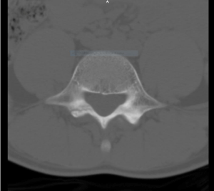
Clinical examination revealed pain with forced lumbar extension, bilateral paravertebral muscle tenderness at the lumbar area during palpation and normal neurological examination of extremities. As diagnostic tools, plain radiographs and then magnetic resonance imaging (MRI) were preceded. MRI revealed spondylolysis of the bilateral L4 pars interarticularis without lysthesis (Figure 1). Patient’s conservative treatment consisted of advising to use nonsteroidal antiinflammatory drugs (NSAIDs), not to participate in high impacted sports activities and also he was placed in lumbosacral orthosis for 4 weeks.

Figure 1:
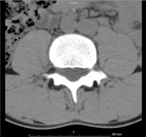
At the 4-week follow-up, the patient’s symptoms significantly decreased with rest and medication. During physical examination he had a minimal pain in lumbar extension. Repeated MRI scan showed a hypo dense line at the level of bilateral L4 pars interarticularis in favor of the lysis defect (Figure 2). The patient was advised core strengthening and stretching exercises after his pain subsided significantly. By the end of the fifth week, the patient was allowed to start mild running activities and the lumbosacral orthosis was removed. At the 8-week follow-up, patient experienced no pain during clinical examination and returned to his previous activity level without any complains.

Figure 2:
Discussion
Low back pain is the most commonly seen complain, particularly in adolescent athletes in 10-15% of cases and spondylolysis is a common diagnosis of low back pain with the presence of radiographic abnormalities [3,4]. Spondylolysis is commonly seen in football, gymnastics, swimmers, divers, weight-lifters, track and field athletes, soccer and volleyball players. An etiological reason that may cause spondylolysis is usually divided into developmental and acquired, in which the former is a genetic predisposition to failure of pars interarticularis and acquired spondylolysis usually occurs from acute or repetitive trauma [5]. Acute trauma occurs most often in contact sports such as football, rugby, and hockey, whereas overuse injuries occur most often in sports with repetitive flexion, extension, and torsion, such as gymnastics, dance, and figure skating. A history of menstrual irregularities, disordered eating, and previous stress fractures may indicate the presence of female athlete triad, which may predispose an athlete to stress fractures [6]. There are some risk factors were reported for spondylolysis, such as Scheuermann’s disease, excessive lordosis, cerebral palsy or spinal bifida and others [6].
Spondylolysis usually occurs at L4-L5 level of vertebra [7]. Some studies suggesting that pars interarticularis of L5 is sheared during extension by the inferior articular process of L4 and the superior articular process of the sacrum acting as a pair of wedges, therefore this mechanism leads to stretching of the pars and eventually to a stress micro fracture [5]. Some other studies report that, due to the sacral angle and the inferior facet of L5 facing anterior with the superior facet of S1 facing posterior causes a large anterior shear on the L5 pars interarticularis. Individuals who stand with an excessive anterior pelvic tilt will greatly increase the anterior shear at the L5 level, for that reason significantly increasing the risk for spondylolysis at L5. The anterior shear along with a skeletal immaturity of young athlete gives a root of making spondylolysis seen more common in the adolescent population [8].
According to study of Micheli and Wood, where 100 adolescent athletes with low back pain were diagnosed, have found that 47% of them have a spondylolysis. The same findings of spondylolysis were detected only in 5% of adults. Therefore authors concluded that for early diagnosis of spondylolysis, physicians should be more precise when diagnosing a low back pain in young patients in compare with adults [9].
The most common complaint in case of spondylolysis is low back pain. The pain typically localized at lumbar area, can be unilateral or bilateral and the level may vary from dull to sharp aggravating with extension, or pain may radiate to the buttock or posterior thigh. The onset can be sudden due to acute trauma or gradual caused by repetitive stress in the posterior elements of the lumbar spine [10]. In some cases radicular symptoms, or pain during the night can be caused by different diagnosis. The differential diagnosis of spondylolysis includes lumbar facet syndrome, spondylolythesis, mechanical low back pain, traumatic fractures and others [4].
Physical examination of the patient includes inspection of coronal and saggital alignment of the lumbar spine, gait assessment and neurovascular examination [3]. Some studies reported that one-legged hyperextension test is pathognomonic to differentiate spondylolysis [11,12]. This test should produce pain on the side of the standing leg in a patient with a symptomatic ipsilateral spondylolytic lesion. However some other researchers claimed that one-legged hyperextension test has no value in diagnosing patients with spondylolysis [13,14]. In addition to the aforementioned to specify and exclude excessive hamstring tightness assessment of the popliteal angle should be performed [15].
Radiographic imaging remains the essential diagnostic tool in assessment of spondylolysis. In most studies and literature the typical “Scotty dog” appearance on oblique view of X-ray is used as initial radiographic instrument [16,17]. Nevertheless some researchers state that spondylolysis in early stages can be negative on plain radiographs. Therefore further radiological imaging may be conducted, if pain is persisting.
Furthermore, radiological imaging computed tomography (CT) may be considered [18]. CT can visualize bony morphology and identify occult fractures. Authors in one study declared that axial CT scans are more precise than oblique plain radiographs [19]. There are no golden standards in choosing radiographic imaging. Single proton emission computed tomography (SPECT) is also preferred as a diagnostic evaluation of spondylolysis [20]. In some cases spondylolysis is asymptomatic, therefore it is essential to verify whether lesions are active or not. On SPECT scans we are able to evaluate stress reactions or sub acute pars injuries, when it’s not seen on X-ray, due to ability to differentiate symptomatic (“hot scan”) from silent (“cold scan”) of spondylolysis [21].
Also MRI is considered as a next step assessment for spondylolysis in athletes [22]. In study where young athletes participating in different sport-related activities, were examined with low back pain complain. 48.5% of athletes showed active spondylolysis on MRI, those that have been missed on X-ray scan [22]. On the other hand, authors of the other study were comparing sensitivity of MRI using SPECT as a gold standard. MRI detected bone stress in 40 out of 50 of the pars interarticularis, in which it was detected by SPECT respectively. Therefore, they stated that despite the advantages of MRI in the form of lack of radiation, SPECT should remain the gold standard for first-line investigation of spondylolysis [14].
Based on the literature most of low-grade spondylolysis treats conservatively. There are some recommendations in treatment protocol, such as resting period, eliminating from sports activities, using non steroidal anti-inflammatory drugs (NSAIDs) for pain relieve [23,24]. Some studies suggest using lumbosacral support that minimize lumbar lordosis, in period of time from 3 to 6 month, which depends on patient’s complains [25,26,1]. Preferred conservative treatment for 58 soccer players who had diagnosis of spondylolysis. They stated that players who had rested from sports activity and wore braces for at least 3 month showed better results compared to players who were not braced and proceeded just to resting period [1]. However, other study stated that conservative treatment with or without orthosis showed positive results in terms of return to play [27].
Also rehabilitation program including dynamic core strengthening exercises, stretching program for tight hip flexors and hamstring are advised [5,28]. One study specifically focused on strengthening of the transverse abdominis, internal oblique, and lumbar multifidus muscles [29]. Hypothesized that muscles that directly attach to the lumbar vertebra have more direct effects on stabilizing it. Authors found a significant improvement in pain and function in comparison with those who had not strengthened that group of muscles [29].
There are limited studies of effectiveness where surgical treatment was chosen in favor of conservative treatment. Nonetheless, the adult athletes and those who failed a conservative treatment, surgical treatment must be considered [30,31,32].
Some physicians recommended optimizing the clinical outcome and taking a 3-month break from sports. A course of physical therapy is essential to ensure proper muscle balance and core strength to prevent future injuries. In rehabilitation regimen, the patients’ goals are to regain full muscle strength and gradually return to full daily and sports activity, without experiencing any pain or complains on the site of the injury. Patients who are asymptomatic without any neurologic deficits can return to sports [1].
Conclusion
We recommend that physicians in sports medicine or in primary care consider obtaining radiographic images with respect to young athletes, particularly the ones who are engaged in contact sports and experience low back pain lasting over two weeks with or without any history of trauma.
References
- El Rassi G, Takemitsu M, Woratanarat P, Shah SA. Lumbar spondylolysis in pediatric and adolescent soccer players. Am J Sports Med. 2005; 33: 1688- 1693.
- Nitta A, Goda Y, Takata Y. Prevalence of Symptomatic Lumbar Spondylolysis in Pediatric Patients. 2016; 39: 434-437.
- Purcell L, Micheli L. Low Back Pain in Young Athletes. Sports Health. 2009; 1: 212-222.
- Miller MD, Thompson SR, Drez D. DeLee & Drez’s Orthopaedic Sports Medicine : Principles and Practice.
- Lawrence KJ, Elser T, Stromberg R. Lumbar spondylolysis in the adolescent athlete. Phys Ther Sport. 2016; 20: 56-60.
- Micheli LJ. Encyclopedia of Sports Medicine. SAGE Publications. 2010.
- Pascal-Moussellard H, Broizat M, Cursolles J-C, Rouvillain J-L, Catonné Y. Association of Unilateral Isthmic Spondylolysis With Lamina Fracture in an Athlete: Case Report and Literature Review. Am J Sports Med. 2005; 33: 591-595
- Leone A, Cianfoni A, Cerase A, Magarelli N, Bonomo L. Lumbar spondylolysis: a review. Skeletal Radiol. 2011; 40: 683-700.
- Micheli LJ, Wood R. Back pain in young athletes. Significant differences from adults in causes and patterns. Arch Pediatr Adolesc Med. 1995; 149: 15-18.
- Tallarico RA, Madom IA, Palumbo MA. Spondylolysis and Spondylolisthesis in the Athlete. Sports Med Arthrosc. 2008; 16: 32-38.
- Syrmou E, Tsitsopoulos PP, Marinopoulos D, Tsonidis C, Anagnostopoulos I, Tsitsopoulos PD. Spondylolysis: a review and reappraisal. Hippokratia. 2010; 14: 17-21.
- Jackson DW, Wiltse LL, Dingeman RD, Hayes M. Stress reactions involving the pars interarticularis in young athletes. Am J Sports Med.1981; 9: 304-312.
- Alqarni AM, Schneiders AG, Cook CE, Hendrick PA. Clinical tests to diagnose lumbar spondylolysis and spondylolisthesis: A systematic review. Phys Ther Sport. 2015; 16: 268-275.
- Masci L, Pike J, Malara F, Phillips B, Bennell K, Brukner P. Use of the onelegged hyperextension test and magnetic resonance imaging in the diagnosis of active spondylolysis. Br J Sports Med. 2006; 40: 940-946.
- Kim HJ, Green DW. Adolescent back pain. Curr Opin Pediatr. 2008; 20: 37- 45.
- Standaert CJ, Herring SA. Spondylolysis: a critical review. Br J Sports Med. 2000; 34: 415-422.
- Brukner P, Khan K. Clinical Sports Medicine. McGraw-Hill; 2007.
- Delavan JA, Stence N V, Mirsky DM, Gralla J, Fadell MF. Confidence in Assessment of Lumbar Spondylolysis Using Three-Dimensional Volumetric T2-Weighted MRI Compared With Limited Field of View, Decreased-Dose CT. Sports Health. 2016; 8: 364-371.
- Saifuddin A, White J, Tucker S, Taylor BA. Orientation of lumbar pars defects: implications for radiological detection and surgical management. J Bone Joint Surg Br. 1998; 80: 208-211.
- Takemitsu M, El Rassi G, Woratanarat P, Shah SA. Low Back Pain in Pediatric Athletes With Unilateral Tracer Uptake at the Pars Interarticularis on Single Photon Emission Computed Tomography. Spine. 2006; 31: 909-914.
- McCleary MD, Congeni JA. Current concepts in the diagnosis and treatment of spondylolysis in young athletes. Curr Sports Med Rep. 2007; 6: 62-66.
- Kobayashi A, Kobayashi T, Kato K, Higuchi H, Takagishi K. Diagnosis of Radiographically Occult Lumbar Spondylolysis in Young Athletes by Magnetic Resonance Imaging. Am J Sports Med. 2013; 41: 169-176.
- Attiah MA, Macyszyn L, Cahill PJ. Management of spondylolysis and spondylolisthesis in the pediatric population: A review. Semin Spine Surg. 2014; 26: 230-237.
- Sakai T, Tezuka F, Yamashita K, et al. Conservative Treatment for Bony Healing in Pediatric Lumbar Spondylolysis. Spine. 2016.
- Logroscino G, Mazza O, Aulisa A, Pitta L, Pola E, Aulisa L. Spondylolysis and spondylolisthesis in the pediatric and adolescent population. Child’s Nerv Syst. 2001; 17: 644-655.
- Sairyo K, Sakai T, Yasui N, Dezawa A. Conservative treatment for pediatric lumbar spondylolysis to achieve bone healing using a hard brace: what type and how long? J Neurosurg Spine. 2012; 16: 610-614.
- Lvarez-Díaz P, Alentorn-Geli E, Steinbacher G, et al. Conservative treatment of lumbar spondylolysis in young soccer players. 2011; 19: 2111-2114.
- Standaert CJ, Herring SA. Expert Opinion and Controversies in Sports and Musculoskeletal Medicine: The Diagnosis and Treatment of Spondylolysis in Adolescent Athletes. Arch Phys Med Rehabil. 2007; 88: 537-540.
- O’Sullivan PB, Phyty GD, Twomey LT, Allison GT. Evaluation of specific stabilizing exercise in the treatment of chronic low back pain with radiologic diagnosis of spondylolysis or spondylolisthesis. Spine. 1997; 22: 2959-2967.
- Scheepers MS, Gomersall JS, Munn Z. The effectiveness of surgical versus conservative treatment for symptomatic unilateral spondylolysis of the lumbar spine in athletes: a systematic review. JBI Database Syst Rev Implement Reports. 2015; 13: 137-173.
- Li Y, Hresko MT. Lumbar Spine Surgery in Athletes: Clin Sports Med. 2012; 31: 487-498.
- Radcliff KE, Kalantar SB, Reitman CA. Surgical Management of Spondylolysis and Spondylolisthesis in Athletes. Curr Sports Med Rep. 2009; 8: 35-40.
