
Abstract
Sudden cardiac death by intrinsic conditions is a leading cause of mortality in athletes during practice.
Purpose: The purpose of this study was to analyze the presence or absence of sinus rhythm in male collegiate soccer players and to determine normality or abnormality in specific Electrocardiography (ECG) segments.
Methods: A 12 lead ECG test was performed with 21 male collegiate soccer players. This study was conducted to analyze possible Electrocardiography (ECG) abnormalities in male collegiate soccer players. Measurements and analysis utilized the following ECG segments: PR Interval, QRS Duration, QT Interval, T Wave, P Wave, and ST Segment. All subjects had their ECG test with sinus rhythm present with 90.5% of bradycardia (>60 beats per minute, b*min.-1).
Results: The data presented no abnormality regarding sinus rhythm. Data was compared between race, ethnicity and soccer position showed differences in some ECG measures. When analyzed by segments and compared by race, ethnicity and position, some abnormalities were present.
Discussion: Differentiating a pathological condition from a physiological adaptation in athletes is very important because the non-diagnosis of underlying heart disease can lead to sudden death during exercise. It is crucial to periodically and adequately analyze athletes with a Sudden Cardiac Death (SCD) prevention strategy. If the standard scanning tests are inadequate, differential diagnoses must be made with further examination methods.
Keywords: Myocardium; Cardiovascular; Sudden Cardiac Death; Electrocardiography; Athletes; Soccer
Introduction
Sudden cardiac death by intrinsic conditions is a leading cause of mortality in athletes during sports practice [1]. In young athletes, Sudden Arrhythmia Death Syndromes (SADS) are genetic myocardial conditions that facilitate sudden death in young, apparently healthy people [2]. High resting heart rate (tachycardia =100 beats per minute) is also associated with mortality [3]. In a longitudinal study (17 years) conducted in Italy, 269 sudden deaths occurred in people between 11 and 35 years old. Of those, 49 were competitive athletes [4]. However, sudden cardiac death among young athletes is rare at 1 to 4/100.000 per year [5].
In a longitudinal study of 12 years, 73% of the subjects observed were engaged in sports, but only 20% were involved in competitions (n=2.112.038). Overall, 349 subjects suffered sudden cardiac death with 52 reported as sports-related deaths and 92.3% of them were males [6]. Most sudden deaths in athletes are due to cardiovascular pathologies [4]. Coronary atherosclerosis is the most common cause of death in athletes over 35 years. Hypertrophic cardiomyopathy is the leading cause of cardiac arrest in young athletes and one third of fatal cases in the United States of America [4]. Additionally, it is reported that deaths occurred during the practice of sport with only 16 of the 40 deaths having Electrocardiographic (ECG) abnormalities or disturbances of rhythm and conduction [4]. In over 1,000 cases of sudden death in athletes in the USA, 690 had their causes precisely defined as ion channel myopathies. Accessory electrical pathways represent the highest percentage of causes of sudden death in young athletes. Moreover, one third did not have a precise diagnostic [7].
High performing athletes often exhibit morphological changes of the myocardium. These structural changes include increased left ventricular chamber, thicker ventricular walls and greater overall mass [8]. These morphological changes are associated with alterations of rhythm and conduction, voltage changes of the QRS complex (caused by ventricular repolarization abnormalities) and myocardial hypertrophy [8]. Common alterations and adaptations typically reflect the autonomic nervous system occuring due to the practice of regular and constant physical activity [9].
Bradycardia and sinus arrhythmia are common and frequently found in senior athletes. Studies have reported that in senior athletes, these changes reflect an increase in cardiac size and an increase of vagal tone [10]. Bradycardia and increased heart volume were seen in a 15-year longitudinal study with endurance athletes in Norway [11]. Over thirty percent (37.1%) of endurance athletes had sinus pause exceeding 2 seconds, as PP interval (the distance between consecutive P waves in the electrocardiogram) [12]. Sinus bradycardia and sinus arrhythmia are found in up to 69% of cases in trained athletes. In addition, sinus pauses are more frequent and longer [13]. A study with athletes (n=140) between 12 and 16 years old found that only two had no sinus rhythm with both being identified as soccer players [14]. When analyzed via computer-based programs, 50.6% of professional soccer players established their ECG test had been categorized as abnormal. Although when the ECG was manually analyzed, only 29.5% had categorized their test as abnormal. A falsepositive interpretation or a missing of a dangerous cardiac condition could lead to needless concern. Misinformation is a primary concern of physicians [15]. Although research in this regard exists, a great amount of information about ECG alterations is focused on senior athletes with minimal data on ECG alterations in junior athletes, in whom sudden death is most common [10]. The purpose of this study was to analyze the presence of sinus rhythm, or the absence of sinus rhythm in male collegiate soccer players, and to determine normality or abnormality in specific ECG segments.
Methods
Participants
Twenty-one male subjects (n=21) participated in the testing. All the subjects were performing athletic training activity (i.e., soccer) for at least six months. This included activities such as weightlifting, running and soccer specific training. Subject’s age (yrs) ranged from 18 to 23 (yrs) and they were assigned in groups by race, such as Native American, Black and White; and by ethnicity, such as Hispanic, Latino and non-Hispanic nor Latino. Additionally, subjects were categorized by position on the field, such as Goalkeepers, Center Backs, Outside Backs, Center Midfielders, Outside Midfielders, and Forwards.
Subjects did not have any serious or traumatic injuries within the past six months prior to participating in this study. Each subject was instructed to keep their normal diet throughout the testing period. All subjects signed a Par-Q Fitness Readiness Questionnaire TM, an Informed Consent and a Waiver of Liability. Prior to data collection, this study was approved by the Midwestern State University Institutional Review Board (IRB).
Protocol
Prior to arriving, subjects fasted overnight, did not consume alcohol or caffeinated beverages and were all measured between the hours of 7 and 9am. Each subject filled out a Medical-Health Questionnaire to determine if previous injury, pathological conditions, heredity or current medications placed them at risk. Age (y), height (cm), and weight (kg.) were recorded on a Healthometer TM height-weight scale prior to testing. The scale was calibrated prior to each subject being tested.
For ECG measurements, the subjects were in the supine position. Subjects had their chest shaved to place the electrodes, to ensure the precordial lead locations and facilitation of conduction. There were 10 electrodes; Right Arm electrode (RA) on the right subscapular fossa, Left Arm (LA) electrode on the left subscapular fossa, Right Leg (RL) will be placed lateral of the rectus abdominis, superior to iliac crest, inferior to the bottom rib, Left Leg (LL) will be placed on lateral of the rectus abdominis, superior of iliac crest, inferior to bottom rib, V1 electrode on the 4th intercostal space just to the right od sternum, V2 electrode on the 4th intercostal space just to left od sternum, V3 electrode on midpoint between V2 and V4 , V4 on the 5th intercostal space in line with midclavicular line, V5 on the 5,sup>th
intercostal space in line with anterior axillary and V6 on the 5th intercostal space in line with midaxillary line (Figure 1). The ECGs was recorded utilizing a SchillerTM Cardiovit AT-1 device at a paper speed of 25mm/s. PR interval (the beginning of the P wave until the beginning of the QRS complex), QRS duration (the onset of ventricular depolarization), QT interval (the time between the start of the Q wave and the end of the T wave in the heart’s electrical cycle), T wave, P wave (the onset of atrial depolarization) and ST segments (the flat isoelectric section of the ECG between the end of the S wave and the beginning of the T wave) was measured in each lead using calipers and a millimeter ruler [16].
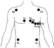
Figure 1: ECG 12 lead location.
Statistical analyses
Descriptive data was determined as means and standard deviations. Comparisons of variables for relationships were analyzed through a Pearson Product R Correlation Coefficient. Differences of groups were determined from an Analysis of Variance (ANOVA) with a pairwise Tukey post hoc analysis. Statistical significance was set a priori at P ≤ 0.05.
Results
This study was conducted to analyze possible Electrocardiography (ECG) abnormalities in male collegiate soccer players. All data is presented as group means and standard deviations. The comparisons of race, ethnicity, and position were analyzed through an Analysis of Variance (ANOVA) with a pairwise Tukey post hoc analysis. Statistical significance was set a priori at P ≤ 0.05.
The participants were male collegiate soccer players, all with previous experience at the competitive level. Twenty-one (n=21) subjects initially volunteered to take part in this study, without anyone being dropped from the study.
The subject data of age, height, weight, and competitive years of experience is as follows. Ages ranged from 18 to 23 years with mean of 20.2 years, height from 167.50 to 193.40 centimeters (cm) with mean of 182.0 cm, weight from 59.80 to 97 kilograms (kg) with mean of 80.6 kg, and the number of competitive soccer years from 6 to 17 years, with mean of 11.8 years. All demographic data collected is presented in Table 1.
![]()
    Variables
    Mean
    Standard    Deviation (Std. Dev.)


    Age (yrs)
    20.2
    1.4


    Competitive    years (yrs)
    11.8
    3.56


    Height    (cm)
    182
    6.72


    Weight    (Kg)
    80.6
    8.41
Table 1: Characteristics of participants.
All subjects had their ECG test with sinus rhythm present with 90.5% of Bradycardia (>60 b*min.-1) at the P=0.01 level of statistical significance. Heart rate ranged from 37.5 to 71.4 b*min.-1 with mean of 49.4 b*min.-1. Comparisons were executed with the objective to answer the purpose of this study, but no abnormalities were statistically significant as observed among the subjects. When compared by race, ethnicity and position, some statistically significant differences were present.
The PR interval when analyzed by position showed that the outside midfielders had PR interval abnormalities compared to all the other positions. Comparisons included the following: Outside midfielders to outside backs (P=0.001), outside midfielders to goalkeepers (P=0.006), outside midfielders to forwards (P=0.003), outside midfielders to center backs (P=0.002), outside midfielders to center midfielders (P=0.001). Among all participants, a total of four had an abnormal PR interval.
The QRS complex duration was significantly different among all the subjects when analyzed by race, ethnicity and position (P <0.001), however the value was within normal QRS duration (mean=0.056 sec). The QT interval varies depending on heart rate therefore the QT interval can be corrected for heart rate, which is called QT corrected, represented by QTc. To correct the QT interval, Bazett’s formula (QTc=QT interval/Square root of RR interval) was utilized and the normal value being less than (<) 0.44 seconds. Some consider a normal QTc to be ≤0.45 seconds in men and ≤0.47 seconds in women. In this study the QTc was significantly different among the subject, but within the normal rate (mean=0.39 sec, P=0.001).
When analyzing the ST segment, a trend in ST segment elevations was observed in 57% of the participants with elevation between 1mm and 2mm, but without statistically significant values. Significant differences among the participants was present when analyzing the length of the ST segment by race (White and Native American P=0.04) and by ethnicity (Hispanic and non-Hispanic nor Latino P=0.03).
Discussion
The goal of the present study was to analyze ECG sinus rhythm in collegiate, competitive male soccer players to determine normality or abnormality in specific ECG segments as a group including subcategorization to race, ethnicity and soccer positions on the field.
The aforementioned data showed no abnormality in regard to sinus rhythm among the participants. However, when the data was compared between race, ethnicity, and position, differences were noted. When the data was analyzed by ECG segments and compared by race, ethnicity and position, some abnormalities were present.
Heart rate
In this present study sinus bradycardia was observed in 90.5 % of the soccer players at the P=0.01 level of statistical significance with a mean of 49.4 b*min.-1. This is in accordance with previous research related to this matter [17]. High intensity training, power-and-sprint events such as soccer, reduces heart rate more than low intensity training and was present in 80% of the athletes tested [17].
Brady-arrhythmia is also a result of the intrinsic physiology of the sinoatrial and Atrioventricular (AV) nodes as well as reduced sensitivity to adrenergic stimulation [18]. Brady-arrhythmias among athletes are considered a benign adaptation to training when symptoms or structural heart disease are not present. This has been contested by several reports among athletes suggesting ventricular standstill caused by brady-arrhythmias can result in brain or cardiac complications, including syncope, angina pectoris, heart failure, embolic complications, or sudden death [19,20].
Longitudinal studies have reported that pauses, blocks, and sick sinus syndrome rarely demonstrate adverse outcomes over long term in athletes with brady-arrhythmias. Two hundred (n=200) athletes with brady-arrhythmias were submitted to several different longitudinal studies and authors reported only two cases of myocardium infarction with two cases of angina over follow-up periods of 3 months to 14.6 years. Of note is a longitudinal study reporting three cases of severe bradycardia where a pacemaker placement was required and one of the athletes had asystolic pauses up to 15 s and bradycardia as low as 17 bpm [11,7].
PR interval
Among all participants in the current study, a total of four had an abnormal PR interval, including a long PR interval noted with the position of outside midfielder. PR interval (>) 0.20 seconds can be interpreted as first degree atrial ventricular block found in 10% of the athletes and may be linked to increased vagal tone [21,9,22,7].
Second degree Mobitz type II block is rarely found in athletes, only in 8%, and may be associated with cardiac conduction system disease [23,4,7]. Despite that, first and second degree Mobitz type I blocks are considered benign among athletes and [17].
QT interval
QT interval variability is directly depended on heartrate, so a correction method is necessary to quantify it in order to determine if it falls within normal range. There are several methods to correct the QT interval; this study used the Bazett’s formula which is QT interval divided by the RR interval square root. The normal range used as reference for this study was time less than (<) 0.44 seconds as a normal value. In this study the QTc was significantly different among the subjects but fell within the normal range (mean=0.39 sec, P=0.001) [24].
These results coincide with previous literature that reported that the long QT syndrome in athletes is prevalent in 1:2,000-5,000 of athletes and seems to be hereditary. Cases of short QT syndrome (<0.34 sec) are even less common and can be associated to ventricular arrhythmia due to accelerate repolarization [1]. A retrospective analysis of sudden unexplained cardiac arrest in the state of Texas in apparently healthy children aged between 1-18 years showed a long QT syndrome was one of the causes [25].
ST segment as an interval and QT interval.
The ST segment length is rarely measured but its duration influences the QT interval duration. Usually when the QT interval is normal, the ST segment is also normal [12].
In the current study, the ST segment interval fell within a normal range with significant differences among the participants only when analyzing the ST segment as an interval by race (White and Native American P = 0.04), and by ethnicity (Hispanic and non-Hispanic nor Latino P=0.03). While not statistically significant, abnormalities with the QT interval (i.e., long QT intervals) were found in 8 players. Of these, all of had a normal ST segment interval, which leads to the conclusion that a long T wave was responsible.
In a previous study of 20 athletes, any QTc abnormality was found with only 1 athlete had QRS duration (>) 120 ms [26]. The duration of cardiac repolarization is cycle length dependent where slower heart rates lead to prolonged repolarization [27].
In contrast, a study with elite soccer players (n=566) showed that 14.1% had a short QT interval and 2.1% had a long QT interval [28]. Yet another study with elite soccer players revealed that depending on the QT interval correction formula the pre-game analyzed QTc was significantly longer compared to a control group [24]. Moreover, prolonged QTc was found, using all correction formulas, utilizing a post-game analysis. It should be noted this occurred only when calculated with the Bazett’s formula allowing a large and significant prolongation in soccer players after the game compared to pregame values. No prolonged QTc of 500 ms were found which would have suggested long QT syndrome [24].
When compared male and female athletes, both the maximum and minimum QTc values were significantly longer in female athletes than in male athletes [29]. In contrast, others reported no difference between men and women [30].
When compared female athletes to female control group, female athletes had significantly shorter QTc. However, no significantly difference was found between male athletes and male control group [29].
ST segment
With ST segment elevations (i.e., variation between 1 and 2 mm) observed in approximately 58% of the participants in the current study, this is in agreement past work. Past studies indicate up to 91% of trained athletes have ST segment elevations characterized by early repolarization as a possible mechanism, as well as athletic activity duration and intensity [31,22,32,33].
Early ventricle repolarization in athletes is considered benign. However, recent studies have suggested that ventricular fibrillation and sudden cardiac death are associated to early repolarization [28,22,32]. Longitudinal studies reveled hypertrophic cardiomyopathy and arrhythmogenic right ventricular cardiomyopathy, dilated cardiomyopathy, and myocarditis were associated with early repolarization abnormalities [11,33,34,35,36-43]. The differentiation between early repolarization and a pathological ST segment elevation includes diffuse ST segment elevation, upward concavity, notching or slurring of the QRS complex, and large amplitude T waves. Brugada syndrome is observed as a down-sloping ST segment whereas athletes may have an up-sloping ST segment. In rare cases a ST segment depression is present in athletes and should be a warning of a pathologic condition 23 [4].
Conclusion
This study was developed to emphasize the importance of ECG screening as a sudden death prevention method. The data showed no abnormality regarding sinus rhythm. Yet when compared between race, ethnicity and soccer position, there were some differences. When the data was analyzed by segments and compared by race, ethnicity, and position, some abnormalities were present.
A trend in ST segment elevations was observed and the duration of the ST segment was significantly different between White and Native American, and between Hispanic and non-Hispanic nor Latino. The outside midfielders had abnormal PR interval and were significantly different than other soccer positions.
Having the ability to differentiate a pathological condition from the physiological process in athletes is very important because the non-diagnosis of underlying heart disease can lead to sudden death during exercise. It is crucial to periodically and adequately analyze athletes with a SCD prevention strategy. If standard scanning tests are not adequate, differential diagnoses must be made with further examination methods.
References
- Drezner JA, Ackerman MJ, Anderson J, Ashley E, Asplund CA, Baggish AL, et al. Electrocardiographic interpretation in athletes: the ‘Seattle criteria’. British journal of sports medicine. 2013; 47: 122-124.
- Drezner JA, Ackerman MJ, Cannon BC, Corrado D, Heidbuchel H, Prutkin JM, et al. Abnormal electrocardiographic findings in athletes: recognizing changes suggestive of primary electrical disease. British journal of sports medicine. 2013; 47: 153-167.
- Jensen MT, Suadicani P, Hein HO, Gyntel berg F. Elevated resting heart rate, physical fitness and all-cause mortality: a 16-year follow-up in the Copenhagen male study. Heart. 2013; 99: 882-887.
- Corrado D, Basso C, Schiavon M, Thiene G. Screening for hypertrophic cardiomyopathy in young athletes. New England Journal of Medicine. 1998; 33: 364-369.
- Menafoglio A, Valention MD, Segatto J, Pezzoli R, Maggi M, Romano GA, et al. Cost and yield of 15-month preparticipation cardiovascular examination with ECG in 1070 young athletes in Switzerland: Implications for routine ECG screening. British Journal of Sports Medicine. 2014; 48; 1157-1161.
- Asatryan B, Vital C, Kellerhals C, Medeiros-Domingo A, Gräni C, Trachsel LD, et al. Sports-related sudden cardiac deaths in the young population of Switzerland. PLOS ONE. 2017; 12: e0174434.
- Maron BJ, Doerer JJ, Haas TS, Tierney DM, Mueller FO. Sudden death in young competitive athletes: analysis of 1866 death in United States. Circulation. 2009; 119: 1085-1092.
- Karakaya O, Sglam M, Barutcu I, Esen AM, Ocak Y, Melek M, et al. Comparison of the predictors for atrial rhythm disturbances between trained athletes and control subjects. The Tohoku Journal of Experimental Medicine. 2005; 207: 165-170.
- Drezner JA, Ashley E, Baggish AL, Börjesson M, Corrado D, Owens DS, et al. Abnormal electrocardiographic findings in athletes: recognizing changes suggestive of cardiomyopathy. British Journal of Sports Medicine. 2013; 47: 137-152.
- Sharma S, Whyte G, Elliott P, Padula M, Kaushal R, Mahon N, et al. Electrocardiographic changes in 1000 highly trained junior elite athletes. British Journal of Sports Medicine. 1999; 33; 319-324.
- Bjornstad HH, Bjornstad TH, Urheim S, Hoff PI, Simith G, Maron BJ. Long term assessment of electrocardiographic and echocardiographic findings in Norwegian elite endurance athletes. Cardiology. 2009; 112; 234-241.
- Viitasalo MT, Kala R, Eisalo A. Ambulatory electrocardiographic recording in endurance athletes. British heart journal. 1982; 47: 213-220.
- Ilic I, Rancovic J, Krstic O, Popovic-Ilic T, Ilic SH, et al. Change of ECG parameters depending on the load level in athletes. Acta Media Medianae. 2007; 51; 57-62.
- Binetoglu FK, Babaoglu K, Altun G, Kayabey O. Effects that different types os sports have on the heart of children and adolescents and the value of twodimensional strain-strain-rate echocardiography. Pediatric Cardiology. 2014; 35; 126-139.
- Berge HM, Steine K, Andersen TE, Solberg EE, Gjesdal K. Visual or computer-based measurements: Important for interpretation of athletes’s ECG. British Journal of Sports Medicine. 2014; 48: 761-767.
- Anderson J, Levine S, Root M, Coyne BJ, Sanford G, Colvin LC. Clinical exercise electrocardiography. Burlington, MA: Jones & Bartlett Publishers. 2015.
- Corrado D, Biffi A, Basso C, Pelliccia A, Thiene G. 12-lead ECG in the athlete: physiological versus pathological abnormalities. British Journal of Sports Medicine. 2009; 43: 669-676.
- Baldesberger S, Bauersfeld U, Candinas R, Seifert B, Zuber M, Ritter M, et al. Sinus node disease and arrhythmias in the long-term follow-up of former professional cyclists. European Heart Journal. 2007.
- Abdon NJ, Landin K, Johansson BW. Athlete’s bradycardia as an embolising disorder? Symptomatic arrhythmias in patients aged less than 50 years. British Heart Journal. 1984; 52: 660-666.
- Ector H, Verlinden M, Eynde EV, Bourgois J, Hermans L, Fagard R, et al. Bradycardia, ventricular pauses, syncope, and sports. The Lancet. 1984; 324: 591-594.
- ACSM. ACSM’s Guidelines for Exercise Testing and Prescription. Philadelphia: Lippincott, Williams &Wilkins. 2010.
- Kasikçioglu E. The incognita of the known: the athlete’s heart syndrome. Anadolu Kardiyol Derg. 2011; 11: 351-359.
- Alasti M, Omidvar B, Jadbabaei MH. Heart and athlete. The journal of Tehran Heart Center. 2010; 5: 1.
- Lengyel C, Orosz A, Hegyi P, Komka Z, Udvardy A, Bosnyák E, et al. Increased short-term variability of the QT interval in professional soccer players: possible implications for arrhythmia prediction. PLOS ONE. 2011; 6: e18751.
- Alapati S, Strobel N, Hashmi S, Bricker JT, Gupta-Malhotra M. Sudden unexplained cardiac arrest in apparently healthy children: a single-center experience. Pediatric Cardiology. 2013; 34: 639-645.
- Brosnan M, La Gerche A, Kalman J, Lo W, Fallon K, Mac Isaac A, et al. Comparison of frequency of significant electrocardiographic abnormalities in endurance versus nonendurance athletes. The American journal of cardiology. 2014; 113: 1567-1573.
- Le´ger L, Gojanovic B, Sekarski N, Meijboom EJ, Mivelaz Y. The Impending Dilemma of Electrocardiogram Screening in Athletic Children. Pediatr Cardiol. 2016; 1-13.
- Bohm P, Ditzel R, Ditzel H, Urhausen A, Meyer T. Resting ECG findings in elite football players. Journal of sports sciences. 2013; 31; 1475-1480.
- Omiya K, Sekizuka H, Kida K, Suzuki K, Akashi YJ, Ohba H, et al. Influence of gender and types of sports training on QT variables in young elite athletes. European Journal of Sport Science, 2014; 14: S32-S38.
- Heffernan KS, Jae SY, Lee M, Mojtahedi M, Evans EM, Zhu W, et al. Gender differences in QTc interval in young, trained individuals with lower spinal cord injury. Spinal cord. 2007; 45: 518-521.
- Farahani, B, Esfahani MP, Abbasi MA, Moradi F, Abbasi A. Prevalence of different electrocardiographic patterns in Iranian athletes. Acta Medica Iranica. 2012; 50: 560.
- Maron BJ. Distinguishing hypertrophic cardiomyopathy from athlete’s heart physiological remodeling: clinical significance, diagnostic strategies and implications for pre-participation screening. Br J Sports Med. 2009; 43: 649- 656.
- Mc Claskey D, Lee D, Buch E. Outcomes among athletes with arrhythmias and electrocardiographic abnormalities: Implications for ECG interpretation. Sports Medicine. 2013; 43: 979-991.
- Papadakis M, Carre F, Kervio G, Rawlins J, Panoulas VF, Chandra N, et al. The prevalence, distribution, and clinical outcomes of electrocardiographic repolarization patterns in male athletes of African/Afro-Caribbean origin. Eur Heart J. 2011; 32: 2304-2313.
- Pelliccia A, Kinoshita N, Pisicchio C, Quattrini F, Dipaolo FM, Ciardo R, et al. Long-term clinical consequences of intense, uninterrupted endurance training in Olympic athletes. J Am Coll Cardiol. 2010; 55: 1619-1625.
- Pelliccia A, Di Paolo FM, Quattrini FM, Basso C, Culasso F, Popoli G, et al. Outcomes in athletes with marked ECG repolarization abnormalities. New England Journal of Medicine. 2008; 358: 152-161.
- Asif IM, Johnson S, Schmieg J, Smith T, Rao AL, Harmon KG, et al. The psychological impact of cardiovascular screening: the athlete’s perspective. British journal of sports medicine. 2014; 48: 1162-1166.
- Cho JH, Selen MA, Kocheril AG. Screening of young competitive athletes for the prevention of sudden cardiac death with a wireless electrocardiographic transmission device: a pilot study. BMC research notes. 2015; 8: 342.
- Drezner JA, Fischbach P, Froelicher V, Marek J, Pelliccia A, Prutkin JM, et al. Normal electrocardiographic findings: recognizing physiological adaptations in athletes. British Journal of Sports Medicine. 2013; 47: 125-136.
- Fuchs T, Torjman A, Galitzkaya L, Leitman M, Pilz-Burstein R. The clinical significance of ventricular arrhythmias during an exercise test in noncompetitive and competitive athletes. Isr Med Assoc J. 2011; 13: 735-739.
- Levine S, Coyne B, Colvin L. Clinic exercise electrocardiography. Burlington, MA: Jones & Bartlett Learning. 2016.
- Maron B J, Pelliccia A. The heart of trained athletes: cardiac remodeling and risk of sports, including sudden death. Circulation. 2006; 114: 1633-144.
- Navalta JW, Stone WJ, Lyons S. Ethical Issues Relating to Scientific Discovery in Exercise Science. International Journal of Exercise Science. 2019; 12: 1.