Case Report
J Stem Cell Res Transplant. 2014;1(1): 1004.
Neuropsychiatric Disorder Tackled by Innovative Cell Therapy-A Case Report in Autism
Sharma A1, Gokulchandran N1, Shetty A3, Kulkarni P2*, Sane H2 and Badhe P1
1Department of Medical Services and Clinical research, NeuroGen Brain and Spine Institute, India
2Department of Research & Development, NeuroGen Brain and Spine Institute, India
3Department of NeuroRehabilitation, NeuroGen Brain and Spine Institute, India
*Corresponding author: Kulkarni P, Department of Research & Development, NeuroGen Brain and Spine Institute-StemAsia Hospital and Research Centre. Plot 19, Sector 40, Nerul, Next to Seawood Station (W), Off Palm Beach Road, Navi Mumbai-400706, India
Received: June 28, 2014; Accepted: Aug 04, 2014; Published: Aug 06, 2014
Abstract
Autism is a pervasive developmental disorder affecting socialization, communication and behavior. Neuropathology of autism spectrum disorders is poorly understood and may involve impaired connectivity in the brain, selectively affecting parts of the brain forming circuits supporting social behavior. The currently available treatment options do not address the core neuropathology of autism. Hence, it is important to develop a treatment modality for autism at the earliest. Cell therapy is recently emerging as a potential treatment option for autism. We hypothesize that it may assist in the repair of the disrupted neuronal circuit. The neural repair may take place not only by cellular restoration but also by paracrine and immunomodulatory effects. Cellular therapy has shown promising results for various other neurological disorders. We administered autologous bone marrow mononuclear cells, intrathecally in an 11 year old boy diagnosed with autism. He was assessed for a follow up period of eight months wherein his autistic features had reduced. This was objectively supported by improvement in scores of CARS (31 to 25), ISAA (130 to 98), CGI-I (6 to 5) and FIM (104 to 110). This case study may hint towards exploring cell therapy as a potential treatment alternative for autism, in addition to standard approach. A longer period of follow up along with functional imaging may further help us understand the repair of the impaired neuronal circuit at cellular level.
Keywords: Autism; Cellular therapy; Autologous; Bone marrow; Mononuclear cells
Abbreviations
CARS: Childhood Autism Rating Scale; ISAA: Indian Scale for Assessment of Autism; CGI: Clinical Global Impression; FIM: Functional Independence Measure; PET CT: Positron Emission Tomography; FDG: Fluorodeoxyglucose; G-CSF: Granulocyte-colony stimulating factor; MNCs: Mononuclear cells; STS: Superior temporal sulcus, FFG: Fusiform gyrus, OFC: Orbital frontal cortex
Case Report
Case Presentation
Herein, we present a case of an 11 year old boy with autism. Mother reported emotional stress throughout the pregnancy and had hypertension in the last 2 months of pregnancy. He was born at full term by normal delivery, cried immediately after birth, with normal birth weight and had no neonatal complications. The motor milestones development was normal, however his speech was delayed.
At the age of 2 years, he was diagnosed with autism as parents noticed poor eye contact along with attention and concentration deficit Over the period of 9 years, the parents observed increased level of hyperactivity, presence of stereotypical and self stimulatory behavior like hand flapping and jumping. Social interaction was poor, presence of odd play behavior, emotional responses were inappropriate to the situation (like he laughed without any reason) and the behavioral issues increased when there was a change in routine. His responses were delayed and the questions needed to be repeated a couple of times for understanding. There was presence of aggressive behaviour like spitting on others or saying foul words. Along with echolalia, his speech was stereotypical and repetitive. He made unusual noises and used meaningless words while talking. Had difficulty in performing fine motor activities and following simple commands. In most of the daily activities of living, he was independent but required assistance in fine motor activities. In spite of regular standard rehabilitation he showed no improvements with respect to eye contact, behavior and social interaction and his autistic symptoms persisted.
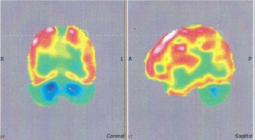
Childhood Autism Rating Scale (CARS) score was 31 which are categorized as mild to moderate autism. We also assessed the case on Indian Scale for Assessment of Autism (ISAA). This scale, based on CARS, has domains such as social relationship and reciprocity; emotional responsiveness; speech, language and communication; behavior patterns; sensory aspects and cognitive component. The items are rated from 1 to 5, increasing score indicating increasing severity of the problem. The content, construct and concurrent validity, internal consistency and test-retest reliability, and sensitivity and specificity of ISAA were studied by the members of the expert committee for the development of assessment tool for autism. ISAA was thus found to be a valid tool, with good reliability and high sensitivity and specificity [1]. On ISAA his score was 130 which is categorized as moderate autism. On Clinical Global Impression (CGI) the severity of index (CGI-I) was 6 which is severely ill. On Functional Independence Measure (FIM) his score was 104. The PET CT scan of the brain showed reduction of FDG uptake in the left cerebellar hemisphere, bilateral amygdale and hippocampi (Figure 1).
Figure 1: PET CT scan brain done before the therapy. The areas in blue indicate reduced FDG uptake.
Treatment
He underwent autologous bone marrow derived mononuclear cell transplantation. Patient selection was based on World Medical Association Helsinki Declaration for Ethical Principles for medical research involving human subjects [2]. The protocol has been reviewed and approved by the Institutional committee for Stem cell Research and Therapy (IC-SCRT). A written informed consent was obtained from the parents of all patients. All the patients included in the study had confirmed diagnosis of autism according to the DSM-V diagnostic criteria for autistic disorder. The exclusion criteria were presence of acute infections such as HIV/HBV/HCV, malignancies, bleeding tendencies, renal failure, severe liver dysfunction and other acute medical conditions such as respiratory infection and pyrexia.
300 mcg of G-CSF injections were administrated 48 hours and 24 hours before bone marrow derived mononuclear cell transplantation, to stimulate CD34+ cells and increase their survival and multiplication. Bone marrow (100ml) was aspirated from the iliac bone. Mononuclear cells (MNCs) were obtained using density gradient separation method. Viable count of the isolated MNCs was taken and was found to be about 98%. The MNCs were checked for CD34+ by FACS analysis. Approximately 56× 106 MNCs were immediately injected post separation, intrathecally in L4-L5 using a lumbar puncture needle and catheter. After the intervention, the patient was given intensive rehabilitation therapy which included occupational therapy (intensive sensory integration therapy, cognitive and perceptual rehabilitation, social skill training) speech therapy and psychological therapy (applied behavior analysis). He was recommended to continue these therapies throughout. He was evaluated at three and eight months based on CARS, ISAA, CGI and FIM.
Results
After the procedure, the patient had no side effects. He showed significant improvements over a period of eight months.
Within a week, there was improvement in his speech i.e. he was able to speak in full sentences and at a faster pace. Awareness about the surroundings had improved along with association and logical thinking. Writing speed had increased and he could eat independently which he wasn’t able to do before.
On follow up of three months, further improvements were observed in his attention and concentration. Hyperactivity and aggressive behaviour had reduced. He maintained more meaningful eye contact. There was an overall increase in level of awareness and now had developed choices. Stereotypical and self stimulatory behaviour had reduced. Also, he became more adaptable to changes in his routine and command following improved. Inappropriate emotional response like laughing without any reason reduced. According to parents, self-talking and making sounds reduced by 10 to 20 % and the frequency reduced to 2 or 3 times in a day. He developed and learnt few new activities like ability to solve 15 pieces puzzles and recite 2 stories.
After 8 months, his reaction time reduced and he could respond faster. There was an overall growth in his school performance. His school teachers reported that his sitting tolerance had improved along with attention span.
On CARS, his scores reduced from 31 to 25. On CGI, severity of illness (CGI-I) pre therapy was 6 i.e. severely ill; post therapy was 5 i.e. markedly ill. Global improvement (CGI-II) was found to be 2 indicating much improved. Efficacy index (CGI-III) was found to be 5 which denote moderate therapeutic effect of intervention without any side effects. His ISAA score reduced from 130 to 98 with improvement in social and emotional reciprocity, emotional responsiveness, speech and language communication, behaviour patterns, sensory aspects and cognition (Table 1).
    Sub-components of    ISAA
    Score Pre treatment
    Score at 6 month    follow up


    A.
    Social    Relationship and Reciprocity
    35
    26


    1
    Poor Eye contact
    5
    3


    2
    Lacks social smile
    5
    3


    3
    Unable to relate to    people
    3
    2


    4
    Unable to respond    to social/ environmental cues
    5
    4


    5
    Unable to take    turns in social interaction
    5
    3


    6
    Does not maintain    peer relationships
    4
    3


    B
    Emotional    Responsiveness
    14
    11


    1.
    Shows exaggerated    emotions
    4
    3


    2.
    Engages in self    stimulating emotions
    5
    3


    C.
    Speech    Language and Communication
    30
    25


    1
    Engages in    stereotyped and repetitive use of language
    4
    3


    2
    Engages in    echolalic speech
    5
    3


    3
    Produces infantile    sequels/unusual noises
    5
    3


    4
    uses pronoun    reversals
    4
    3


    5
    Unable to grasp    pragmatics of communication (real meaning)
    4
    3


    D.
    Behaviour    Patterns
    27
    18


    1
    Engages in    stereotyped and repetitive motor mannerisms
    4
    3


    2
    Shows attachment to    inanimate objects
    5
    3


    3
    Shows hyperactivity    and restlessness
    5
    3


    4
    Exhibits aggressive    behavior
    5
    3


    E.
    Sensory    Aspects
    11
    10


    1
    Unusually sensitive    to sensory stimuli
    4
    3


    F.
    Cognitive    Component
    12
    8


    1
    Inconsistent    attention and concentration
    5
    3


    2
    Shows delay in    responding
    5
    3
Table 1: Table giving details of the improvements in different sub components on ISAA at follow up of 6 months.
Discussion
Autism spectrum disorder is a group of complex neuropsychiatric disorders which according to DSM V, are diagnosed by early symptoms of social communication/interaction, and restricted and repetitive behaviors. The etiology of autism involves a number of different environmental and genetic factors. The model of Autism Spectrum Disorder suggests early failure to develop the specialized functions of one or more set of neuro-anatomical structures which help in social information processing i.e. the social brain [3]. The social brain is the complex network of areas which help in recognizing other individuals and evaluate their mental status (e.g., intentions, dispositions, desires, and beliefs). Functional imaging of patients with autism has shown disruptions in connectivity, selectively affecting parts of the brain forming circuits supporting social behavior [4]. These disturbances in the connectivity may also give rise to communication issues, restricted interests, repetitive behaviors, difficulty in recognizing other agents and their actions, difficulty in perceiving the emotional states of others, analyzing the intentions and dispositions of others, sharing attention with one another, and representing another person’s perceptions and beliefs. In 1990, Brothers emphasized the contribution of the superior temporal sulcus (STS), fusiform gyrus (FFG), orbital frontal cortex (OFC), and amygdala to social perception [5]. In animal studies, it is found that the STS region has reciprocal connections to the amygdala which is connected to the OFC region. The STS region is also connected to the OFC. The OFC is connected to prefrontal cortex, which is further connected to motor cortex and the basal ganglia, thus completing what Allison and colleagues (2000) described as a pathway from perception to action. Interruption in this pathway may affect the way subjects perceive the surroundings and organize their actions [6].
Extensive research has been carried out in the field of regenerative medicine for neurological disorders [7-9]. Various types of cells have been explored such as bone marrow cells, umbilical cord blood cells, olfactory ensheathing cells, adipose tissue cells, embryonic cells, etc. [10]. Bone marrow cells have shown promising results. They are easily obtainable, safe and have no ethical issues.
Inflammation, immune dysfunction and hypoxia are postulated etiology of autism. Bone marrow cells are capable of carrying out the neural repair process not only by cell restoration but also by paracrine and immunomodulatory effects. They stimulate angiogenesis and lead to reperfusion. It is also observed that reversal of hypoperfusion leads to neural proliferation and self repair by restoring neural connections. These cells modulate the immune system and repair the altered brain organization. They restore the imbalance of the immune system by inhibiting the proliferation of CD8+ and CD4+ T lymphocytes and natural killer cells, suppress the immunoglobulin production by plasma cells, and inhibit the maturation of dendritic cells and the proliferation of regulatory T cells [11].
A clinical study including 32 cases of autism has shown that autologous bone marrow mononuclear cell transplantation is safe and improves the quality of life of the patients with respect to ability to perform activities of daily living independently [12]. Similar results were obtained in a study carried out by Lv et al. [13] using human umbilical cord mesenchymal stem cells (hUC-MSCs) and human cord blood mononuclear cells (hCB-MNCs) transplantation in patients with autism. Also, in our previous study on pediatric incurable neurological disorders including autism, autologous mononuclear cells led to improved functional outcome [14]. We hypothesize that with multiple mechanisms, as described above, cell therapy along with rehabilitation stimulated restoration of neural circuitry in the affected areas of brain that was reflected as clinical improvements [15].
Conclusion
This case study has demonstrated that cell therapy has a positive outcome in case of autism. Hence, it should be given due importance and studied extensively to prove its long term benefit in treating incurable disorders like autism.
References
- Natrajan P, Kumar A, Goyal H, et al. Scientific report on research project for development of Indian Scale for assessment of autism.
- https://www.thenationaltrust.co.in/nt/index.php?option=com_content&task=view&id=30&Itemid=130, 2008.
- World Medical Association. World Medical Association Declaration of Helsinki: Ethical Principles for Medical Research Involving Human Subjects. JAMA. 2013; 310: 2191-2194.
- Volkmar FR. Understanding the social brain in autism. Dev Psychobiol. 2011; 53: 428-434.
- Gotts SJ, Simmons WK, Milbury LA, Wallace GL, Cox RW, et al. Fractionation of social brain circuits in autism spectrum disorders. Brain. 2012; 135: 2711-2725.
- Brothers L. The social brain: A project for integrating primate behaviour and neurophysiology in a new domain. Concepts in Neuroscience. 1990; 1: 27-51.
- Von Hofsten C, Rosander K. Perception-action in children with ASD. Front Integr Neurosci. 2012; 6: 115.
- Sharma A, Sane H, Badhe P, Kulkarni P, Chopra G, et al. Autologous Bone Marrow Stem Cell Therapy shows functional improvement in hemorrhagic stroke- a case study. Indian Journal of Clinical Practice. 2012; 23: 100-105.
- Politis M, Lindvall O. Clinical application of stem cell therapy in Parkinson's disease. BMC medicine. 2012; 10: 1.
- Sharma A, Kulkarni P, Sane H, Gokulchandran N, Badhe P, et al. Positron Emission Tomography- Computed Tomography scan captures the effects of cellular therapy in a case of cerebral palsy. Journal of clinical case reports. J Clin Case Rep. 2012; 2: 195.
- Yoo J, Kim HS, Hwang DY. Stem cells as promising therapeutic options for neurological disorders. J Cell Biochem. 2013; 114: 743-753.
- Hoogduijn MJ, Popp F, Verbeek R, Masoodi M, Nicolaou A, et al. The immunomodulatory properties of mesenchymal stem cells and their use for immunotherapy. International Immunopharmacology. 2010; 10: 1496–1500.
- Sharma A, Gokulchandran N, Sane H, Nagrajan A, Paranjape A, et al. Autologous bone marrow mononuclear cell therapy for autism – an open label proof of concept study. Stem Cell Int. 2013; 2013: 623875.
- Lv YT, Zhang Y, Liu M, Qiuwaxi JN, Ashwood P, et al. Transplantation of human cord blood mononuclear cells and umbilical cord-derived mesenchymal stem cells in autism. J Transl Med. 2013; 11: 196.
- Sharma A, Gokulchandran N, Chopra G, Kulkarni P, Lohia M, et al. Administration of autologous bone marrow derived mononuclear cells in children with incurable neurological disorders and injury is safe and improves their quality of life. Cell Transplantation. 2012; 21: S79–S90.
