
Case Report
Austin Surg Case Rep. 2025; 10(1): 1066.
Gorlin Gotz Syndrome: Multisystematic Disorder Case Report
Benwadih Sarra*
Maxillofacial Surgery Department, Hospital of Specialties, Mohammed V University, Rabat 10100, Morocco
*Corresponding author: Benwadih Sarra, Maxillofacial Surgery Department, Hospital of Specialties, Mohammed V University, Rabat 10100, Morocco Tel: 00212662100484; Email: sarrabenwadih045@gmail.com
Received: July 22, 2025 Accepted: August 04, 2025 Published: August 07, 2025
Abstract
Nevoid basal cell carcinoma syndrome (“Gorlin–Goltz syndrome”) is a heritable condition and thought to be due to a genetic defect which is a mutation of the “Patched” tumor suppressor gene. It is inherited in an autosomal dominant manner, but sporadic cases have been described. The syndrome shows high penetrance and variable expressivity. It is a multisystem disorder with multiple pigmented basal cell carcinomas, jaw keratocysts, palmar and/or plantar pits, and calcification of the falx cerebri. Other less prominent features have also been noted, such as skeletal, dermatologic, and neurologic abnormalities. Very aggressive basal cell carcinomas and other malignant neoplasms have been seen in some cases. Since the oral and maxillofacial manifestations of this syndrome are of utmost importance, their characteristics must be understood for diagnosis, early preventive treatment, and genetic counseling. Here we summarize the principal clinicopathologic features and treatment options associated with this syndrome.
Keyworlds: Gorlin–Goltz; Patched; Basal cell carcinomas; Palmar pits; Keratocysts; Falx cerebri
Introduction
Gorlin syndrome (Gorlin-Goltz syndrome, basal cell nevus syndrome [BCNS], nevoid basal cell carcinoma syndrome) is an inherited autosomal dominant cancer syndrome. It is characterized by the occurrence of multiple basal cell carcinomas (BCCs), as well as skeletal, ocular and neurological anomalies. These neoplasms start to manifest in childhood.
Case Presentation
We present the case of the first patient, a 50-year-old man, who was hospitalized in the Dermatology Department in October 2024 for suspected Gorlin-Goltz syndrome. His medical history showed skin lesions on his face and scalp that appeared in 2010 and were treated by excision. He had undergone surgery for a left mandibular cyst. However, detailed information on the procedure or the histology of the removed lesions was not available.
Examination revealed numerous (more than 40) papules, nodules, and plaques, primarily on his face, but also on his scalp and upper chest, reaching up to 2.5 cm in diameter (Figure 1).
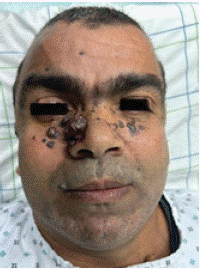
Figure 1: Front photograph showing skin lesions.
Some of these nodules were pigmented and firm, dome-shaped, while others had pearly borders and were covered with a crust. The plaques were erythematous and located mainly retroauricularly (Figure 2).
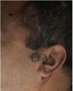
Figure 2: Side profile photograph showing retroauricular lesions.
Dermoscopy of the nodule on the right side of the nose revealed: large arborescent vessels, ovoid nests, and shiny white structures (chrysalides) suggestive of basal cell carcinoma (Figure 3).
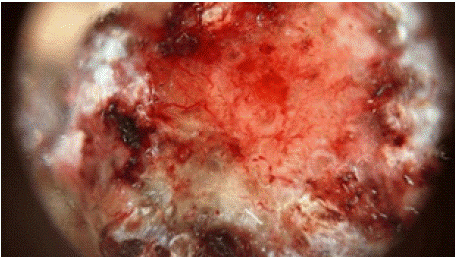
Figure 3: Dermoscopic image of the nodule.
Physical examination revealed macrocephaly, frontal bossing, depressed nasal bridge andhypertelorism.
Palmar pits were brown coloured and measuring 1-3 mm in diameter (Figure 4).
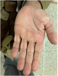
Figure 4: Palmar pits lesions.
No other anomalies of the skeletal, cardiovascular, or central nervous system were present.
An orthopantomogram was realised showing two large mandibular cysts (Figure 5).
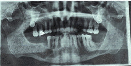
Figure 5: Large mandibular cysts.
The second case is a 51-year-old patient with a history of corticotropic insufficiency who had presented with multiple facial lesions, predominantly jugo-temporo-nasal, for 8 years.
Physical examination revealed frontal bossing, depressed nasal bridge and hypertelorism. Dermatological examination revealed papules and nodules scattered across the front of the nose, nose, and around the eyes (Figure 6).
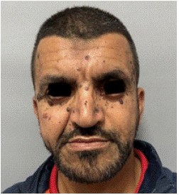
Figure 6: Front photograph showing skin lesions (case 2).
A biopsy of one of the nasal lesions suggested basal cell carcinoma.
No other anomalies of the skeletal, cardiovascular, or central nervous system were present.
A cT scan was performed showing bilamellar calcification of the falx cerebri (Figure 7).
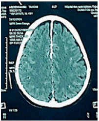
    Figure 7: Axial computed tomography brain image showing bilamellar
calcification of the falx cerebri.
The skin lesions were removed under general anesthesia.
The patients were closely observed with follow-ups every 6 months.
No signs of tumor recurrence.
A genetic study was conducted on the patients’ families, but none presented a mutation.
Discussion
Gorlin syndrome is caused by a mutation in patched 1 (PTCH1), a tumor suppressor gene located on chromosome 9q [3].
Gorlin-Goltz syndrome initially consisted of triads of: basal cell carcinomas, jaw cysts, and skeletal anomalies [1].
Diagnosis of BCNS can be established when two major or one major and two minor features are present [2].
Major criteria include: multiple (>2) BCCs or 1 BCC by =20 years of age, histologically proven OKCs of the jaws, palmar or plantar pitting (three or more), bilamellar calcification of the falx cerebri, bifid/fused/splayed ribs, first-degree relative with BCNS.
Minor criteria include: medulloblastoma, macrocephaly, congenital malformations (frontal bossing, coarse facies, cleft lip/ palate, coarse face, moderate or severe hypertelorism), other skeletal abnormalities (Sprengel deformity, marked pectus deformity, marked syndactyly of the digits), radiologic abnormalities (bridging of the sella turcica, vertebral anomalies, fusion or elon, modeling defects of the hands and feet, or flame-shaped lucencies of the hands or feet), ovarian fibroma [2-4].
Our first patient had four major features: multiple BCCs, palmar pits, two PKCs in the jaw, and lamellar calcification of the falx cerebri, as well as minor features such as macrocephaly and hypertelorism.
Our second patient had three major features, such as multiple BCCs, palmar pits, PKCs in the maxillary bone, as well as minor features such as hyperthorism.
Genetic testing for PTCH1 is necessary in the following cases [4]: diagnosis confirmation in patients lacking sufficient clinical diagnostic features [5]; predictive testing for patients at risk with an affected family member but not presenting clinical features; [6] prenatal testing if there is a known familial mutation.
On the therapeutic level, there is currently no specific treatment for this condition. Their management is symptomatic of specific oncological monitoring, involving the intervention of different specialists. Sun protection is imperative, and patients must be checked regularly; in particular, a dermatological examination must be carried out every two to three months. Genetic investigation is essential for the detection of new cases in the family. A neurological examination is required annually in children up to seven to detect medulloblastoma [7].
Conclusion
Gorlin-Goltz syndrome is a rare, genetic disorder. It is classically defined by the triad of basal cell nevi, maxillary keratocysts and skeletal malformations.
This syndrome is due to mutations in a tumor suppressor gene.
The carcinological potential of this syndrome is serious, justifying early detection and regular surveillance.
Written informed consent was obtained from the patient for publication of this case report and accompanying images. A copy of the written consent is available for review by the Editor-in-Chief of this journal on request.
References
- Chetan A Pol, Suvarna K Ghige, Ritesh R Kalaskar, Suchitra R Gosavi. Contemp Clin Dent. 2013; 4: 547–550.
- Gorlin Syndrome. Alison M. Spiker; Todd Troxell; Michael L. Ramsey. 2023.
- Shevchenko A, Durkin JR, Moon AT. Generalized basaloid follicular hamartoma syndrome versus Gorlin syndrome: A diagnostic challenge. Pediatr Dermatol. 2018; 35: e396-e397.
- Hazemann G, Michel C, Mahé A, Lipsker D, Cribier B. [Histopathological study of basaloid follicular hamartoma]. Ann Dermatol Venereol. 2019; 146: 181-191.
- Scalia AC, Farulla A, Fiocchi F, Alboni C, Torricelli P. Imaging features of uterine and ovarian fibromatosis in Nevoid Basal Cell Carcinoma Syndrome. J Radiol Case Rep. 2018; 12: 21-30.
- Hasan A, Akintola D. An Update of Gorlin-Goltz Syndrome. Prim Dent J. 2018; 7: 38-41.
- Mohamed Rachid Ghailan, Hanane Benhalima, Ghafour Rabeh, Abdelilah Oujilal, Anas Benbouzid, Razika Bencheikh, et al. Gorlin-Goltz syndrome: report of a case medecine buccale chirurgie buccale. 2007; 13: 97-101.