
Special Article - Subtalar Dislocation
Austin Surg Case Rep. 2017; 2(2): 1020.
Subtalar Dislocation: Case Report and Review of Literature
Dinesh Dhar*
Senior Specialist Orthopedics, Nizwa Regional Referral Hospital, Oman
*Corresponding author: Dinesh Dhar, Senior Specialist Orthopedics, Nizwa Regional Referral Hospital, Oman
Received: November 20, 2017; Accepted: December 04, 2017; Published: December 11, 2017
Abstract
Subtalar Dislocations (SD) is unusual injuries usually associated with fractures of talus, calcaneum and fifth metatarasal. Isolated subtalar dislocation is very rare. Four types have been described medial, lateral, posterior and anterior. A rare case of closed lateral subtalar dislocation in a 23 years old male patient following motor vehicle accident is reported. It was treated successfully by closed reduction and immobilization in below knee cast with good functional outcome. The management of the case with recent review of literature on the subject has been discussed.
Keywords: Subtalar; Dislocation; Lateral; Isolated
Introduction
Isolated subtalar dislocation is very rare injuries and very few cases have been reported in the English literature. Strong ligament connecting talus with calcaneum along with strong joint capsule and biomechanics of ankle joint make this injury unusual [1]. Subtalar dislocation involves simultaneous disruption of talocalcaneal and talonavicular joints while tibiotalar and calcaneocuboid joints remain intact. Patients with bony or soft tissue aplasia around ankle joint are more likely to suffer isolated subtalar dislocation [1]. The foot along with calcaneum can dislocate medially, laterally, posteriorly or anteriorly in relation to talus. This case report describes the mechanism, pattern of injury and management of case, highlighting good functional outcome after closed reduction immobilization and good postoperative rehabilitation programme.
Case Presentation
23 years male patient involved in motor vehicle collision was referred from peripheral health centre with trauma to left ankle. He was seen in accident and emergency by on call orthopedic team and noticed to have gross swelling all around left ankle and foot with deformity. Dorsalis pedis and posterior tibial pulsations were not felt but patient had capillary circulation of toes with intact sensations. Radiographs (AP, Lateral) of ankle, foot revealed dislocation of talaonavicular and talocalcaneal joints with prominence of talar head dorsomedially and calcaneum laterally without any associated fractures and diagnosis of isolated lateral subtalar dislocation was made (Figure 1,2,3).
Under sedation successful closed reduction of subtalar dislocation was done in emergency by on call orthopedic team and left foot and ankle was immobilized initially in posterior plaster slab for period of 05 days, followed by below knee scotch cast for 05 weeks (Figure 4,5). After reduction posterior tibial and dorsalis pedis pulsations were well felt. The patient went on to start partial weight bearing at 05 weeks with active and passive range of motion exercises of left ankle under physiotherapist guidance and full weight bearing by 9 weeks post injury (Figure 6).
Discussion
Subtalar dislocations are very rare injuries and account for 1-2 percent of all foot dislocations [2]. Majority of cases are reported in third decade of life with M: F ratio of 6:1. The mode of injury is commonly high energy traumas motor vehicle accidents, sport injuries [3]. Associated fractures of talus or fifth metatarsal have been reported. Subtalar dislocation was classified initially by Broca (1853) into three types: Medial, lateral and posterior dislocations. Malgaigne and Buerger (1856) described an additional anterior dislocation. Medial dislocation comprises 80-85% followed by lateral 15-20%, posterior and anterior 1-2%.
The medial subtalar dislocation also referred to as acquired clubfoot results from forceful inversion of forefoot with resultant stress on lateral collateral ligaments which along with talocalcaneal and talonavicular ligaments rupture resulting in subtalar dislocation with talus remaining in its normal position. The lateral subtalar dislocation also referred to as acquired flatfoot results from forceful eversion of foot with talus pivitoning on anterior calcaneus process. The posterior and anterior types results from extreme forceful plantar flexion or dorsiflexion of forefoot [1]. Lateral subtalar dislocation results from high energy trauma and have poor prognosis in the long run compared to medial subtalar dislocation [2]. Associated osteochondral shearing injuries to articular surface of talus, calcaneus or navicular are common up to 45% however they are difficult to be seen on plain radiographs.
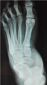
Figure 1: AP view of left foot showing talonavicular dislocation.
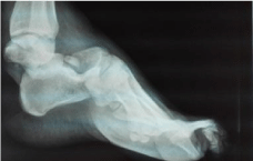
Figure 2: AP and Lateral view of left ankle showing talocalcaneal dislocation.
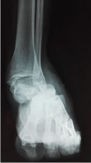
Figure 3: AP and Lateral view of left ankle showing talocalcaneal dislocation.
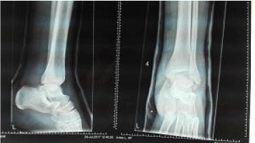
    Figure 4: Post reduction AP and Lateral views of left ankle showing well
reduced subtalar dislocation.
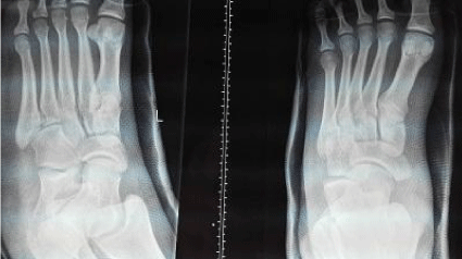
    Figure 5: Post reduction AP and oblique views of left foot showing well
aligned talonavicular dislocation.
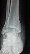
Figure 6: AP view of ankle after cast removal at 09 weeks.
Subtalar dislocations can be treated by prompt closed reduction and immobilization with below knee cast. Duration of immobilization remains controversial with some authors recommending immobilization for 02 weeks, partial weight bearing after 02 weeks and full weight bearing at 03 weeks post injury [4]. However majority of authors recommend immobilization for 04 weeks period to allow enough time for healing [5].
In 10% of medial and 15% of lateral subtalar dislocations closed reduction and manipulation may not be successful due to soft tissue interposition requiring open reduction [2].
In our case the patient was involved in high energy motor vehicle accident resulting in lateral subtalar dislocation. Prompt closed reduction of the dislocation and immobilization for 05 weeks prevented any soft tissue, neurovascular complications with excellent functional outcome at 06 months follow up. Close follow up for long time is needed for manifestations of any late complications as a vascular necrosis of talus or post traumatic arthritis.
Conclusion
There is a very low incidence of isolated subtalar dislocation which may be easily missed in multiple injured patients. Early prompt reduction of dislocation can lead to better functional outcome. Adequate immobilization time depending on type of dislocation should be allowed followed by supervised active and passive range of motion of ankle with gradual mobilization progressing from partial weight bearing to full weight bearing to achieve good results.
References
- Giannoulis D, Papadopoulos DV, Lykissas MG, Koulouvaris P, Gkiatas I, Mavrodontidis A. Subtalar dislocation without associated fractures. Case report and review of literature. World J Orthop. 2015; 6: 374-379.
- Kulambi V, Gaurav M. Lateral subtalar dislocation: A case report. The foot and ankle online journal. 2014; 7.
- Carvera TP. Subtalar dislocation Case report and literature review. Global Journal of Medical Research. 2016; 16: 1.
- James D. Functional outcome after early mobilization in isolated subtalar dislocation. Case report and review of current evidence. J Orthop Traumatol Rehabil. 2017; 9: 50-52.
- Hotouras A, Sandiford N, Rao S. Medial subtalards location: A case report. Internet Journal of Orthopedic Surgery. 2006; 5: 1-4.