Special Article - Pediatric Surgery
Austin Surg Case Rep. 2017; 2(2): 1021.
Accessory Caudate Liver Lobe Torsion in a 7-Year Old Boy: Case Report
Ashjaei B* and Noveiry BB
Department of Pediatric Surgery, Children Medical Center, Tehran University of Medical Sciences, Iran
*Corresponding author: Ashjaei B, Department of Pediatric Surgery, Children Medical Center, Tehran University of Medical Sciences, Iran
Received: December 04, 2017; Accepted: December 15, 2017; Published: December 22, 2017
Keywords
Torsion; Caudate; Accessory
Introduction
Acute blood flow loss of an organ will eventually lead to hypoxia and severe pain. Abdominal organs torsion will cause acute abdomen in this way. Caudate Lobe (CL) of the liver is derived from the right liver lobe anatomically on the inferior and posterior surface of liver; however, it’s functionally separated from that [1]. Here is a case report of a seven-year old boy presenting with acute abdomen, following torsion in accessory caudate lobe of the liver.
Case Presentation
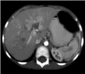
A seven-year old boy with chief complaint of sustained non colicky pain in right upper quadrant of abdomen without radiation and irrelevant to neither a feeding or defecation, from three days prior to admission, was brought to the emergency service. He had also anorexia accompanied by recurrent biliary vomiting. No history of constipation, diarrhea, fever, reflux, jaundice or itching, previous trauma or polluted water and food intake he had. Nothing remarkable was in past medical history. He had no medication taken before admission. In physical exam he was moderately ill in general appearance but not toxic with stable vital signs. Abdominal tenderness, without rebound tenderness, in Right Upper Quadrant (RUQ) and epigastria was observed. Also a mass was palpated in RUQ. Bowel sounds were slightly diminished. Complete blood count showed WBC of 13.02*103/ml (neutrophil 72.2%, lymphocyte 17.4%). Hematocrit was 41.2% and hemoglobin was 14.2g/dl. Platelets count was 324*103 and coagulation tests were normal. Ultrasound imaging revealed a hypoechoic mass with a central echogenic zone, in the lower part of the liver, 35*18*16 mm in dimensions. Falciform ligament, in the vicinity of Gallbladder (GB) and duodenum was thickened, suggesting a thrombotic umbilical vein. No free fluid or paraaortic lymphadenopathy was detected. Spiral CT-scan with contrast media showed normal size and density in liver and patent main portohepatic veins (Figure 1). Contracted GB with severe wall edema noted a low attenuated well defined lesion close to GB and intersegment ligament of left lobe, suspecting hematoma or small collection (Figure 2).
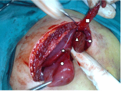
With the diagnosis of hepatic mass, patient was taken to operation room by general anesthesia in supine position. Laparatomy with Kocher incision was done. Inflammated round ligament was seen and resected. Beside round ligament, accessory caudate lobe had ischemic appearance, resting on a long and slim peduncle with clockwise tortuosity (Figure 3). Accessory CL was resected by clamping, and the origin of pedicle was ligated by vicryl 2.0 and hemostasis was performed. Neither free fluid nor blood was detected in peritoneal cavity. Other lobes of the liver were normal in appearance and size and color (Figure 4). GB was in normal anatomic region with normal appearance. Nothing abnormal or pathologic was seen at the other regions of peritoneal cavity. Abdomenoraphy after gauze counting was done. Biopsy showed normal congested hepatic tissue without any cystic appearance or clue to malignancy. In the next two days, no complication occurred and child was discharged in good general condition, eating food without problem.
In next week follow-up, the child had well general condition, and liver function tests (ALT, AST and ALP) and bilirubin were within normal ranges.
Discussion
This case was presenting torsion in caudate lobe of liver as a rare cause of acute abdominal pain. It was previously reported in some animal cases [2-6]. It seems to that rabbits are more susceptible for CL torsion than the other animals. To our knowledge, this is the first ever case report of CL liver torsion in human. Accessory Liver Lobe (ALL) torsion has been reported in approximate 20 cases ahead of this [7-10]. One accessory liver lobe torsion originating from the CL process has been reported [11]. Ectopic liver detachment leading to acute abdomen was another rare entity [12]. In our patient and within all accessory liver lobe cases, and animal caudate torsions, the congested mass had originated from a thin and tall stalk; making it susceptible to torsion. Thus it’s not far beyond the rationality, to excise any thin stalked mass originating from the liver; if it was seen incidentally in laparatomies so as to ban further probable torsion.

Figure 1: CT scan of patient, showing congested mass with blood inflow but no outflow.
▲ Accessory caudate lobe with a long stalk
♦ Left liver lobe
● Gall bladder
* Right lobe

    Figure 2: ■ Falciform ligament
▲ Accessory caudate lobe with a long stalk
♦ Left liver lobe
● Gall bladder
* Right lobe

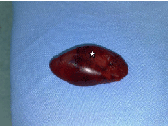
    Figure 3: Resected tortuous accessory caudate lobe of liver (*) with focal
ischemic zones.

Figure 4: Torsion of accessory caudate lobe of the liver (on the surgeon's finger).
Accessory caudate lobe of liver is reported in codover in some articles [13-15], but torsion of it in a live patient is a rare condition.
Most of the previous reports, had ineffective diagnostic approach to specify underlying cause of abdominal pain; leading to variable diagnosis, from appendicitis to gallbladder problems. Sonography seems to be a logic 1st line imaging. Computed Tomography (CT) scan helps localizing the hepatic mass effectively. Using contrast media may be beneficial for more careful assessment, if the patient is not allergic to the substance. Magnetic resonance imaging was not preceding CT-scan for the diagnosis. Localizing the hepatic mass can help the surgeon for a better approach in laparotomy. Fine needle aspiration or core needle biopsy don’t seem to be a logic procedure in these patients as they have sustained pain, and also no diagnostic clue seems to be achieved by them in advantage. Liver function tests were not assessed in this child preoperatively as no sign or symptom was elucidating hepatic dysfunction; although modest increase could be detected as it has been before.
References
- SS. Gray's Anatomy: Churchill Livingstone. 2008.
- Saunders R, Redrobe S, Barr F, Moore AH, Elliott SC. Liver lobe torsion in rabbits. J Small Anim Pract. 2009; 50: 562.
- Wenger S, Barrett EL, Pearson GR, Sayers I, Blakey C, Redrobe S. Liver lobe torsion in three adult rabbits. Journal of Small Animal Practice. 2009; 50: 301-305.
- Downs MO, Miller MA, Cross AR, Selcer BA, Abdy MJ, Watson E. Liver lobe torsion and liver abscess in a dog. J Am Vet Med Assoc. 1998; 212: 678-680.
- Schwartz SG, Mitchell SL, Keating JH, Chan DL. Liver lobe torsion in dogs: 13 cases (1995-2004). J Am Vet Med Assoc. 2006; 228: 242-247.
- Weisbroth SH. Torsion of the caudate lobe of the liver in the domestic rabbit (Oryctolagus). Vet Pathol. 1975; 12: 13-15.
- Hundal R, Ali J, Korsten M, Khan A. Torsion and Infarction of an Accessory Liver Lobe. Zeitschrift für Gastroenterologie. 2006; 44: 1223-1226.
- Umehara M, Sugai M, Kudo D, Hakamada K, Sasaki M, Munakata H. Torsion of an accessory lobe of the liver in a child: Report of a case. Surgery Today. 2009; 39: 80-82.
- Carrabetta S, Piombo A, Podestà R, Auriati L. Torsion and infarction of accessory liver lobe in young man. Surgery. 2009; 145: 448-449.
- Negru D, Tudorache C, Marinescu M, Grigoras M, Crumpei F, Georgescu S. [Torsion and infarction of an accessory liver lobe]. Rev Med Chir Soc Med Nat Iasi. 2007; 111: 442-445.
- Koumanidou C, Nasi E, Koutrouveli E, Theophanopoulou M, Hilari S, Kakavakis K. Torsion of an accessory hepatic lobe in a child: Ultrasound, computed tomographic, and magnetic resonance imaging findings. Pediatr Surg Int. 1998; 13: 526-527.
- Elsayes H, Elzein M, Razik A, Olude I. Torsion of an ectopic liver in a young child. Journal of Pediatric Surgery. 2005; 40: E55-E58.
- Chhabra N, Shrivastava T, Garg L, Mishra BK. An anatomic variant caudate lobe in a cadaver. International Journal of Research in Medical Sciences. 2014; 2: 759-761.
- Soni G, Boddeti RK. Accessory caudate lobe. International Journal of Anatomy and Research. 2016; 4: 3309-3311.
- Pryakhin A, Yukhimets S, Chernomortseva E, Ainory P. Gesase. Accessory Lobes, Accessory Fissures and Prominent Papillary Process of the Liver. Anatomy Journal of Africa. 2015; 4: 611-616.