Clinical Image
Austin Surg Case Rep. 2018; 3(1): 1026.
Sacrococcygeal Teratoma in the Neonate with Clinical and Operative Photos
Pradyumna Pan*
Pradyumna Pan, Departments of Pediatrics Surgery, Ashish Hospital and Chirayu Hospital, Jabalpur, India
*Corresponding author: Pradyumna Pan, Departments of Pediatrics Surgery, Ashish Hospital and Chirayu Hospital, Jabalpur, India
Received: August 07, 2018; Accepted: August 24, 2018; Published: August 31, 2018
Clinical Image
Sacrococcygeal teratomas are the most common type of germ cell tumors diagnosed in neonates, infants, and children younger than 4 years [1].
SCTs are classified morphologically according to their relative extent outside and inside the body:
- Altman type I - entirely outside, sometimes attached to the body only by a narrow stalk
- Altman type II - mostly outside
- Altman type III - mostly inside
- Altman type IV - entirely inside; this is also known as a presacral teratoma or retro rectal teratoma.
The incidence of malignancy in SCTs increases with age. By the age of 9 months, the incidence of malignancy is around 70% [2]. Early diagnosis and complete excision with removal of the coccyx is associated with good prognosis [3]. Recurrence is related to tumor spillage during excision, incomplete resection, and leaving the coccyx behind and requires long-term follow up.

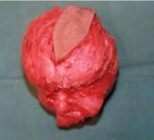
Figure 1:

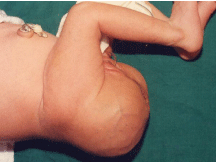
Figure 2:

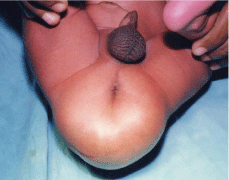
Figure 3:
References
- Moore SW, Satgé D, Sasco AJ, Zimmermann A, Plaschkes J. The epidemiology of neonatal tumours. Report of an international working group. Pediatr Surg Int. 2003; 19: 509-519.
- Hager T, Sergi C, Hager J. Sacrococcygeal teratoma - a single center study of 43 years (1968-2011) including follow-up data and histopathological reevaluation of specimens. Eur Surg. 2012; 44: 255-266.
- Rescorla FJ, Sawin RS, Coran AG, Dillon PW, Azizkhan RG. Long-term outcome for infants and children with sacrococcygeal teratoma: A report from the Childrens Cancer Group. J Pediatr Surg. 1998; 33: 171-176.