
Case Report
Austin Surg Case Rep. 2019; 4(1): 1030.
Endoscopic En-Bloc Resection of Calcific Tendinitis of Rectus Femoris: A Surgical Advance
Torres-Eguia R1, Abd El-Radi M1,2*, Trujillo LB1 and Guntin AM1
¹Department of Orthopedics and Traumatology, Chief of Hip Unit, Spain
²Department of Orthopedics and Traumatology, Assiut University Hospitals, Egypt
*Corresponding author: Mohamed Abd El-Radi Abd El-Salam, Department of Orthopedics and Traumatology, Assiut University Hospitals, Assiut, Egypt and Member of Joint Supervision Mission of Egyptian High Ministry of Education for M.D degree in Spain, Spain
Received: August 27, 2019; Accepted: September 27, 2019; Published: October 04, 2019
Abstract
Purpose: The aim of this study is to show effectiveness of en-bloc resection technique as a treatment for rectus femoris calcific tendinitis.
Method: Five patients who underwent hip arthroscopy as treatment for calcific tendinitis of rectus femoris were retrospectively evaluated for pre and postoperative function and pain. Follow up ranged from 9 to 28 months (mean: 15.2 months). Mean age was 32.4 years, all patients were practicing football at recreational basis and right side was affected in four cases (80%).
Results: Mean scores for Western Ontario and McMaster Universities (WOMAC) and Hip disabilities and Osteoarthritis Outcome Score (HOOS), evolved from 74.4 to 91.7 points and from 77.7 to 91.2 points respectively.
Conclusion: En-bloc resection technique ensures complete resection of the calcific portion of rectus femoris and allows minimally invasive joint access offering rapid recovery and lower complication rate.
Keywords: Rectus Femoris; Calcification; Hip Pain; Hip Arthroscopy
Introduction
Calcific tendinitis results from deposition of calcium hydroxyapatite crystals in tendinous tissue [1]. Multifactorial etiology including traumatic [1], genetic and metabolic issues [2,3] have been addressed as underlying causes of calcific tendinitis.
Calcific tendinitis presents mechanical pain with decreased range of motion and occasionally tenderness and warmness at affected joint [4]. In spite of it commonly affecting rotator cuff tendons, it is described in other periarticular regions such as elbow, wrist, knee and foot [5-7]. In hip region, chronic tendinopathy of the gluteus muscles is a well-recognized condition and is considered the second most frequently affected region after shoulder region [7-9]. However, calcification of rectus femoris was firstly described by King and Vanderpool as a rare disease in 1967 [8,9]. Later, a few cases of rectus femoris calcific tendinitis were reported with different treatment lines [4,7,10].
Conservative treatment is the first option including different methods such as rest, anti-inflammatory drugs or sometimes local steroid injections could be recommended. Surgical management is indicated when conservative treatment fails or symptoms are intractable and includes open removal through anterior approach [11] or endoscopic removal [7].
Arthroscopic resection is considered a minimally invasive technique to treat this condition, showing a rapid recovery. The aim of this study is to show our results when performing endoscopic enbloc resection to treat calcific tendinitis of the rectus femoris with a mean follow up 15.2 months.
Methods
Five patients were programmed for hip arthroscopy to treat calcific tendinitis of rectus femoris between August 2014 and February 2016. All patients were informed of procedure and possible complications with informed consents taken and IRB/Ethics Committee decided approval was not required for this study. They were evaluated retrospectively in our hip unit.
Patient’s ages ranged from 27 to 37 years (mean 32.4 years). All patients (100%) were male. The right side was affected in four patients (80%). All patients were regular football players on recreational basis.
All patients presented associated cam deformity (100%), one patient had pincer deformity (20%) and three patients had associated labral tear (60%).
Diagnosis was made based on clinical history, physical examination, and imaging procedures, which include plain radiographs, computerized topography and magnetic resonance imaging.
All patients referred chronic hip pain (more than 6 months duration), especially after sport practice. One patient (20%) had clear history of noted injury in the form of avulsed anterior inferior iliac spine 6 months before his visit to our unit.
Preoperative pelvic radiographs showed calcifications of the proximal part of rectus femoris (Figure 1AB). Computerized topography with three dimensional reconstruction was applied to assess shape of the lesion (Figure 2AB). Magnetic resonance arthrography were indicated in all patients to address associated lesions such as labral tears, chondrolabral injuries and cartilage damage as well as femoroacetabular impingement.

    Figure 1A, 1B: Plain radiographs (Anteroposterior pelvic view and axial
view of right hip) show calcification at origin of rectus femoris of the right hip
preoperatively.
Surgical Technique
All patients were placed in supine position on a traction table under general anesthesia. Traction was applied on affected hip until no less than 1 cm of joint distraction was achieved. Surgical site was draped and an anterolateral portal was performed under radioscopic guidance. A 70°C scope was used primarily to evaluate central compartment. A mid-anterior portal was set under direct visualization. An interportal capsulotomy was performed using banana blade and radiofrequency ablation device. Primarily, correction of pincer deformity and reattachment of torn labarum were performed.
Anterior standard portal was established to assist in resection of calcified portion of rectus femoris. The use of a third portal has the great advantage of clarifying margins of the calcified portion with assistance of shaver and radiofrequency ablation devices in order to take it out as one piece. At this point, alternation of visualizing and working portals is the key point to isolate the calcified portion of rectus femoris. As the limits of the lesion are identified (Figure 3AB), a curved osteotomy was applied to detach calcified fragment entirely from the bone (Figure 4A). With the help of grasper, the calcified portion was withdrawn en-bloc through the anterior portal as the osteotomy was applied through the mid-anterior portal (Figure 4BC). Finally, traction was released and correction of cam deformity using 5-mm bur was performed.
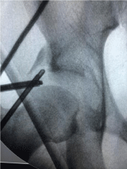
Figure 3A: Fluoroscopic localization of calcified portion of rectus femoris.
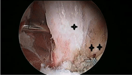
    Figure 3B: Arthroscopic Localization of calcified portion of rectus femoris
(One asterisk) showing the resected pincer deformity and repaired labrum
(Two asterisks) of right hip.
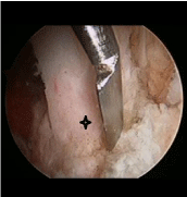
    Figure 4A: A curved osteotome applied to detach calcified fragment (One
asterisk) entirely from the bone.
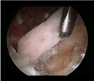
    Figure 4B: Calcified portion with drawn en-bloc with grasper through the
anterior portal.
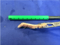
    Figure 4C: Calcified portion of rectus femoris resected en-bloc of 6-7
centimeters length.
The interportal capsulotomy was closed using 2 non-absorbable stitches. Portals were closed with absorbable sutures, after which sterile bandages were applied.
Follow up ranged from 9 to 28 months, with an average of 15.2 months. All arthroscopies were performed by the same surgeon.
Results
Patients were discharged on the following day with partial weight bearing for 3 weeks assisted by crutches. No patient received antiinflammatory drugs post-operatively. A program to achieve full range of motion in the affected hip had been started third week postoperatively. Exercise to regain strength was delayed until seventh post-operative week especially selected rectus femoris and quadriceps muscles exercises. It is recommended not to return to contact sports at least until the sixth post-operative month.
All patients showed clinical and radiological improvement (Figure 1CD).On physical examination, pain on flexion and internal rotation is reduced in all patients. WOMAC score ranged from 70.3 to 78.9 points preoperatively (mean 74.4 points) and from 73.4 to 98.4 in the postoperative period (mean 91.7 points). HOOS score ranged from 77.5 to 78.3 points preoperatively (mean 77.7 points) and from 87.5 to 93.1 in the postoperative period (mean 91.2 points).

    Figure 1C, 1D: Plain radiographs show resection of the calcified portion
postoperatively (Anteroposterior pelvic view and axial view of right hip).
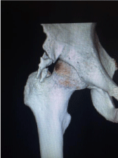
Figure 2A: Three dimensional Computerized Topography (C.T) scan.
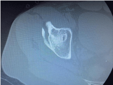
    Figure 2B: Axial C.T scan show calcification of the reflected head of rectus
femoris of the right hip.
Among the arthroscopic findings were evidence of articular cartilage flap lesion at the acetabulum side managed by debridement and micro fractures in one patient (20%). He had the above mentioned post-operative protocol with maintained partial weight bearing for 6 weeks. No complications have been observed at the moment of writing.
Discussion
Although calcification of rectus femoris is a rare condition, reflected head is more commonly affected [12-14] causing hip pain. Other causes of hip pain that relate to rectus femoris include traumatic avulsion, so acetabula and myositis ossificans [13].
Diagnostic tools of this condition are based on clinical and radiological assessment including: Chronic hip pain, positive iliac spine (Ely’s) test and calcifications appearing in both anteroposterior pelvic view and axial view of the affected hip on plain radiographs [4].
Conventional treatment approaches are rest, anti-inflammatory drugs and physical therapy. Adjunct procedures include local steroid injections. Surgical treatment is indicated when other treatment methods fail and includes open or endoscopic removal of calcific lesions [11,15].
Peng et al [16] reported 3 cases treated effectively by hip arthroscopy for rectus femoris calcification. All cases were calcification of reflected head using adequate capsulotomy parallel to labral sulcus with favorable short-term follow up. Comba et al, [17] published a completely extra-articular endoscopic technique to resect ossifications of rectus femoris tendinitis avoiding capsulotomy of hip joint. Zini et al [18] described results of 6 high level football players at mean 1 year follow up showing 3 patients returned to their level of activity and the other three returned to 80%of their previous sport level. All of their patients had a satisfactory outcome dealing with daily activities. Recently, Comba et al [7] reported 9 patients with heterotopic calcification of rectus femoris treated with peripheral compartment access technique [19] showing improvement in outcome and 90% return to full activity. He found one recurrence in their cases at fourth month postoperative but continued asymptomatic at 5-year follow up.
To this 0 day, our study is the first one reporting en-bloc resection technique to treat calcific tendinitis of rectus femoris. On the same surgery, we addressed other associated pathologies: Resecting pincer deformity, suturing the torn labrum and resecting cam deformities. We performed microfractures in one case in which cartilage flap lesion was present. Assessment of both central and peripheral compartments is the key point to success as calcific tendinitis might not be the only pathology causing hip pain in those patients.
Conclusion
Hip arthroscopy is a well-established method to treat femoroacetabular impingement in young highly active patients. Endoscopic resection has proven to be an effective and safe procedure when treating calcific tendinitis of rectus femoris. En-bloc resection technique ensures complete resection of the calcified portion of rectus femoris reducing and liberating fewer particles articularly and periarticularly reducing risk of heterotopic ossification. We conclude that this technique allows for minimally invasive joint access, complete lesion resection while offering rapid recovery and lower complication rate.
References
- Holt PD, Keats TE. Calcific tendinitis: a review of the usual and unusual. Skeletal radiology. 1993; 22: 1-9.
- Cannon RB, Schmid FR. Calcific periarthritis involving multiple sites in identical twins. Arthritis & Rheumatology. 1973; 16: 393-396.
- Uhthoff HK, Sarkar K, Maynard JA. Calcifying tendinitis: a new concept of its pathogenesis. Clinical orthopaedics and related research. 1976; 118: 164- 168.
- Braun-Moscovici Y, Schapira D, Nahir AM. Calcific tendinitis of the rectus femoris. JCR: Journal of Clinical Rheumatology. 2006; 12: 298-300.
- Cox D, Paterson F. Acute calcific tendinitis of peroneus longus. J Bone Joint Surg Br. 1991; 73: 342.
- Dilley D, Tonkin M. Acute calcific tendinitis in the hand and wrist. The Journal of Hand Surgery: British & European Volume. 1991; 16: 215-216.
- Comba F, Piuzzi NS, Oñativia JI, Zanotti G, Buttaro M, Piccaluga F. Endoscopic Extra-articular Surgical Removal of Heterotopic Ossification of the Rectus Femoris Tendon in a Series of Athletes. Orthopaedic Journal of Sports Medicine. 2016; 4: 2325967116664686.
- King J, Vanderpool D. Calcific tendonitis of the rectus femoris. The American journal of orthopedics. 1967; 9: 110-111.
- Gondos B. Observations on periarthritis calcarea. The American journal of roentgenology, radium therapy, and nuclear medicine. 1957; 77: 93-108.
- Kim YS, Lee HM, Kim JP. Acute calcific tendinitis of the rectus femoris associated with intraosseous involvement: a case report with serial CT and MRI findings. European Journal of Orthopaedic Surgery & Traumatology. 2013; 23: 233-239.
- Kandemir U, Bharam S, Philippon MJ, Fu FH. Endoscopic treatment of calcific tendinitis of gluteus medius and minimus. Arthroscopy: The Journal of Arthroscopic & Related Surgery. 2003; 19: 4.
- Gamradt SC, Brophy RH, Barnes R, Warren RF, Byrd JT, Kelly BT. Nonoperative treatment for proximal avulsion of the rectus femoris in professional American football. The American journal of sports medicine. 2009; 37: 1370-1374.
- Sarkar J, Haddad F, Crean S, Brooks P. Acute calcific tendinitis of the rectus femoris. J Bone Joint Surg Br. 1996; 78: 814-816.
- Pierannunzii L, Tramontana F, Gallazzi M. Case report: calcific tendinitis of the rectus femoris: a rare cause of snapping hip. Clinical Orthopaedics and Related Research®. 2010; 468: 2814-2818.
- Randelli F, Pierannunzii L, Banci L, Ragone V, Aliprandi A, Buly R. Heterotopic ossifications after arthroscopic management of femoroacetabular impingement: the role of NSAID prophylaxis. Journal of Orthopaedics and Traumatology. 2010; 11: 245-250.
- Peng X, Feng Y, Chen G, Yang L. Arthroscopic treatment of chronically painful calcific tendinitis of the rectus femoris. European journal of medical research. 2013; 18: 49.
- Comba F, Piuzzi NS, Zanotti G, Buttaro M, Piccaluga F. Endoscopic surgical removal of calcific tendinitis of the rectus femoris: surgical technique. Arthroscopy techniques. 2015; 4: 365-369.
- Zini R, Panascì M, Papalia R, Franceschi F, Vasta S, Denaro V. Rectus femoris tendon calcification: arthroscopic excision in 6 top amateur athletes. Orthopaedic journal of sports medicine. 2014; 2: 2325967114561585.
- Domb BG, Philippon MJ, Giordano BD. Arthroscopic capsulotomy, capsular repair, and capsular plication of the hip: relation to atraumatic instability. Arthroscopy: The Journal of Arthroscopic & Related Surgery. 2013; 29: 162- 173.