Case Report
Austin Surg Case Rep. 2020; 5(1): 1033.
Osteochondroma-Cases Presentation
Bunjaku I1, Gjonbalaj N1, Lenjani B2*, Rashiti P2, Krasniqi B3, Bunjaku G3 and Bunjaku M3
¹Department of Radiology Clinic, University Clinical Center of Kosova, Kosova
²Department of Emergency Clinic, University Clinical Center of Kosova, Kosova
³Department of Medical Sciences Rezonanca, Royal University Iliria, Kosovo
*Corresponding author: Lenjani B, Department of Emergency Clinic, University Clinical Center of Kosova, Kosova
Received: December 03, 2019; Accepted: January 07, 2020; Published: January 14, 2020
Abstract
Definition: Developmental osseous anomaly resulting in exophytic outgrowth on surface of bone. Osteochondromas account for about 12% of bone tumors. In morphologic view we separate as pedunculate and sessile form. Clinical are usually painless for a few years, the pain appear when nerves and blood vessels are touched by osseous growth. The diagnosis id done using x-ray, CT, MRI, Ultrasound, angiography, scintigraphy bone scan and histological examination. X-ray is first method of examination and gives us a many information about bone pathology. Later we can evaluate bone pathology with other methods.
Conclusion: In this case, we have described a patient 15 year old with pain and limited movements. We used X-ray, CT, MRI, Scintigraphy and histological results for lesion assessment. After these results our patient is operated by orthopedist. In Histopathology department, they take material of surgery and the result was osteochondroma. Finally, we want to say that early diagnosis is very important to except malign transformation or nerves and blood vessels complitations.
Keywords: Osteochondroma; Pedenculate; Sessile; Diagnostic Methods; Histopathology Department
Background
Osteochondroma is a process that develops as an anomaly that results in increased growth on the surface of long bone. Osteochondrosis appears in the metaphyses of long bones about knee insertion, the proximal part of the humerus, rarely seen in the scapula skull, the clavicle, the bones of the laca as well as in the rings processors etc. Morphologically seen as peduncular mass or as sessil lesion.
Epidemiology
Osteochondrosis is considered as a developed lesion rather than as a true neoplasm. Osteokondrosis by WHO, is defined as an osteocartilaginous exostosis with continuity in the cortical and medullary part. Osteochondromial ethylogenic factors they are unknown, but it is thought that the nose tumor is associated with an abnormality on the growth plate, causing bone flaconism that rises far away from the bone. Osteochondromas has intercourse with mutations of EXT1 and EXT2 genes that are related to the biosynthesis of heparan sulfate proteoglycans, which may favor epiphyseal growth. Generally, Osteochondromines appear in the first and third decades of life, appear especially in children, teenagers, without a sex preference. as well as diagnostic procedures; ray-x, CT. RM, HP as well as other diagnostic methods.
Case Report
Patients, A.B. 15 years old is admitted to the clinic because of the restriction of motion on the right knee. The main complaints have started before 6 weeks with pain and edema of the distal part of the right femoral. Patients are initially subjected to laboratory blood tests and their outcome is at the limit of reference values. After the anlizas is also the art radiography. right knees where it is presented as degrading: After clinical examinations of Rtg, CT, MRI and laboratory tests, indications for operative intervention, operation and extractable material are sent for biopsy. After the operation, a rehabilitation treatment was applied.
Discussion
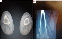
Osteochondroma is an unusual hereditary disease that occurs in long bone metaphs, around the knee articulation, the proximal knee, rarely patches on the ribs, the scalpel, the clavicle and the bones of the bone. This disease occurs in the first decade to the third of the life. The reported case is typical of the localization and for two main reasons: the age of the disease for 15 years based on the worldly likelihood as well as the disease and the localization. Osteochondria develops faster in younger age than in the elderly. Localization of Osteochondromes in the knee region of the diseased may give rise to obstructions in motion, degenerative arthritis, and neurovascular suppression by shaking even more health problems. At the young age degenerative arthritis is more rare than in the elderly. CT of the right knee, axial and sagittal plan, Changes in images obtained from 3mm MDCT images show changes in the meta-diaphysic region of the femoral artery in the distal front of the femoral arm coupled with cardiac cap and minimal cortical destruction .
MRI
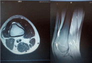
Exophytic mas lesion (30×10) in anterior aspect of distal metaphysio-diaphyseal region of right femur. T2-hyperintensity overlyng lesion is cartilage cap.
There is impression for cortical irregularities in anterior edge of lesion. It is consistent with sessile ostechondroma. Maximum cartilage cap thickness is measured as 6 mm. MRI up is recommended
Histopathology. Tumoral tissue has zoned architecture, the superficial part is constructed of well-differentiated chondrocyte, located in the background of the abundant chondrocyte matrix, the cartilaginous depth of the tissue is replaced by the osteoid trabeculae with regular osteoblasts.

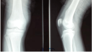
    Figure 1: Projection and latero-lateral. The broad base of bone lesion
in relation to the cortex in the distal front part of the metaphysia-diaphysial
region of the femur has been identified, with relatively clear boundaries; this
is well noted in the lateral projection.

    Figure 2: Osteochondroma, right knee joint, axial plan. (A) and sagittal
(B), changes presented in images obtained by MDCT with 3mm cutting
changes recorded in the meta-diafizal region of the femur, ekzostoze in
the anterior distal femoral be coupled with the cap of cartilage and minimal
cortical being destructive.

    Figure 1: MRI of the distal femur: Examination Technique MRI images of
the right knee were obtanined using TSE/PDW + T2W (with fat saturation)
sequence in axial, coronal,sagittal planes and TSE /T1W sequence in
coronal plane. Findings: There is exophytic mass lesion (30×10 mm) in
anterior aspect of distal metaphysio –diaphysal region of right femur.
T2-hyperintensity overlying lesion cartilage cap. It is consistent with
sessile ostechondroma. Maximum cartilage cap thickness is measured as
6 mm.There is impression for cortical irregularities in anterior edge of lesion.
Otherwis cortical and distal femura and patella are normal.No periostal
reaction.
Anterior and posterior cruciale ligaments are normal in ttheir course
and linear in their contour. There is abnormal signal associated with
these ligaments. Quadriceps tendon is normal. All muscles within imaging
field are normal in morphology and signal. Fascial planes are intact.
MRI. Exophytic mass lesion (30×10) in anterior aspect of distal metaphysiodiaphyseal
region of right femur. T2-hyperintensity overlyng lesion is cartilag
cap.There is impression for cortical irrgularities in anterior edge of lesion. It is
consistent with sessile ostechondroma. Maximum cartilage cap thickness is
measured as 6 mm. MRI follow up is recommended.

    Figure 4: Histopathological report; Tumoral India has zonal architecture:
the superficial part is constructed of well differentiated chondrocyte, placed
in the background of the abundant chondrocyte matrix; the cartilage depth
is replaced by the osteoid trabeculae demarcated with regular osteoblasts.
Among osteoarthritis trabeculae is the yellow palette built from sebaceous
indigo.
Among osteoarthritis trabeculae is the yellow palette built from sebaceous indigo. According to world literature, about 40-50% of cases may occur at any age, especially at the age of 10 to 30, while most often affect males. 70% of the bone metaphors are 60-70% long. Whereas cases of malignancy are rarely seen, and the recurrences are around 5-10%. Laboratory analyzes are not convincing for the Osteochondromes’ identification, but were within the limits of the reference values, but radiological diagnostics with CT and RMI are convincing methods for diagnosis.
Conclusion
Osteochondria appear in the first and third decades of life, especially in children, adolescents, without a sex preference, and in the event of the occurrence of pain and limited motion, the sufferer should be told to the doctor for further diagnosis and treatment. Radiology diagnostic approaches are of great importance to native radiography, CT, MRI and histopathological examination and with a few diagnostic laboratories.
After the diagnostic operative intervention is required. After the operation should begin the rehabilitation phase and treatment with cryotherapy and kinesiotherapy, should also be recommended ambulatory treatment.
References
- Passanise AM, Mehlman CT, Wall EJ, Dieterle JP. Ra-diographic evidence of regression of a solitary osteo-chondroma: a report of 4 cases and a literature review. J Pediatr Orthop. 2011; 31: 312-316.
- Volders D, Vandevenne JE, Van de Casseye W. Trevor’s disease and wholebody MRI. Eur J Radiol. 2011; 79: 363-364.
- Masquijo JJ, Willis B. Dysplasia epiphysealis hemimelica (Trevor’s disease). Arch Argent Pediatr. 2010; 108: e20-23.
- Araujo CR Jr, Montandon S, Montandon C, Teixeira KI, Moraes FB, Moreira MA. Best cases from the AFIP: dysplasia epiphysealis hemimelica of the patella. Radio-graphics. 2006; 26: 581-586.
- Magnetna Rezonaca Muskuloskeletnog sistema Robert Semnic Novi Sad. 2013; 492-494.
- Semiologjia Imazherike dhe Bazat e Imazherise Diagnostike. 2010; 468-469.
- Diagnostic Imaging Orthopaedics Stoller – Tirman –Bredella, 2004; 8: 23-24.
- Costeira O. Farmoquímica; Rio de Janeiro: 2001. Termos e expressões da prática médica. acetabulum: a case report and review. Iowa Orthop J. 2005; 25: 60-65.
- Wiart E, Budzik JF, Fron D, Herbaux B, Boutry N. Bilateral dysplasia epiphysealis hemimelica of the talus associated with a lower leg intramuscular cartilaginous mass. Pediatr Radiol. 2012; 42: 504-506.
- Bahk WJ, Lee HY, Kang YK, Park JM, Chun KA, Chung YG. Dysplasia epiphysealis hemimelica: radiographic and magnetic resonance imaging features and clinical outcome of complete and incomplete resection. Skeletal Radiol. 2010; 39: 87-89.
- Andrea, CE; Reijnders CM; Kroon HM; De Jong D; Hogendoorn PC; Szuhai K; Bovée JV. “Secondary peripheral chondrosarcoma evolving from osteochondroma as a result of outgrowth of cells with functional EXT”. Oncogene. 2012; 31: 1096-1098.
- Sekharappa V, Amritanand R, Krishnan V, David KS. “Symptomatic solitary osteochondroma of the subaxial cervical spine in a 52-year-old patient”. Asian Spine J. 2014; 8: 84-88.
- EL-Sobky TA, Samir S, Atiyya AN, Mahmoud S, Aly AS, Soliman R. “Current paediatric orthopaedic practice in hereditary multiple osteochondromas of the forearm: a systematic review”. 2018.