
Clinical Image
Austin Surg Case Rep. 2022; 7(1): 1046.
Chondrosarcoma of the Right Iliopubic Branch Incidentally Discovered: about a Case
Mokhchani Y1,2*, Rabbah A1, Rachdi A1,2, Boukhriss J1,2, Chafry B1,2 and Boussouga M1,2
1Department of Orthopedic Surgery and Traumatology II, Mohammed V Military Teaching Hospital, Rabat, Morocco
2Faculty of Medicine and Pharmacy, Mohammed V University, Rabat, Morocco
*Corresponding author: Youness Mokhchani, Department of Orthopedic Surgery and Traumatology II, Mohammed V Military Teaching Hospital, Rabat, Morocco; Faculty of Medicine and Pharmacy, Mohammed V University, Rabat, Morocco
Received: April 07, 2022; Accepted: April 29, 2022; Published: May 06, 2022
Clinical Image
Chondrosarcoma is the most common primary malignant bone tumor after osteosarcoma, characterized by an anarchic endo and exo bony proliferation of malignant chondrocytes, producing tumor cartilage. It has a clear male predominance especially between 40 and 60 years. The pelvis is a frequent location (40%). Vascular complications, which may be venous or arterial, are rare. We report a case of chondrosarcoma developing at the expense of the right iliopubic branch, incidentally discovered during a radiological assessment.
This is a 64-year-old patient, who consults for pain in the right hip occurring after a domestic accident (following a slip in the bathroom) which led to forced separation of the thighs. In addition, the clinical examination found pain during mobilization of the hip and especially on palpation of the right groin region. In view of these clinical data, an X-ray of the pelvis was performed, which revealed an image of osteolysis located at the level of the right iliopubic branch, blowing the corticals without breaking them (Figure 1). Magnetic resonance imaging (MRI) revealed a large polylobed tumor mass with a cartilaginous cuff with endopelvic extension very suggestive of a chondrosarcoma (Figure 2 and 3). The indication for a surgical biopsy was retained because of the radiological aspects of the tumor which are in favor of malignancy. The anatomopathological study of the biopsy specimen found elements in favour of a well-differentiated chondrosarcoma of low grade of malignancy (grade I of O’NEAL and ACKERMAN). The extension assessment was without anomaly.
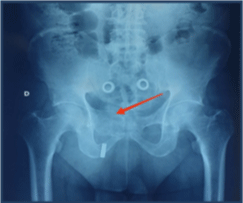
Figure 1: X-ray of the pelvis showing an osteolytic image of the right iliopubic brace.
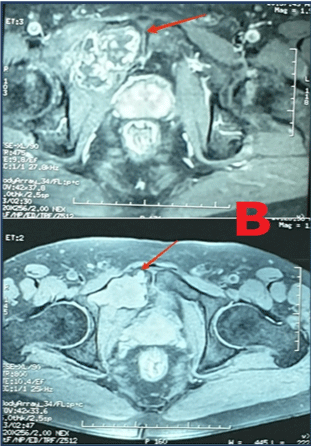
    Figure 2: MRI: Axial slice, tumor in T2 hypersignal with right latero-vesical

endo-pelvic extension.

Major axis diameter: 72mm.
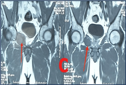
Figure 3: MRI: Axial section, showing the tumor in hyposignal T1.
An extra-marginal resection of the tumor was performed in collaboration with a visceral surgeon. The approach was centered on the region of the right groin and directed downwards and inwards to become elective on the tumour. The dissection was careful to avoid any lesion of the femorocutaneous nerve and the external iliac vessels on the lateral side, and the right spermatic cord on the medial side. Resection removed the right iliopubic branch after protection of the endopelvic organs with a retractor (Figure 4). The closure was carried out plane by plane on the suction Redon drain after placement of a wall reinforcement plate (Figure 5).
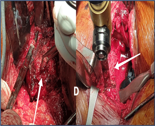
Figure 4: Intraoperative images showing the tumor and level of resection.
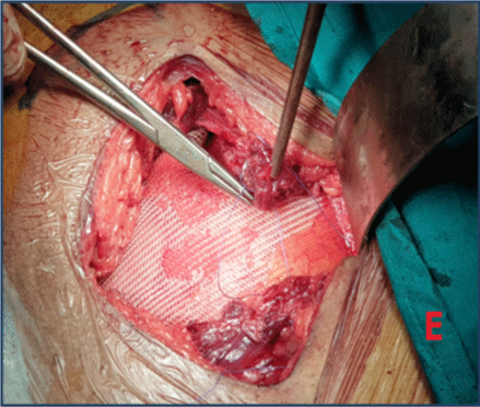
    Figure 5: Intraoperative image showing the placement of a wall reinforcement

plate.
The anatomo-pathological study of the excised specimen found healthy resection limits (Figure 6 and 7). The postoperative followup was simple. Clinical and radiological controls after two years of follow-up did not find any recurrence.

Figure 6: The excision piece measuring 96mm at the major axis.
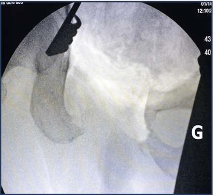
Figure 7: Postoperative image intensifier control showing the resection area.