
Case Report
Austin Surg Case Rep. 2022; 7(1): 1047.
Revisions of Total Hip Prostheses: About 03 Cases and Review of the Literature
Mokhchani Y1,2*, Abderrafia R1,2, Rabbah A1,2, Boukhriss J1,2, Chafry B1,2 and Boussouga M1,2
1Department of Orthopedic Surgery and Traumatology II, Mohammed V Military Teaching Hospital, Rabat, Morocco
2Faculty of Medicine and Pharmacy, Mohammed V University, Rabat, Morocco
*Corresponding author: Youness Mokhchani, Department of Orthopedic Surgery and Traumatology II, Mohammed V Military Teaching Hospital, Rabat, Morocco; Faculty of Medicine and Pharmacy, Mohammed V University, Rabat, Morocco
Received: April 07, 2022; Accepted: April 30, 2022; Published: May 07, 2022
Abstract
Hip arthroplasty is a reliable means in the treatment of hip conditions. By restoring its mobility, stability and indolence. However, this prosthetic surgery exposes to the risk of the occurrence of complications that can affect the functional prognosis. The most common complications are dislocations, fractures, loosening, and infections. These complications may require surgical revision of the total hip prosthesis (THA). We present three cases of patients who required revision THA, and we present the therapeutic recommendations for each of the complications in the literature, to ensure adequate management, and the recovery of a painless, mobile and functional hip.
Keywords: Prosthesis; Hip; Arthroplasty; Revision; Complications
Introduction
The revision of total hip prosthesis is a surgical procedure which aims to replace all or part, femoral or acetabular, of the total hip prosthesis (THA) [1]. It is becoming more and more frequent, and represents about 15% of all prostheses placed. This is explained by an increase in the implantations of primary prostheses from the 1980s and a longer life expectancy in patients whose functional demands are increasingly high. Here we present interesting observations from 3 patients who had different reasons for revision surgery for THA [2].
Case Presentation
First patient
This is a 66-year-old patient, with a history of repeated head trauma during public road accidents, and who is being monitored in neurology for epilepsy and memory disorders. This patient was admitted to our training for a classified right femoral neck fracture (Garden 4) following a fall from the stairs during a “Grand mal” epileptic seizure, with landing on the right hip. The patient was operated on with a non-cemented total right hip prosthesis, dual mobility. The postoperative course was simple and satisfactory. In addition, the patient was lost to sight 3 months after the intervention for family reasons. At his first consultation, we noticed a stiffness of the hip in the absence of functional rehabilitation. The evolution was marked by the persistence of stiffness with the appearance of some periprosthetic ossifications, hence the prescription of antiinflammatories such as “indomethacin” with patient awareness of the need for physiotherapy. Given the non-cooperation of the patient, he was lost sight of once again, to return after 9 months of the surgical gesture accusing pains of the right hip with cutaneous fistula. The standard X-ray showed loosening of the acetabular implant with the constitution of a true periprosthetic bone bridge. Computed tomography (CT) of the right hip better visualized the loosening and the bony bridge between the antero-inferior part of the acetabulum and the trochanteric massif, and showed the communication of the fistulous course with the joint without collection image. After a preoperative and infectious assessment, the patient was admitted to the operating room, where the same postero-external incision was made (according to MOORE), taking away the fistula in orange wedge, and we proceeded to the excision of the fibrosis around the fistulous path, then the bone bridge and prosthetic implants were removed with bacteriological samples taken. We ended with an abundant wash and placement of a spacer which was done using a 30mm pin and surgical cement with antibiotics. Postoperatively, the patient was put on analgesics, anticoagulants, and parenteral bi-antibiotic therapy adapted to the data of the antibiogram for 6 weeks. Six months later, the patient underwent surgical revision, with ablation of the spacer and reconstruction of the acetabulum with a bone allograft and placement of a screwed RING, on which the acetabulum implant was cemented, then placed in places a long cemented femoral stem. The medium-term evolution was favorable with the resumption of a functional and painless mobile hip without infectious recurrence (Figure 1-5).
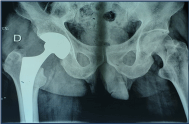
    Figure 1: X-ray of the pelvis showing a total prosthesis of the right hip after a

fracture of the neck of the right femur.
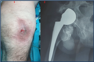
    Figure 2: At 9 months: 1) Cutaneous fistula next to the surgical scar; 2) X-ray

of the right hip showing the loosening with the peri-prosthetic calcifications.
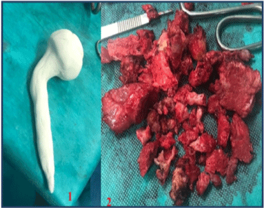
    Figure 3: 1) Spacer prepared from a pin and surgical cement; 2: Excised

periprosthetic calcifications.
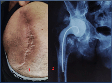
    Figure 4: 1) Skin condition after placement of the Spacer; 2) X-ray of the right

hip showing the spacer in place.
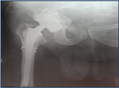
Figure 5: X-ray of the right hip showing a revision THA in place.
Second patient
This is a 70-year-old woman, operated in 2005 for a fracture of the neck of the right femur by a cemented total right hip prosthesis, with simple mobility (metal-polyethylene couple), and who has been reporting for some months of right hip pain with significant reduction in walking distance. A standard X-ray was performed, showing loosening of the acetabular implant with acetabular protrusion, as well as a border of more than 3mm between the cement and the bone on the femoral side. The patient underwent revision surgery, with acetabular reconstruction using a bone allograft and a targeted KERBOULL cross, on which a total hip prosthesis, dual mobility, cemented and with a long stem, was placed. The evolution was marked by the recovery of a mobile, painless and functional hip (Figure 6 and 7).
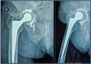
Figure 6: X-ray of the right hip showing a loose THA.
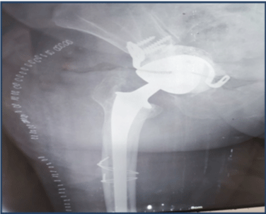
    Figure 7: X-ray of the right hip showing revision THA, with acetabular

reconstruction by allograft and the KERBOULL cross + femoral cerclage.
Third patient
This is a 65-year-old patient, known to be diabetic, for whom we placed in 2006, a total prosthesis of the left hip, simple mobility and cemented following a fracture of the neck of the femur. The patient has been reporting pain in her left hip for several weeks following a fall from her height. The standard radiograph of the left hip showed prosthetic loosening with a periprosthetic shear line. The acetabular reconstruction was performed using a bone allograft and a screwed RING on which the acetabular implant was cemented, then a long cemented femoral stem was placed. Finally, we completed a diaphyseal cerclage. Postoperatively, functional rehabilitation was started with verticalization without support for 2 months. Full functional recovery was obtained after 3 months (Figure 8).
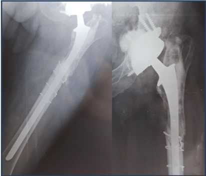
    Figure 8: X-ray of the left hip showing revision THA with acetabular

reconstruction by allograft and a RING + femoral cerclage.
Discussion
Revisions of total hip prostheses represent approximately 15% of their total number. These are surgical interventions that aim to replace all or part of the total hip prosthesis (THA) [3,4]. The usual causes of revision of THA are aseptic loosening, dislocations, fractures and infections. The problematic prosthesis is manifested by persistent or intermittent pain. Sitting at the level of the hip or the gluteal region, accompanied by lameness, loss of autonomy, and stiffness of the joint. Signs suggestive of an infection are fever, redness, swelling, and fistula in the skin next to the joint [5].
Before any surgical revision, a complete imaging assessment, an infectious assessment and an operability assessment must be carried out to specify and analyze the extent of the lesions and plan the therapeutic modalities for the replacement of the prosthesis [6-8].
The surgery is performed under general or locoregional anesthesia. Usually by reworking the old incision. In the event of aseptic loosening, the replacement of the prosthesis will be associated with a bone graft, the volume of which depends on the extent of the loss of bone substance (autograft or allograft) [9]. The revision can be done with implants identical to the first intervention or with specific implants (long femoral implant, screwed acetabular implant, etc). In addition, additional parts can be used to reinforce the hold of the new prosthesis (the KERBOULL cross, BURCH SCHNEIDER ring, etc.)
In the event of recurrent dislocation, revision may consist of a simple change of position of the original parts or the fitting of a new prosthesis [10].
In the event of a fracture on prosthesis, the intervention may be limited to fixing the fracture with osteosynthesis material (screwed plate, hook, and strapping) or changing the old prosthesis if it is loosened at the same time as the repair of the femur [11].
Septic loosening is the most complex situation. The prosthesis change can be done in one or two stages. The removal of the infected prosthesis, the cleaning of the infected surfaces and the fitting of a new prosthesis can be carried out during the same intervention or in two operations separated by several weeks. Depending on the age of the infection, the characteristics of the germ and the general condition of the patient. In all cases, the operation will be followed by antibiotic therapy for several weeks. A suction drain is usually left to limit the formation of a hematoma [12].
Lifting and pressing on the limb are authorized after a delay which depends on the intervention carried out. In cases of bone grafting or fracture repair, support is often prohibited or partial with the use of canes for at least 6 weeks. Hip rehabilitation using antiluxating movements is necessary to avoid any stiffness [13-15]. The resumption of normal activity depends on the type of intervention. If the intervention consisted of a simple change of prosthesis, the delays will be 6 to 8 weeks. If the intervention included bone grafts, the resumption of normal activity may require 3 to 6 months of convalescence. The lifespan of a total hip prosthesis is currently 15 years minimum in the absence of complications.
Conclusion
The replacement of a Total Hip Prosthesis (THA) is a complex intervention, longer and more difficult than the installation of the first prosthesis (reconstruction by bone grafts and means of osteosynthesis). The follow-up is longer and the results may be less satisfactory than those of the first prosthesis, especially if the damage is severe, with an intervention carried out too late. Finally, preoperative and postoperative complications are more frequent than for a first THA and their prevention is based on a rigorous anesthetic, biological and imaging assessment.
References
- Yu R, Hofstaetter JG, Sullivan T, Costi K, Howie DW, Solomon LB. Validity and reliability of the Paprosky acetabular defect classification. Clin Orthop Relat Res. 2013; 471: 2259-2265.
- Vastel L, Lemoine CT, Kerboull M, Courpied JP. Structural allograft and cemented long stem prosthesis for complex revision hip arthroplasty: use of a trochanteric claw plate improves final hip function. Int Orthop. 2007; 31: 851-857.
- Sakellariou VI, Babis GC. Management bone loss of the proximal femur in revision hip arthroplasty: Update on reconstructive options. World J Orthop. 2014; 5: 614-622.
- Lautmann S, Rosset P, Burdin P. Reconstruction acétabulaire par anneau de soutien dans les prothèses totales de hanche. Annales orthopédiques de l’Ouest. 1998; 30: 129-135.
- Rosenberg AG. Cementless acetabular components: the gold standard for socket revision. J. Arthroplasty. 2003; 18: 118-120.
- Hunter GA, Welsh RP, Cameron HU. The results of revision of total hip arthroplasty. J Bone Joint Surg. 1979; 61B: 419-422.
- Solomon DH, Losina E, Baron JA, Fossel AH, Guadagnoli E, et al. Contribution of hospital characteristics to the volume outcome relationship: dislocation and infection following total hip replacement surgery. Arthritis Rheum. 2002; 46: 2436-2444.
- Lortat Jakob. Prothèse totale de hanche infectée. Cahier d?enseignement de SOFCOT. 1998.
- Jenny JY, Boeri C. Les reprises des prothèses totales de hanche infectée: étude bactériologique. Symposiume de SOFCOT. 2001; 1S: 164-165.
- Mertl P, Vrenois J, Meunier W, Havet E, Massy S. Infection chronique: résultats de réimplantation en 2 temps. Symposium de SOFCOT. 2001; 1S: 174-178.
- Masaoka T, Yamamoto K, Shishido T, Katori Y, et al. Study of hip joint dislocation after total hip arthroplasty. Int Orthop. 2006; 30: 26-30.
- Wroblewski BM. Dislocation of the hip arthroplasty. In: Revision surgery in total hip arthroplasty. Londres: Springer Verlag. 1990: 29-46.
- Morrey BF. Difficult complications after hip joint replacement - Dislocation. Clin Orthop Relat Res. 1997: 179-187.
- Amstutz HC, Kody MH. Dislocation and subluxation. New York, Tokyo Melbourne: Churchill Livingston. 1991: 78-80.
- Hunten D Langlais. Luxations et subluxations des prothèses totales de la hanche-Prothèse totale de la hanche: les choix. Cahiers d’enseignement de la Sofcot. 2007; 90: 371-413.