
Clinical Image
Austin Surg Case Rep. 2022; 7(1): 1048.
Cholecysto-Hydatid Cyst Fistula: Images in Medicine
Youssef El Mahdaouy*, Noureddine Njoumi, Mohamed El Fahssi, Mbarek Yaka, Abderrahman Hjouji and Abdelmouaim AIT Ali
Department of Surgery, Service of Visceral Surgery 2, Military Hospital Mohamed V, Rabat, Morocco
*Corresponding author: Youssef El Mahdaouy, Department of Surgery, Service of Visceral Surgery 2, Military Hospital Mohamed V, Rabat, Morocco
Received: May 27, 2022; Accepted: June 14, 2022; Published: June 21, 2022
Keywords
Hepatichydatidcyst; Gallbladder; Fistula.
Visual Case Discussion
Human hydatid disease is a zoonosis caused by the larval stage of the Echinococcus species. It is endemic in sheep and cattle raising region such as Morocco. The liver is the most frequently affected organ in echinococcosis (first filter). Cyst rupture into the biliary tree is the most common complication of hepatic hydatid cyst, seen in 5% to 15% of cases. However, fistulization between gallbladder and hydatid cystis rare. The number of cases in the literature is below 10 [1].
Ultrasonography and computed tomography may show the communication but endoscopic retrograde cholangio pancreatography can detect the fistulization in detail. However, the diagnosis, in some cases, can only be made by laparotomy [1].
Here in, we report a case of a 28-year-old man, with no particular pathological history, who was admitted to our service for abdominal pain in the right hypochondrium of 5 month’s duration without any other signs. The clinical examination revealed a patient in good general condition, with no abdominal tenderness or palpable mass. The hydatid serology was positive and the other laboratory findings were normal. The abdominal computed tomography showed welldefined heterogeneous solid cyst with peripheral calcification of approximately 5×7×8 cm in segment V of the liver. A communication between the hydatid cyst and the gallbladder was suspected (Figure 1). The patient was taken to open surgery, which confirmed the imaging finding. The treatment consisted of partial pericystectomy along with cholecystectomy following sterilization maneuvers for cyst hydatid (Figure 2). Then, an investigation of the common bile duct and biliary lavage followed by transcystic biliary drainage were carried out. The postoperative recovery was uneventful. The anatomopathological analysis of the specimen confirmed the diagnosis by highlighting the fistula between the gallbladder and the hydatid cyst. The patient was discharged from hospital 5 days after surgery. A postoperative Albendazole therapy was prescribed for 3 months and a percutaneous transcystic cholangiography was planned in a month.
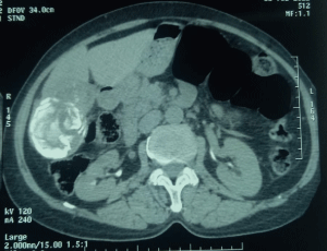
    Figure 1: Computed tomography scan of the communication between the

gallbladder and the hydatid cyst (arrow).
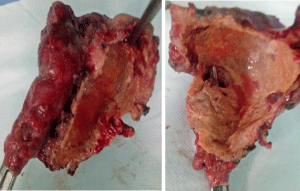
    Figure 2: Specimen of partial pericystectomy along with cholecystectomy.

The gallbladder was tunneled by a surgical instrument from its cystic duct to

the fistula with the hydatid cyst.
Conflict of Interest and Authorship Confirmation
All authors have participated in (a) conception and design, or analysis and interpretation of the data; (b) drafting the article or revising it critically for important intellectual content; and (c) approval of the final version.
The authors have no affiliation with any organization with a direct or indirect financial interest in the subject matter discussed in the manuscript.
References