Case Report
Austin Surg Case Rep. 2022; 7(2): 1051.
Short-Term Efficacy of Lars Artificial Ligament Reconstruction for Spontaneous Quadriceps Tendon and Contralateral Patellar Tendon Rupture in a Chronic Hemodialysis Patient: A Case Report
Fuyuan Deng1#, Juncai Liu2,3* and Zhong Li2,3*
¹Department of Orthopaedics, People’s Hospital of Deyang City, China
²Department of orthopedics, The Affiliated Hospital of Southwest Medical University, China
³Sichuan Provincial Laboratory of Orthopaedic Engineering, China
#These authors contributed equally to this work
*Corresponding author: Zhong Li, Department of orthopedics, The Affiliated Hospital of Southwest Medical University, China
Juncai Liu, Sichuan Provincial Laboratory of Orthopaedic Engineering, China
Received: July 18, 2022; Accepted: August 17, 2022; Published: August 24, 2022
Abstract
Background: Spontaneous rupture of the quadriceps and patellar tendons is a very rare injury that usually occurs in middle-aged to elderly patients. It’s often associated with chronic metabolic disorders, such as diabetes, hyperparathyroidism, gout, chronic renal failure, or long-term steroid use.
Case Presentation: This article describes a 43-year-old man who has been on hemodialysis for 8 years due to uremia. The patient occurred pain without an obvious cause in both knees, difficulty in walking and limited extension of both knees for more than 4 months, and aggravation for more than 1 month. Conservative treatment for him was not valid. The rupture of the quadriceps tendon and the patellar tendon was confirmed according to the patient’s clinical symptoms and Magnetic Resonance Imaging (MRI). We used Ligament Advanced Reinforcement System (LARS) artificial ligament to reconstruct the patellar tendon and quadriceps tendon. The tendon and knee joint function recovered well after surgery. No infection or tendon rupture was found during follow-up.
Conclusion: We report a case of the femoral quadriceps tendon and contralateral patellar tendon rupture during long-term hemodialysis. After the failure of conservative treatment, LARS artificial ligaments were used to reconstruct the ruptured tendon, and good results were obtained.
Keywords: Chronic renal failure; Hemodialysis; Tendon rupture; Artificial ligament
Abbreviations
MRI: Magnetic Resonance Imaging; LARS: Ligament Advanced Reinforcement System; WBC: White Blood Cell; HGB: Hemoglobin; Crea: Creatinine; UA: Uric Acid; Ca: Calcium; P: Phosphorus; ALP: Alkaline Phosphatase
Background
Spontaneous rupture of the quadriceps or patellar tendon is very rare in clinical practice, and there are only a few cases reported abroad. Such diseases are considered secondary to systemic diseases such as chronic renal failure [1,2,6,10,15], diabetes [8], hyperparathyroidism [5], osteogenesis imperfecta [4], Etc. It is easy to miss or misdiagnose clinically. There is related literature that the early misdiagnosis rate can reach more than 50% during the reporting period [5], often the best treatment time is missed, which affects the recovery of knee function after surgery. Some scholars believe that early surgical treatment leads to better prognosis [12], and delayed repair may lead to poor functional recovery in the later period [3]. In this article, a patient with long-term hemodialysis of uremia complicated with rupture of the right knee quadriceps tendon and contralateral patellar tendon rupture. Due to unclear early diagnosis, it developed a chronic injury. We used LARS artificial ligament to reconstruct the patellar tendon and the four femoral heads. The tendon and knee joint function recovered well after surgery. The report is as follows.
Case Presentation
A 43-year-old male patient was admitted to the hospital with “restriction of knee joint straightening for more than 4 months and aggravation for more than 1 month. Four months ago, the patient presented pain and difficulty in walking with no obvious cause in both knees and received analgesic medication with poor effect. Therefore, he went to a local hospital and was recommended to our hospital for treatment. In our hospital, he was admitted with right knee quadriceps tendon rupture and left knee patellar tendon rupture. Parathyroidectomy was performed more than 4 months ago for “secondary hyperparathyroidism”, and no history of trauma was denied. The patient has a history of uremia for more than 8 years and he is undergoing regular hemodialysis treatment in the hospital 3 to 4 times a week. Physical examination after admission: no obvious swelling of the knee joints, no varicose veins in the lower extremities, no surgical scars, depressions can be touched below the patella of the left knee and above the patella of the right knee, no obvious tenderness in the right knee, and the knees cannot be actively extended Straight, both knees active and passive flexion functional activity is acceptable, both legs are equal in length. A lateral radiograph of the right knee joint and MRI showed a quadriceps rupture (Figure 1A-B), and a lateral radiograph of the left knee joint and MRI showed a patellar tendon rupture (Figure 1CD). Laboratory tests: White Blood Cell (WBC) count 6.54×10⁹/L, Hemoglobin(HGB) 120g/L, Urea 25.83mmol/L, Creatinine(Crea) 120.1μmol/L, Uricacid(UA) 506.6μmol/L, Calcium(Ca) 2.36mmol/L, Phosphorus(P) was 2.77mmol/L, and Alkaline Phosphatase(ALP) was 91.6U/L. Hemodialysis was performed 1 day before the operation, and antibiotics were used prophylactically for 30 minutes before the operation. On the second day, after the patient’s creatinine, ions and other indicators reached the surgical requirements, the LARS artificial ligament was used to reconstruct the patellar tendon and quadriceps tendon under general anesthesia.

    Figure 1A: X-ray film of the left knee shows the extension of the distance from the lower extremum of the patella to the tibial tubercle, indicating patella Alta B.

This MRI demonstrates the discontinuity of the patellar ligament C. X-ray of the right knee shows the patella descending, indicating patella Baja D. MRI shows a

continuous discontinuity of the signal of the quadriceps tendon of the right knee, severe contracture.
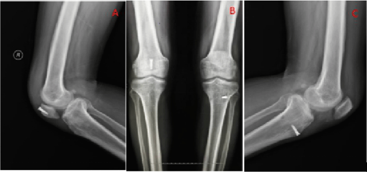
After anesthesia under general anesthesia, the patellar tendon was reconstructed. The anterior median patellar incision was made, and the skin and subcutaneous tissues were cut down and across the tibial tubercle. Patellar tendon rupture was observed intraoperatively (Figure 2A). Firstly, the LARS artificial ligament was passed under the torn patellar tendon, and then the Kirschner needle was used to penetrate the posterior medial side from the lower tibial plateau to expand the bone tunnel. Then, the steel wire after being shaped was pulled into the medial side from the lateral side and passed out. The LARS artificial ligament was guided through the wire through the bone tunnel. After the metal squeeze screw was fixed, The patella track was good when knee flexion and extension were measured, and the patella height was normal when measured by fluoroscopy during the operation. Then carry out the reconstruction of the right quadriceps tendon of the right knee, and similarly take the anterior midline approach, about 9cm in length, and cut through each layer of tissue in order. During the operation, the right knee femoral quadriceps tendon attachment was completely abraded and the tear extends to both sides of the tendon. The stump extended on both sides, severely shrunk and formed adhesions, and poor blood supply (Figure 2B). After the degenerative and necrotic tendon is excised and the stump tendon tissue is trimmed and shaped, a curette is used to decorate the cortices at the top of the patella and let it bleed slightly. First, a 6mm diameter bone tunnel was drilled longitudinally on the upper pole of the patella, and a 6mm diameter LARS artificial ligament was introduced into the patella tunnel. The ligament line was included in the longitudinal patella tunnel and the broken quadriceps tendon was reset and fixed. Then, the quadriceps tendon and LRAS artificial ligament and surrounding soft tissue were reinforced and sutured. During the operation, the knee joint was flexed and stretched, and patella activity and movement track were observed to return to normal. No fracture of the quadriceps tendon was viewed. Two years after the operation, the patella height of the left knee was restored to normal, patella position of the right knee was slight lower (Figures 3A-C), but the flexion and extension function of both knees recovered well. The patient had no ligament rupture again, pain, knee stiffness with limited flexion and extension activities. The Lysholm score of the left knee was 20 before surgery and 90 after surgery. The Lysholm score of the right knee was 20 before surgery and 85 after surgery.

    Figure 2A: Intraoperative photo showing the left knee patellar ligament rupture B. Intraoperative photo showing the right knee femoral quadriceps tendon rupture

and stump atrophy.

    Figure 3A-C: Metal compression screws can be seen on the lateral and lateral radiographs of both knees. A. The lateral radiograph of the right knee indicates a

lower patella but is better than before. C. The radiograph of the left knee indicates that the patella is normal.
The most common complications after knee extensor repair are decreased knee flexion and quadriceps atrophy. The patient should support the crutches within 3 weeks after the operation, carry the appropriate weight within the tolerable range, and abandon the crutches after the quadriceps strength is enhanced. The patient wore a knee brace within 6 weeks after surgery and walked under the protection of the brace. The range of knee joint movement from 0° to 45° was allowed to increase to 0° to 90° after 3 weeks, and encourage early straight leg elevation(Wear a locked brace before the delayed extension of the knee extension) to strengthen the quadriceps and prevent the quadriceps atrophy that is common practice after surgery. Functional training should be reinforced after 7 weeks, and confrontational exercise should be started 10 to 12 weeks after surgery. After3 months, the patient can gradually go back to exercise. By strengthening the initial range of motion and the strength of the quadriceps, you can achieve Full strength recovery.
Discussion
The extensor device of the knee joint includes the quadriceps tendon, the patella, and the patellar tendon. Any damage to the structure will lead to weakness and even loss of function of the extensor device. Injuries of unilateral extensor devices usually occur from trauma, while injuries of bilateral extensor devices usually occur from certain chronic metabolic diseases, such as chronic renal failure [1,2,6,10,15], diabetes [8], Hyperparathyroidism [5], osteogenesis imperfecta [4] or long-term use of steroids [16]. In 1962, Preston and Adicoff reported the first hemodialysis patient with chronic renal failure who had quadriceps tendon avulsion at the same time [23]. Since then, many authors have reported spontaneous bilateral quadriceps ruptures after hemodialysis due to uremia [2,7,9], bilateral patellar tendon ruptures [13], ipsilateral quadriceps, and three brachial Muscle rupture [6] and so on.
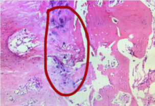
Tendon rupture is related to the duration of renal failure and the duration of hemodialysis treatment [14]. Although the exact mechanism of injury is unknown, most researchers believe that secondary hyperparathyroidism caused by repeated dialysis treatments plays an important role in the pathogenesis of tendon rupture [11]. Phosphorus retention caused by decreased glomerular filtration rate in uremia patients leads to hypocalcemia and increased reactive parathyroid hormone. Hyperparathyroidism stimulates osteoclast activity, especially in the subtended area, and weakens the bone cortex [5]. Finally, repeated mild avulsion fractures lead to a total tendon rupture with mild trauma [17]. Malnutrition, chronic acidosis, and accumulation of uremic toxins can all lead to tendon damage [18]. We performed a pathological biopsy of the patient’s stump tendon: “ossifying myositis”, calcium deposits can be seen under the microscope (Figure 4), which will cause the quality of the tendon to deteriorate and tendon rupture.

    Figure 4: Pathological biopsy prompts: “ossifying myositis” with visible

calcium deposits.
In the acute phase of knee extension rupture, it usually complains of pain in the knee joint with limited knee extension or difficulty in walking upright [22]. Physical examination showed that the patient could not actively extend the knee and the depression of the upper or lower poles of the patella. The knee pain in the knee can not actively extend the knee and the depressions on the condyle are specific signs of complete rupture of the quadriceps tendon. However, in some cases, Hematoma in the area of acute injury and scar tissue in the joint of chronic injury may make the depression difficult to detect, especially when the injury is bilateral, and a contrast physical examination cannot be performed, which may cause misdiagnosis [19]. Siwek and Rao [20] Studies in 1981 showed that 28% of ruptures were not initially diagnosed in thirty-six ruptures of the quadriceps tendon and thirty-six ruptures of the patellar ligament .n. Perfitt et al. [21] published a study showing that 67% of patients with quadriceps femoris rupture are misdiagnosed at their first visit, which may lead to poor clinical outcomes. Therefore, in addition to clinical diagnosis, some imaging methods are also needed to improve the diagnosis rate. Quadriceps femoris rupture and patellar tendon rupture can be diagnosed by plain radiographs of the knee showing patella Alta or patella Alta [24]. MRI is the most effective tool for diagnosing chronic or suspicious ligament rupture and excluding other intra-articular injuries. Preoperative MRI can accurately determine the location and extent of tendon rupture, which is very helpful for clinical diagnosis and preoperative planning. And the last postoperative MRI can show the healing and continuity information of the repaired quadriceps tendon [1]. Ultrasound is a fast and simple way to determine the rupture of the patellar ligament. The location of the tear can be determined by the hypoechoic or non-echoic area, but accurate ultrasound results require an experienced operator [25].
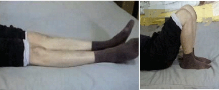
The destruction of the knee extensor mechanism should be treated in time to achieve the best functional results. Delaying treatment for more than 2 weeks may lead to poor functional results [3,20]. Surgical treatment methods include simple suturing or suturing and wire band fixation, patella drilled suture to preserve the semitendinosus or gracilis sutures to reconstruct the ligament, Patellofemoral tunnel patelloid-tendon anastomosis, Suture with thread anchors, etc. However, as a whole, there is no gold standard for the treatment of tendon ligament rupture. A meta-analysis by Neubauer [26] showed that transpatellar tunnel quadriceps tendon repair is currently the most commonly used and effective repair technique. In the case, we reported, the patient came to the hospital 4 months after spontaneous quadriceps tendon and contra lateral patellar tendon rupture. The doctor did not use the auto graft of the patient, because the patient’s tendon and bone conditions are poor. At the same time, there is a contracture deformity of the tendon. We used the LARS artificial ligament to reconstruct the broken tendon. Due to the poor quality of autogenous tendons and large tendon defects, sufficient quantity and quality are needed for tendon reconstruction, and LARS ligament can solve this problem. LARS ligament strength is sufficient, which can meet patients’ early functional exercise and promote the recovery of knee joint function. After 4 years of follow-up, the patient’s flexion and extension of both knee joints had a good recovery (Figure 5). However, the right knee quadriceps tendon was severely contracture due to the long rupture time, and the problem of the right knee lower patella still existed after the operation. The knee joint function of the patient was not affected much in the short term, and the long-term efficacy still needs further follow-up.

Figure 5: Four years after surgery, the patient’s knee joint extension, and flexion function returned to normal.
Conclusion
In recent years, more and more uremia patients need long-term hemodialysis, tendon rupture of patients may be more frequent. For this kind of patient, combined with MRI, knee ultrasound, and other examination methods can improve the diagnosis rate of the disease and reduce the rate of misdiagnosis. Early surgery can improve the function of the knee. For patients with delayed surgery, the use of artificial ligament reconstruction can achieve good results. During surgery, lateral x-rays or fluoroscopy should be used to ensure normal patella height before tension adjustment and repair, thus avoiding patella Alta or patella Baja.
Consent for Publication
Written informed consent was obtained from the patient for publication of this case report and any accompanying images.
Availability of Data and Materials
The datasets used and/or analysed during the current study are available from the corresponding author on reasonable request.
Competing Interests
The authors declare that they have no competing interests.
Funding
This work was supported by the Applied Basic Research Project of Science and Technology Department of Sichuan Province (2020YJ0265), Sichuan University-Luzhou Government Strategic Cooperation Project (No. 2019CDLZ-17).
Authors’ Contributions
ZL and JCL designed this study. YL and FYD wrote this study. ZL, JCL performed the surgery.
References
- Kim YH, Shafi M, Lee YS, Kim JY, Kim WY, Han CW. Spontaneous and simultaneous rupture of both quadriceps tendons in a patient with chronic renal failure. Knee Surgery, Sports Traumatology, Arthroscopy. 2005; 14: 55-59.
- Seng C, Lim YJ, Pang HN. Spontaneous disruption of the bilateral knee extensor mechanism: a report of two cases. J Orthop Surg (Hong Kong). 2015; 23: 262-6.
- Zribi W, Zribi M, Guidara AR, Jemaa MB, Abid A, Krid N, et al. Spontaneous and simultaneous complete bilateral rupture of the quadriceps tendon in a patient receiving hemodialysis: A case report and literature review. World Journal of Orthopedics. 2018; 9: 180-184.
- Figueroa D, Calvo R, Vaisman A. Spontaneous and simultaneous bilateral rupture of the quadriceps tendon in a patient with osteogenesis imperfecta: a case report. Knee. 2006; 13: 158-60.
- Wu W, Wang C, Ruan J, Wang H, Huang Y, Zheng W, et al. Simultaneous spontaneous bilateral quadriceps tendon rupture with secondary hyperparathyroidism in a patient receiving hemodialysis. Medicine. 2019; 98: e14809.
- Moerenhout K, Gkagkalis G, Benoit B, Laflamme GY. Simultaneous Ipsilateral Quadriceps and Triceps Tendon Rupture in a Patient with End-Stage Renal Failure. Case Reports in Orthopedics. 2018; 2018: 1-5.
- Gao MF, Yang HL, Shi WD. Simultaneous bilateral quadriceps tendon rupture in a patient with hyperparathyroidism undergoing long-term haemodialysis: a case report and literature review. J Int Med Res. 2013; 41: 1378-83.
- Torkaman A, Gomrokchi AY, Elahifar O, Barmayoon P, Shojaei SF. Simultaneous bilateral rupture of patellar tendons in diabetic hemodialysis patient: A case report. Caspian Journal of Internal Medicine. 2018; 9: 306- 311.
- Kayali C, Agus H, Turgut A, Taskiran C. Simultaneous bilateral quadriceps tendon rupture in a patient on chronic haemodialysis. (Short-term results of treatment with transpatellar sutures augmented with a quadriceps tendon flap). Ortopedia, traumatologia, rehabilitacja. 2008; 10: 286-91.
- Kim BS, Kim YW, Song EK, Seon JK, Kang KD, Kim HN. Simultaneous Bilateral Quadriceps Tendon Rupture in a Patient with Chronic Renal Failure. Knee Surgery & Related Research. 2012; 24: 56-59.
- Malta LMA, Gameiro VS, Sampaio EA, Gouveia ME, Lugon JR. Quadriceps tendon rupture in maintenance haemodialysis patients: results of surgical treatment and analysis of risk factors. Injury. 2014; 45: 1970-1973.
- Hassani ZA, Boufettal M, Mahfoud M, Elyaacoubi M. Neglected rupture of the quadriceps tendon in a patient with chronic renal failure (case report and review of the literature). The Pan African Medical Journal. 2014; 18.
- Cherrad T, Louaste J, Kasmaoui EH, Bousbaä H, Rachid K. Neglected bilateral rupture of the patellar tendon: A case report. Journal of clinical orthopaedics and trauma. 2015; 6: 296-299.
- Wani NA, Malla HA, Kosar T, Dar IM. Bilateral quadriceps tendon rupture as the presenting manifestation of chronic kidney disease. Indian Journal of Nephrology. 2011; 21: 48.
- Tasoglu O, Ekiz T, Yenigün D, Akyüz M, Özgirgin N. Bilateral quadriceps and triceps tendon rupture in a hemodialysis patient. Hemodialysis International. 2016; 20: E19-E21.
- Lewis AC, Purushotham B, Power DM. Bilateral simultaneous quadriceps tendon rupture in a bodybuilder. Orthopedics. 2005; 28: 701-702.
- Malta LMA, Gameiro VS, Sampaio EA, Gouveia ME, Lugon JR. Quadriceps tendon rupture in maintenance haemodialysis patients: results of surgical treatment and analysis of risk factors. Injury. 2014; 45: 1970-1973.
- Ribbans WJ, Angus PD. Simultaneous bilateral rupture of the quadriceps tendon. Br J Clin Pract. 1989; 43: 122-5.
- Calvo E, Ferrer A, Robledo AG, Alvarez L, Castillo F, Vallejo C. Bilateral simultaneous spontaneous quadriceps tendons rupture. A case report studied by magnetic resonance imaging. Clinical imaging. 1997; 21: 73-76.
- Siwek CW, Rao JP. Ruptures of the extensor mechanism of the knee joint. The Journal of bone and joint surgery. American volume. 1981; 63: 932-937.
- Perfitt JS, Petrie MJ, Blundell CM, Davies MB. Acute quadriceps tendon rupture: a pragmatic approach to diagnostic imaging. European Journal of Orthopaedic Surgery & Traumatology. 2013; 24: 1237-1241.
- Maffulli N, Wong J. Rupture of the Achilles and patellar tendons. Clinics in sports medicine. 2003; 22: 761-776.
- PRESTON FS, ADICOFF A. Hyperparathyroidism with avulsion of three major tendons. Report of a case. The New England journal of medicine. 1962; 266: 968-971.
- Nance EP, Kaye JJ. Injuries of the quadriceps mechanism. Radiology. 1982; 142: 301-307.
- LaRocco BG, Zlupko G, Sierzenski P. Ultrasound diagnosis of quadriceps tendon rupture. J Emerg Med. 2008; 35: 293-5.
- Neubauer Y, Wagner M, Potschka T, Riedl M. Bilateral simultaneous rupture of the quadriceps tendon: a diagnostic pitfall? Report of three cases and meta – analysis of the literature. Knee Surg Sports Traum Arthros. 2007; 15: 43-53.