
Case Report
Austin Surg Case Rep. 2022; 7(2): 1052.
Cerebral Infarction and Myocardial Depression after Cardiac Surgery Caused by Extensive Vasospasm of Carotid Artery, Vertebral Artery and Coronary Artery
Ko TY¹, Cho SH¹* and Lee Y²*
¹Department of Thoracic and Cardiovascular Surgery, Kosin University Gospel Hospital, Kosin University College of Medicine, Korea
²Department of Pschiatry, Kosin University Gospel Hospital, Kosin University College of Medicine, Korea
*Corresponding author: Seong Ho Cho, Department of Thoracic and Cardiovascular Surgery, Kosin University Gospel Hospital, Kosin University College of Medicine, 262 Gamcheon-ro, Seo-gu, Busan 49267, South Korea
Yunna Lee, Department of Pschiatry, Kosin University Gospel Hospital, Kosin University College of Medicine, 262 Gamcheon-ro, Seo-gu, Busan 49267, South Korea
Received: August 09, 2022; Accepted: August 24, 2022; Published: August 31, 2022
Abstract
Postoperative cerebral infarction is associated with significant morbidity and mortality. In this study, we describe a case of postoperative (Mitral valve plasty with MAZE procedure) cerebral infarction and myocardial depression caused by extensive vasospasm of Common Carotid Artery (CCA), Vertebral Artery (VA) and coronary artery. The 59-year-old male patient was treated with Intraarterial isosorbide dinitrate injection. After treatment, there are no recurrences of cerebral or myocardial infarction. But he is attending the outpatient clinic because of the sequelae of Lt. sided hemiplegia.
Introduction
Cerebral infarction occurs in 1.5% to 5.2% of patients after cardiac surgery and it may have increased postoperative mortality and morbidity, as well as admission days and costs [1]. Cerebral embolism arised from aorta and hypoperfusion are regarded as the major factors of perioperative cerebral infarction [2]. Here in, we report a extremely rare case of vasospasm-associated postoperative cerebral infarction and myocardial depression.
Case Report
59-year-old male presented to the cardiology outpatient clinic with dyspnea on exertion. After admission, physical exam findings revealed a blood pressure of 162/106 mmHg, atrial fibrillation with rapid ventricular response and Transesophageal Echocardiogram (TEE) showed moderate to severe mitral valve regurgitation. Mitral valve plasty with MAZE procedure was performed as an elective surgery. Mitral valve repair was conducted by prolapsed site folding technique and modified Cox-Maze procedure was performed. Mitral anuuloplasty was done using 30mm complete ring. The saline test for evaluation of residual mitral regurgitation and intraoperative TEE confirmed minimal regurgitation. The Cardiopulmonary Bypass (CPB) time was 112 minutes. After epicardial defibrillation, cardiac rhythm was recovered, however weaning from Cardiopulmonary Bypass (CPB) took difficult step because cardiac contraction was depressed despite of high dose of inotropics and vasodilators.
The patient was transferred to Intensive Care Unit (ICU) after successful weaning from CPB. On the arrival to the ICU, the vital signs were stable with adequate dose of inotropics. As the patient was being monitored, a seizure episode was noticed and diffusion weighted brain Magnetic Resonance Imaging (MRI) revealed acute infarction in right Parietal-Occipital-Temporal lobe and left cerebellum (Figure 1). Immediately performed Transfemoral Cerebral Angiography (TFCA) of the right brachiocephalic trunk showed stenosis of both the right Vertebral Artery (VA) and proximal to distal part of the Common Carotid Artery (CCA) (Figure 2). Right common carotid angiography also showed severe stenosis of the external, internal carotid artery and internal cerebral artery (Figure 2). Likewise, stenosis of the left CCA, VA was observed. Cerebral infarction in this case was found to be caused by these flow restricting events. Then the decision was made to perform chemical angioplasty to dilatate the CCA and VA on both sides. Angiocatheter was placed at the origin of the affected CCA and VA, and vasodilating agent nimodipine and nicardipine was infused through the arteries. Despite the attempt, the inner diameter of the carotid artery showed no signs of dilatation. Intraarterial isosorbide dinitrate was effective in increasing the vessel diameter, thus continuous injection was performed. Procedure was discontinued at the point when the diameter of the CCA and the VA increased to normal providing sufficient Middle Cerebral Artery (MCA), anterior cerebral artery (ACA) perfusion (Figure 3). The patient’s cerebral perfusion improved as the diameter of internal carotid artery. The diameter of the internal carotid artery increased approximately 1.5mm. (4.10mm -> 5.60mm) The patient’s cerebral perfusion improved and returned to normal vascular diameter (Figure 4).
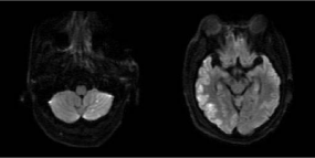
    Figure 1: Brain MRI shows infarction of right Parietal-Occipital-Temporal lobe

and left cerebellum.
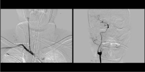
    Figure 2: Right brachiocephalic angiography shows vasospasm of whole

common-carotid artery with right cerebral artery and vertebral artery.
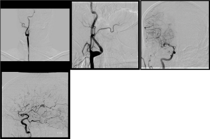
Figure 3: It is observed that the inner diameter of the internal carotid artery and right cerebral artery is recovered.
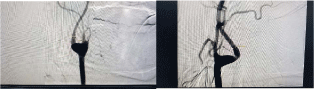
Figure 4: It is observed that the inner diameter of internal carotid artery widened 4.10mm to 5.60mm.
Comment
Cerebral infarction is one of the important complications following cardiac surgeries and significantly affects postoperative course of the patient. In this case, cerebral infarction was the result of acute vasospasm other than thromboembolism. Absence of a dissection flap in the aorta, carotid artery on Computed Tomography (CT) and MRI ruled out a possibility of carotid artery dissection from aortic dissection as the cause of the cerebral infarction. Pre-operative chest CT showed no atherosclerotic plaque in aorta and carotid artery and the carotid artery diameter of 7mm was measured in both sides. For these results, it is also unlikely that intra-arterial atheroma was detached during the surgical procedure causing cerebral infarction.
In the literature review, there were very few reports of simultaneous vasospasm in the carotid artery and coronary artery. A similar case of cerebral and myocardial infarction caused by carotid and coronary artery vasospasm has been reported and termed ‘idiopathic carotid and coronary vasospasm syndrome (ICCV) by Yoshimoto [3]. Known symptoms of visual disturbance and migraine preceding the Infarction could not be confirmed since the patient was sedated for post-operative ventilation. Takeuchi et al. [4] reported a case of focal extracranial Internal Carotid Artery (ICA) vasoconstriction in a patient presenting with recurrent transient hemiplegia who has a history of distal Left Anterior Descending artery (LAD) stent insertion due to coronary artery spasm. The patient in this case was subsequently treated with a CCB plus an anticoagulant and had no improvement in recurrent hemiplegia. Then, with steroid pulse therapy (methylprednisolone 1000 mg/d for 3 d) and subsequent oral corticosteroid therapy (prednisolone 60 mg/d), resolution of vasospasm and relief of symptoms had occurred. The cause for cerebral vasospasm is not established yet, but Kuzumoto Y et al. [5], reported a case of which the disease appeared to be associated with smoking. As for this case report, the patient’s smoking history, intra- and post-operative adrenergic drug use, and accumulation of inflammatory mediators due to cardiopulmonary bypassing might have contributed to the vasospasm of carotid and coronary arteries. In previously reported cases, focal and recurrent vasospasm of the carotid arteries led to cerebral hypoperfusion and it was treated with carotid artery stenting. This case was unique in that the vasospasm was seen throughout the whole areas of carotid arteries and there has been no recurrence. Instead of carotid artery stenting, rapid arterial dilatation and increased cerebral perfusion could be obtained through isosorbide dinitrate injection. Thereafter the patient was transferred to the department of neurology for subsequent treatment and currently attending the outpatient clinic without any recurrence of cerebral or myocardial infarction except the sequelae of Lt. sided hemiplegia. Cerebral infarction after cardiac surgery is most often caused by the embolus detached from a thrombus or an atheroma. Follow-up treatment with an anticoagulant, anticonvulsant administration or thrombectomy should be considered when the diagnosis is made. If, as in this case, cerebral infarction due to vasospasm is suspected, it can be treated with direct CCB or isosorbide dinitrate injection to the lesions. In case of postoperative cerebral infarction patient who have difficulty with CPB weaning, the possibility of vasospasm-associated infarction may be considered.
Acknowledgments
This work was supported by a grant from the National Research Foundation of Korea (NRF) (No. 2019M3E5D1A02070866).
References
- Hogue CW, Gottesman RF, Stearns J. Mechanisms of cerebral injury from cardiac surgery. Critical care clinics. 2008; 24: 83-98.
- Hogue CW, Palin CA, Arrowsmith JE. Cardiopulmonary Bypass Management and Neurologic Outcomes: An Evidence-Based Appraisal of Current Practices. Anesthesia & Analgesia. 2006; 103: 21-37.
- Yoshimoto H, Matsuo S, Umemoto T, Kawakami N, Moriyama T. Idiopathic Carotid and Coronary Vasospasm: A New Syndrome?. Journal of Neuroimaging. 2011; 21: 273-276.
- Takeuchi M, Saito K, Kajimoto K, Nagatsuka K. Successful Corticosteroid Treatment of Refractory Spontaneous Vasoconstriction of Extracranial Internal Carotid and Coronary Arteries. The Neurologist. 2016; 21: 55-57.
- Kuzumoto Y, Mitsui Y, Ueda H, Kusunoki S. [Vasospastic cerebral infarction induced by smoking: a case report]. No to shinkei = Brain and nerve. 2005; 57: 33-6.