Case Report
Austin Surg Case Rep. 2022; 7(2): 1053.
Role of CT Scan in Constrictive Pericarditis Presenting with Refractory Ascites – Review of Literature
Rajaram Sharma¹*, Vikash Sharma², Tapendra Tiwari¹, Saurabh Goyal¹
¹Assistant Professor, Pacific Institute of Medical Sciences, Umarda, Udaipur, Rajasthan, India
²Resident Doctor, Pacific Institute of Medical Sciences, Umarda, Udaipur, Rajasthan, India
*Corresponding author: Rajaram Sharma, Assistant Professor, Radio-Diagnosis, Pacific Institute of Medical Sciences (PIMS), Umarda, Udaipur, Rajasthan, India
Received: August 18, 2022; Accepted: September 15, 2022; Published: September 22, 2022
Abstract
Constrictive Pericarditis (CP) is a rigid, thick noncompliant, fibrotic, and/or calcific pericardium. Early diagnosis of CP is difficult due to its vague symptoms, deceptive course and absence of usual cardiopulmonary signs. In this case report, we represent a 27-year-old male who came to our hospital with a distended abdomen and breathlessness. Because of predominant symptoms of abdominal distention and breathlessness, the patient was advised for Contrast-Enhanced Computed Tomography (CECT) of the chest and abdomen. Interestingly, the CT scan raised the possibility of constrictive pericarditis as the underlying aetiology for all of his symptoms which was confirmed using echocardiography and right heart catheterisation CP. The patient underwent surgical pericardiectomy, and the post-surgery period was uneventful; he did not require further paracentesis and was discharged in stable condition.
Keywords: Constrictive pericarditis; Unexplained ascites; Gross pleural effusion; MRI; CECT; Echocardiography
Background
Constrictive Pericarditis (CP) is an entity in which the heart is shelling into a rigid hard pericardium with adhesion or dense fibrosis. This causes a high diastolic cardiac function workload [1]. The patients with CP may present with two types of sickness: first related to fluid overload, ranging from peripheral oedema to generalised oedema, and second related to low cardiac output response to fatigue. Unexplained elevation in Jugular Venous Pressure (JVP) with appropriate history suggests pericardial constriction. The most common cause of this disease is viral pericarditis or idiopathic. Other causes include trauma, cardiac surgery, and irradiation with mediastinum, septic infections, non-suppurative infection (tuberculosis) malignancies, systemic lupus erythematosus, rheumatoid arthritis, histoplasmosis and chronic kidney disease patients on long term dialysis [2]. Cardiac computed tomography and Magnetic Resonance Imaging (MRI) can detect pericardial thickening and calcification with high accuracy [3]. Echocardiography is a beneficial tool to differentiate between CP and restrictive cardiomyopathy. Cardiac catheterisation is the gold standard and confirmatory procedure for the diagnosis. Pericardiectomy is the most definitive treatment of CP and should be as done as soon as possible. The physicians sometimes neglect the diagnosis of CP because of the non-specific signs and symptoms; the physicians may attribute the symptoms to another disease process. This case exemplifies the issues in diagnosing this condition; the essential investigation, outcomes or follow-up of prompt treatment, and a discussion of the disease.
Case Presentation
A 27-year-old man was presented with complaints of breathlessness, pleuritic chest pain, weakness, fatigue feeling, distension of abdomen, and peripheral oedema for about one year. The patient complained of pain and progressive abdominal distention in the past ten days. The patient was hemodynamically stable, but JVP was significantly elevated. Heart sounds were muffled, and a reduction of the sounds was found at the right lung base. Significant right pleural effusion, mild hepatomegaly with ascites and peripheral oedema were noted in the clinical examination. Primary laboratory evaluations were unremarkable. Ascitic fluid examination revealed high protein content (4.1 g/dL) and a raised serum-ascites gradient (1.6 g/dL).
Investigations
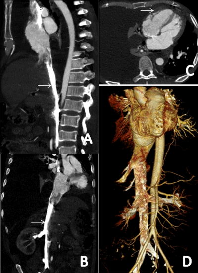
A Contrast-enhanced CT (CECT) scan of the chest and abdomen was advised for further evaluation, which revealed significant right pleural effusion, gross ascites, and dilated Inferior Vena Cava (IVC) & hepatic veins. (Figure 1A & B) It also showed that the heart was overall smaller in size and was pushed towards the left side. The pericardium was thickened, and it measured 5 mm in maximum thickness. The right atrium was dilated with an early enhancement of IVC and hepatic veins in the arterial phase. (Figure 1C & D) The right atrium enlargement, inferior vena cava dilation, and septal bouncing were found in the conducted echocardiography. All of these features were suggestive of CP. Thereafter, the right heart catheterisation was performed for confirmation of the diagnosis. The odd finding to this particular case was gross unilateral pleural effusion which could not be explained. The patient underwent corrective surgery, during which a complex thickened pericardium was found for which pericardiectomy was performed. The pathological assessment showed pericardial tissue infiltrated with chronic inflammatory cells like lymphocytes, cyst macrophages in the background of fibroblastic tissue proliferation and dilated/ congested blood vessels. No necrosis or granulomatous were identified (The commonest aetiology in India). The final histological diagnosis of chronic non-specific pericarditis was made.

    Figure 1: Contrast-Enhanced Computed Tomography (CECT) scan of the

chest including upper abdomen, arterial phase: (A) Sagittal reformatted

images demonstrate descending aorta and inferior vena cava. Regurgitation

of the contrast is noted in inferior vena cava. (white arrow) (B) Coronal oblique

reformatted image demonstrates a dilated inferior vena cava enhancing

early in the arterial phase (white arrow). Gross ascites is also visible. (C)

The axial image of the CT chest reveals thickening of the pericardium (white

arrow) and significant right-sided pleural effusion. Heart size appears normal

with normal-sized heart chambers (D) Arterial phase, 3D Volume rendering

technique image in coronal plane shows regurgitation of contrast in IVC. (The

figures have been created by the authors).
Differential Diagnosis
It is difficult to differentiate between CP and restrictive cardiomyopathy clinically. It is essential to distinguish among CP and restrictive cardiomyopathy as the earlier benefits from pericardial stripping. In this case, a thickened pericardium along with significant ascites, right side pleural effusion, normal-sized cardiac chambers and finding of tricuspid regurgitation on CT suggested the diagnosis of constrictive pericarditis.
Treatment
After reaching the diagnosis, the surgery was planned. Pericardiectomy was performed, and the patient was discharged.
Outcome and Follow-Up
On one-month follow-up, the patient reported a dramatic improvement in exercise tolerance with complete resolution of ascites and pleural effusion. Presently he is on follow up with our hospital, which has been uneventful till now.
Discussion
CP is a unique clinical entity that can cause gest diagnostic problems. A high clinical suspicion is required for the diagnosis of CP. The gold standard for diagnosing the CP is a cardiac catheterisation with analysis of the intracavitary pressure curves, which are typically high and, in end-diastole, equal in all chambers.
The jugular venous pressure is critical when ascites are present since it can frequently separate cardiac and non-cardiac causes. Elevated JVP can be challenging to detect, even when experienced clinicians make the assessment. The overall resemblance between clinical assessment of the JVP and direct measurement of Central Venous Pressure (CVP) by central venous catheterisation is deficient; an overall accuracy of 56% has been reported in classifying the central venous pressure as low, usual, or high, with a sensitivity for detection of a high CVP (>10 cm of water) of less than 60% [4].
This case demonstrated the difficulty and delay in diagnosing and managing a patient with CP presented with chronic severe refractory ascites and pleural effusion. The relationship between CP, hepatic dysfunction, and ascites is well recognised. The clinical presentation of CP is typically related to fluid overload evident by an elevated JVP, pleural effusions, lower extremity oedema, ascites, and a pulsatile liver. The most crucial consistent finding in patients with constrictive pericarditis is an elevation of JVP.
A CT scan should not be used as the first imaging modality for constrictive pericarditis; however, in specific clinical scenarios such as end-stage calcific pericardial constrictions, the CT scan is essential in evaluating the location and extent of pericardial calcification. Also, CECT is very helpful in patients with prior cardiac surgery and radiation heart disease because it assesses the lung parenchymal disease and the proximity of cardiovascular structures to the sternum. It should be claimed that the absence of pericardial thickening/ calcification on CECT does not exclude CP [5]. Cardiac MRI is also highly sensitive for diagnosing constrictive pericarditis; anatomical findings such as pericardial thickening can be evaluated with T1- weighted black blood imaging [6]. Cardiac MRI can provide a beneficial complementary role to echocardiography as it can visualise early septal flattening, a feature of constrictive physiology, using realtime cine imaging with free breathing [7].
In summary, this case demonstrated chronic unrecognised CP as the cause of chronic severe ascites. The clinicians need to have a high index of suspicions for CP when evaluating patients with unexplained ascites. The most consistent clinical finding is the detection of an elevated JVP. Echocardiography is the initial diagnostic test of choice in evaluating patients with suspected CP; even if echocardiography is reported to be unremarkable, CP may still be a possibility. The complementary role of the CECT and the cardiac MRI can often provide diagnostic certainty to confirm the diagnosis and initiate the treatment.
Most patients with CP require surgical pericardiectomy. Removal of the densely adherent pericardium is usually successful but sometimes can be tremendously challenging. Moreover, recovery may be delayed for several weeks, and patients in whom the constriction has increased delayed stage or at the point of abnormal ventricular function, severely decreased cardiac output, cachexia, or end-organ dysfunction derive a negligible benefit from the procedure [8], an observation that underscores the importance of prompt diagnosis and treatment. The diagnosis of CP in this patient was probably delayed for two reasons: the rarity of the diagnosis and fewer cardiac symptoms on initial examination. This case reminds us that reconsidering clinical information from a different angle can facilitate the diagnostic process in patients with complex conditions.
1. Learning points/Take home messages constrictive pericarditis should be implicated in any patient with refractory ascites with no findings suggestive of heart failure or myocardial dysfunction.
2. Echocardiography or cardiac MRI and CT scan are essential to detect the anatomical features and characterise hemodynamic issues such as secondary valvular regurgitation, pulmonary hypertension and calcification. Cardiac catheterisation may be confirmatory when echocardiography, cardiac MRI, and CT provide equivocal results or a mixed cardiac pathology that requires further evaluation.
3. CT can be imaging investigation in the preoperative planning of patients with known constriction to rule out the degree of calcification and proximity to critical vascular structures in patients with previous cardiac surgery.
References
- RBH Myers, DH Spodick. Constrictive pericarditis: clinical and pathophysiologic characteristics. American Heart Journal. 1999; 138: 219–232.
- LH Ling, JK Oh, HV Schaff, Danielson GK, et al. Constrictive pericarditis in the modern era: evolving clinical spectrum and impact on outcome after pericardiectomy. Circulation. 1999; 100: 1380–1386.
- R Rienmuller, M Gurgan E Erdmann, BM Kemkes, E Kreutzer, C Weinhold. CT and MR evaluation of pericardial constriction: a new diagnostic and therapeutic concept. Journal of Thoracic Imaging. 1993; 8: 108–121.
- DJ Cook. Clinical assessment of central venous pressure in the critically ill. American Journal of the Medical Sciences. 1990; 299: 175–178.
- DR Talreja, WD Edwards, GK Danielson, HV Schaff, AJ Tajik, HD Tazelaar, et al. Constrictive pericarditis in 26 patients with histologically normal pericardial thickness Circulation. 2003; 108: 1852-1857.
- K Yared, AL Baggish, MH Picard, U Hoffmann, J Hung. Multimodality imaging of pericardial diseases JACC Cardiovasc Imaging. 2010; 3: 650-660.
- MA Bolen, P Rajiah, K Kusunose, P Collier, A Klein, ZB Popovic, et al. Cardiac MR imaging in constrictive pericarditis: multiparametric assessment in patients with surgically proven constriction. Int J Cardiovasc Imaging. 2015; 31: 859-866.
- SC Bertog, SK Thambidorai, K Parakh, Schoenghagen P, Ozduran V, et al. Constrictive pericarditis: etiology and cause-specific survival after pericardiectomy. Journal of the American College of Cardiology. 2004; 43: 1445–1452.