
Case Report
Austin Surg Case Rep. 2024; 8(2): 1062.
Rare Tumors of the Oesophagus: Two Cases
Pikivaca T1; Vrancic M2; Madarac G2; Seiwerth F3; Batelja-Vuletic L1,4*
1Clinical Department of Pathology and Cytology, University Hospital Centre Zagreb, Croatia
2Clinical Department of Thoracic surgery, University Hospital Centre Zagreb, Croatia
3Clinical Center for Pulmonary Diseases Jordanovac, University Hospital Centre Zagreb, Croatia
4Department of pathology, School of Medicine, University of Zagreb, Zagreb, Croatia
*Corresponding author: Lovorka Batelja Vuletic Clinical Department of Pathology and Cytology, University Hospital Centre Zagreb, Croatia. Email: lbatelja@mef.hr
Received: February 19, 2024 Accepted: March 19, 2024 Published: March 26, 2024
Abstract
Gastrointestinal Stromal Tumors (GISTs) are rare neoplasms of the gastrointestinal tract and, at the same time, also the most common mesenchymal tumors of the gastrointestinal tract. The most often localisation of the gastrointestinal stromal tumors is the stomach and small intestine, while esophageal GISTs are rare.
We report two cases of esophageal GISTs.
The first case is about a 61-year-old woman who was diagnosed with tumor of the distal oesophagus on CT during treatment of auditory hallucinations. The tumor was surgically removed afterwards. Histology of the tumor and immunohistochemical staining resulted in a diagnosis of a low-risk Gastrointestinal Stromal Tumour (GIST), a spindle cell type. The patient is disease-free five years after diagnosis.
Another case is about a 73-year-old woman presented to emergency unit with hematemesis and dysphagia. An X-ray showed an oval shadow of the paracardial right in a diameter of 6.3 cm, and oesophagogastroduodenoscopy demonstrated an exulcerated tumor that occupies two thirds of the esophageal circumference. Biopsy was done. Histology and immunohistochemical profile resulted in a diagosis of high grade Gastrointestinal Stromal Tumour (GIST), epithelioid cell type.
On multislice computed tomography (MSCT) of the thorax, abdomen and pelvis partially necrotic tumor in a diameter of >11 cm, which occupied most of the circumference and reduced the lumen of the oesophagus with spreading into the posterior mediastinum, was observed. Medical council concluded it was inoperabile stage and started neoadjuvant therapy. Two years after diagnosis patient is still alive and without significant change of tumor size and clinical status.
Keywords: Esophageal GIST; Spindle cell type; Epithelioid cell type; Low and high grade GIST
Case Presentation
A 61-year-old woman underwent a CT scan during psychiatric treatment for auditory hallucinations (schizoid disorder). CT scan revealed tumor in the central part of the esophagus. A complete gastroenterological treatment (esophageal X-ray, endoscopic ultrasound, puncture) was performed, but the etiology was not clarified. The patient did not complain of respiratory problems or subjective problems by the digestive tract such as dysphagia, regurgitation, vomiting, loss of appetite or weight loss. The patient is a non-smoker and does not consume alcohol. She was on aripiprazole and quetiapine therapy. There weren´t any deviations in the patient's physical examination. An indication for surgical treatment has been set. On the April of 2018 a muscle-sparing right thoracotomy was performed through the 6th intercostal space. In the distal esophagus, just below the bifurcation of the trachea, a spherical, lobulated tumor of the esophageal wall of hard-elastic consistency, has protruded. The tumor has been gradually dissected, completely separated from the esophageal wall and removed. A whitish tumor measuring 4.5:3.5:2 cm was received for pathohistological analysis. Morphologically, the tumor was made up of intertwined bundles of relatively unimorphic spindle cells, and was bounded by a connective capsule. Perinuclear vacuoles were seen in places. No necrosis was observed (Figure 1). The tumor was DOG1, CD117, vimentin and caldesmon positive (Figure 2) and SMA and desmin negative. The overall clinical, pathohistological and immunohistochemical features were consistent with a Gastrointestinal Stromal Tumour (GIST), a spindle cell type, low grade.
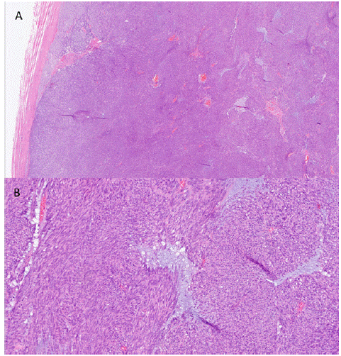
Figure 1: Histologically, the tumor was made up of intertwined bundles of relatively unimorphic spindle cells of the connective tissue, and was bounded by a connective capsule. Perinuclear vacuoles were seen in places. No necrosis was observed. (A: H&E 2x; B: H&E 10x).
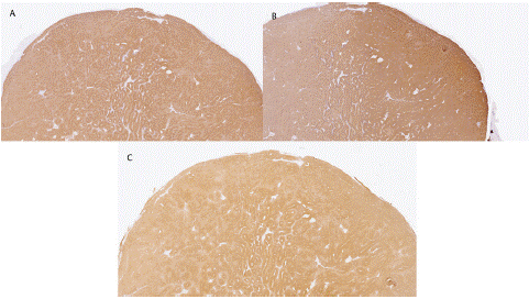
Figure 2: The tumor was DOG1 (A; 4x), CD117 (B; 4x), vimentin and caldesmon (C; 4x) positive and SMA and desmin negative.
The postoperative course undergone well.
The surveillance protocol consisted of CT scans and laboratory findings 3 months after surgery, followed by one CT scan six months after and then a CT scan was performed once a year afterwards. Abdominal ultrasounds were performed every six months. In the meantime, colonoscopy was performed once and oesophagogastroduodenoscopy three times.
At the last check-up in November 2023, it was determined that the patient is disease-free five years after diagnosis. It was recommended to do laboratory tests, ultrasound of the abdomen and, depending on the clinical picture, possibly an oesophagogastroduodenoscopy once a year.
Another case is 73-year-old woman who presented to emergency unit with hematemesis in December of 2021. She has been complaining of dysphagia, impaired appetite, loss of weight and mild umbilical pain. She was afebrile and did not complain of any respiratory problems. She has been on antihipertensive therapy. There weren´t any deviations in the patient's physical examination.
Among the previous diseases, melanoma of the left lowerleg, cerebrovascular stroke that occurred 17 years ago, arterial hypertension, GERD and hiatal hernia stood out.
An X-ray of the heart, lungs and abdomen showed an oval shadow of the paracardial right in a diameter of 6.3 cm, which primarily corresponded to a hiatal hernia.
Oesophagogastroduodenoscopy demonstrated an exulcerated tumor that occupies two thirds of the esophageal circumference on 36-40 cm from the incisors. A biopsy was done.
Two slightly larger and few very small tissue samples up to 0.2 cm in diameter were received for pathohistological analysis. Histologically, tumor tissue was composed of medium-sized epitheloid cells of lighter cytoplasms and moderately polimorphic nuclei, partly with a visible nucleolus. Focally, tumor cells formed smaller clusters surrounded by a thin reticulin layer. Mitotic activity was moderate (4/10 VVP), pathological mitosis were not found (Figure 3).
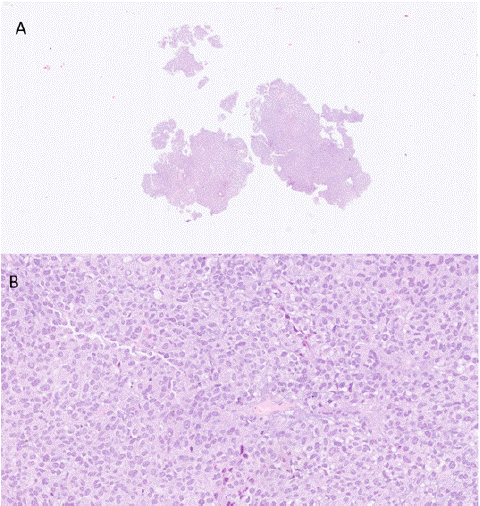
Figure 3: A) Two slightly larger and few very small tissue samples up to 0.2 cm in diameter were received for pathohistological analysis (H&E). B) A tumor tissue was composed of medium-sized epitheloid cells of lighter cytoplasms and moderately polimorphic nuclei, partly with a visible nucleolus. Focally, tumor cells formed smaller clusters surrounded by a thin reticulin layer. Mitotic activity was moderate (4 / 10 VVP), pathological mitosis were not found. (H&E, 20x).
Immunohistochemically, tumor tissue was positive for CD 34, CD117, DOG1, sparsely focal for bcl-2, and STAT 6 (nonspecific), and negative for CK AE1/3, CK5/6, CK7, p63, p40, CDX2, TTF- 1, NSE, chromogranin, synaptophysin, S100, SOX10, CD99 and desmin (Figure 4).
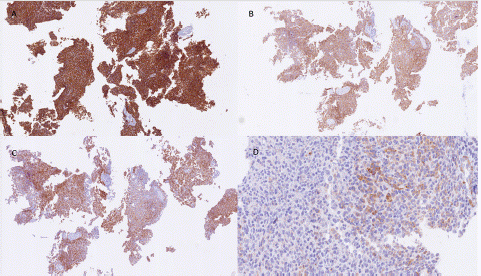
Figure 4: Immunohistochemically, tumor tissue was positive for CD 34 (A;4x), CD117 (C;4x), DOG1 (B;4x), sparsely focal for bcl-2 (D;40x), and STAT 6 (nonspecific), and negative for CK AE1/3, CK5/6, CK7, p63, p40, CDX2, TTF- 1, NSE, chromogranin, synaptophysin, S100, SOX10, CD99 and desmin.
The overall clinical, pathohistological and immunohistochemical features were consistent with a high grade Gastrointestinal Stromal Tumour (GIST), an epitheloid cell type.
On Multislice Computed Tomography (MSCT) (Figure 5) of the thorax, abdomen and pelvis a thickening of the wall in the middle third of the esophagus was observed. It occupied most of the circumference and reduced the lumen of the esophagus. Heterogeneous necrotic tumor measuring 11.4 x 11.1 x 7.5 cm was inseparable from the esophageal wall. The tumor spreaded to the posterior mediastinum and it seemed well limited. No other significant pathology was visualised within the thorax, the abdomen or the pelvis.
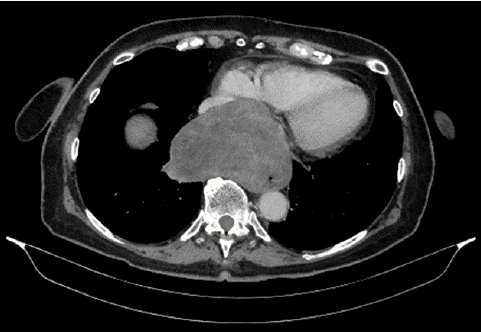
Figure 5: On multislice computed tomography (MSCT) of the thorax, abdomen and pelvis a thickening of the wall in the middle third of the esophagus was observed. It occupied most of the circumference and reduced the lumen of the oesophagus. Heterogeneous and necrotic tumor formation measuring 11.4 x 11.1 x 7.5 cm was inseparable from the oesophageal wall. The tumor formation spreaded to the posterior mediastinum. It seemed well limited, pushing the right atrium towards the ventral.
The patient was referred to a medical council, which determined that she was not a candidate for radical thoracosurgical treatment and that she should be referred to an oncologist to start oncological treatment.
Given that the patient still tolerates oral intake of porridge, at this time medical council have not decided on the possible placement of PEG/stent, which of course can be planned later, depending on the development of dysphagic disorders and response to treatment.
The patient has been receiving imatinib regularly since February 2022. A CT scan is performed every 6 months. At the last check-up in April 2023 there was no significant change in the size of the necrotic tumor.
Discussion
Gastrointestinal Stromal Tumors (GISTs) are the most common mesenchymal neoplasms of the digestive tract [1]. GISTs often arise in the stomach and small intestine, while esophageal GISTs are extremely rare (fewer than 5% of all GISTs) [2]. According to this fact clinicopathological data on esophageal GISTs are trully limited.
Although Lott's study data suggests that esophageal GIST occurs significantly more frequent in men as well as in patients younger than 60 at diagnosis in comparison to gastric GIST [3], in both presented cases patients were female and older than 60 years.
About 18% of patients are asymptomatic [4], as presented patient with spindle cell type GIST. In the case of epitheloid cell GIST patient was presented with the most frequent symptoms of esophageal GIST - hematemesis and dysphagia. Other symptoms could be weight loss (20%), chest pain (8–15%), and bleeding (1–10%) [5].
Leiomyomas are the most common mesenchymal tumors of the esophagus [6] and the first differential diagnosis of an esophageal GIST. Leiomyomas and GISTs appear similar on CT and EUS [7], so it is impossible to distinguish those two entities on diagnostic imaging. Other diferential diagnoses of an esophageal GIST includes hemangioma, schwannoma, leiomyosarcoma, and papillary epithelioma [1].
Final diagnosis is based on pathological analysis; morphology and most important-imunohistochemical profil of the tumor.
GISTs can be pathologically classified into three types: spindle cell, epithelioid cell, and mixed cell type [8] An immunohistochemical panel including KIT (CD117), DOG1, CD34, Smooth Muscle Actin (SMA), desmin, and S100 protein is used for distinguishing GISTs from other tumors [9,10] GISTs are positive for c-KIT, DOG 1 and CD 34 [11].
According to ESMO guidelines, surgical resection is considered to be the only potentially curative treatment for localized GISTs [12]. Enucleation of esophageal GISTs is optimal for smaller tumors (diametar 2–5 cm) [13,14], whereas esophagectomy is recommended for GISTs above 9 cm in size [13].
Surgical treatment was also treatment of choice in presented case of low-grade GIST, spindle cell type. In the case of patient with locally advanced high-grade GIST, epithelioid cell type, treatment of choice was neoadjuvat therapy (imatinib). Kang et al. [9] suggested that neoadjuvant imatinib treatment can be considered in patients with GISTs with high mitotic rates and/or larger tumor sizes. In the study of Feng et al. [5], 5-year survival rate was 65.1%. Both of our patients are alive. The patient with low grade GIST, spindle cell type is disease-free five years after diagnosis, and the patient with high grade GIST, epithelioid cell type, is without significante change of tumor size and clinical status two years after diagnosis.
Author Statements
Conflicts of Interest
The authors have no conflicts of interest to declare.
References
- Zhang FB, Shi HC, Shu YS, Shi WP, Lu SC, Zhang XY, et al. Diagnosis and surgical treatment of esophageal gastrointestinal stromal tumors. World J Gastroenterol. 2015; 21: 5630-4.
- Miettinen M, Lasota J. Gastrointestinal stromal tumors: review on morphology, molecular pathology, prognosis, and differential diagnosis. Arch Pathol Lab Med. 2006; 130: 1466-78.
- Lott S, Schmieder M, Mayer B, Bruns DH, Knippschild U, Agaimy A, et al. Gastrointestinal stromal tumors of the esophagus: evaluation of a pooled case series regarding clinicopathological features and clinical outcome. Am J Cancer Res. 2014; 5: 333-43.
- Soreide K, Sandvik OM, Soreide JA, Giljaca V, Jureckova A, Bulusu VR. Global epidemiology of gastrointestinal stromal tumours (GIST): A systematic review of population-based cohort studies.Cancer Epidemiol. 2016; 40: 39-46.
- Feng F, Tian Y, Liu Z, Xu G, Liu S, Guo M, et al. Clinicopathologic Features and Clinical Outcomes of Esophageal Gastrointestinal Stromal Tumor: Evaluation of a Pooled Case Series. Medicine (Baltimore). 2016; 95: e2446.
- Miettinen M, Sarlomo-Rikala M, Sobin LH, Lasota J. Esophageal stromal tumors: a clinicopathologic, immunohistochemical, and molecular genetic study of 17 cases and comparison with esophageal leiomyomas and leiomyosarcomas. Am J Surg Pathol. 2000; 24: 211-22.
- Dendy M, Johnson K, Boffa DJ. Spectrum of FDG uptake in large (>10 cm) esophageal leiomyomas. J Thorac Dis. 2015; 7: E648-51.
- Dei Tos AP, Laurino L, Bearzi I, Messerini L, Farinati F, Gruppo Italiano Patologi Apparato Digerente (GIPAD), et al. Gastrointestinal stromal tumors: the histology report. Dig Liver Dis. 2011; 43: S304-9.
- Kang GH, Srivastava A, Kim YE, Park HJ, Park CK, Sohn TS, et al. DOG1 and PKC-0 are useful in the diagnosis of KIT-negative gastrointestinal stromal tumors. Mod Pathol. 2011; 24: 866-75.
- Hirota S, Isozaki K, Moriyama Y, Hashimoto K, Nishida T, Ishiguro S, et al. Gain-of-function mutations of c-kit in human gastrointestinal stromal tumors. Science. 1998; 279: 577-80.
- Parab TM, DeRogatis MJ, Boaz AM, Grasso SA, Issack PS, Duarte DA, et al. Gastrointestinal stromal tumors: a comprehensive review. J Gastroint Oncol. 2019; 10: 144-154.
- ESMO/European Sarcoma Network Working Group. Gastrointestinal stromal tumours: ESMO Clinical Practice Guidelines for diagnosis, treatment and follow-up. Ann Oncol. 2014; 25: iii21-6.
- Jiang P, Jiao Z, Han B, Zhang X, Sun X, Su J, et al. Clinical characteristics and surgical treatment of oesophageal gastrointestinal stromal tumours. Eur J Cardiothorac Surg. 2010; 38: 223-7.
- Nemeth K, Williams C, Rashid M, Robinson M, Rasheed A. Oesophageal GIST-A rare breed case report and review of the literature. Int J Surg Case Rep. 2015; 10: 256-9.