Surgical Technique
Austin J Surg. 2014;1(3): 1012.
Hepaticoduodenostomy in Hepatectomy for Perihilarcholangiocarcinoma: A Preliminary Report
Hiroshi Yoshida1*, Yasuhiro Mamada2, Nobuhiko Taniai2, Hiroshi Makino1, Tadashi Yokoyama1, Hiroshi Maruyama1, Atsushi Hirakata1, Masahiro Hotta1 and Eiji Uchida2
1Department of Surgery, Nippon Medical School Tama Nagayama Hospital, Japan
2Department of Surgery, Nippon Medical School, Japan
*Corresponding author: Hiroshi Yoshida, Department of Surgery, Nippon Medical School Tama Nagayama Hospital, 1-7-1, Nagayama, Tama-city, Tokyo, 206-8512, Japan
Received: February 08, 2014; Accepted: April 18, 2014; Published: April 21, 2014
Abstract
A Roux–en–Y anastomosis fashioned from the jejunum (i.e., hepaticojejunostomy) is usually used to reconstruct the biliary system in hepatectomy. In this study, we review our experience with hepaticoduodenostomy (HD) as an alternative to Roux–en–Y biliary anastomosis in patients undergoing hepatectomy for perihilarcholangiocarcinoma, report our preliminary findings in 2 patients, and speculate on future applications. Laparotomy was performed using a Kent retractor. Wide kocherization of the duodenum was done to provide a tension–free anastomosis to the hepatic duct. The Kent retractor was released transiently, and the anastomosis was confirmed to be free of tension. Hepatectomy and excision of the common bile duct were performed. In patients with short distances between the hepatic ducts, a hepaticoplasty was performed. A 10–Fr silicon drain with channels along the sides, approximately 20 mm in length, was used as an internal stent. HD was performed with a single–layer anastomosis with continuous sutures. No complication occurred after HD. Our initial experience suggests that HD may be a viable alternative to Roux–en–Y biliary anastomosis in patients undergoing hepatectomy for perihilarcholangiocarcinoma.
Keywords: Hepaticoduodenostomy; Hepatectomy; Perihilarcholangiocarcinoma
Introduction
A Roux–en Y anastomosis fashioned from the jejunum (i.e.,hepaticojejunostomy [HJ]) is usually used to reconstruct the biliary system in patients undergoing hepatectomy for perihilarcholangiocarcinoma [1]. Recently, reconstruction by hepaticoduodenostomy (HD) or choledochoduodenostomy has been recommended instead of reconstruction by HJ or choledochojejunostomy [2–14]. Complications such as biliary leakage and cholangitis are well documented after HJ and choledochojejunostomy [15–19]. Moreover, the biliary tree is difficult to access endoscopically in patients undergoing enteric reconstruction using a Roux–en–Y anastomosis to the jejunum [20]. HD offers the possible advantage of simple postoperative access to the biliary system by endoscopy and avoids the complications associatedwith HJ [2].
In this study, we review our experience with HD as an alternative to Roux–en–Y biliary anastomosis in patients undergoing hepatectomy for perihilarcholangiocarcinoma, report our preliminary findings in 2 patients, and speculate on future applications.
Case 1
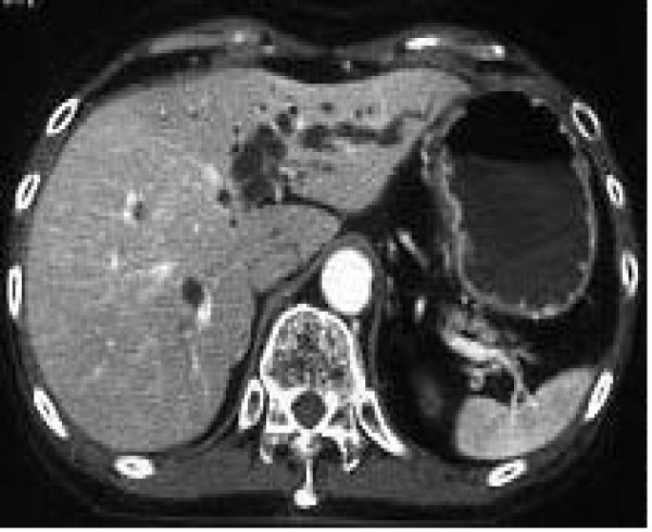
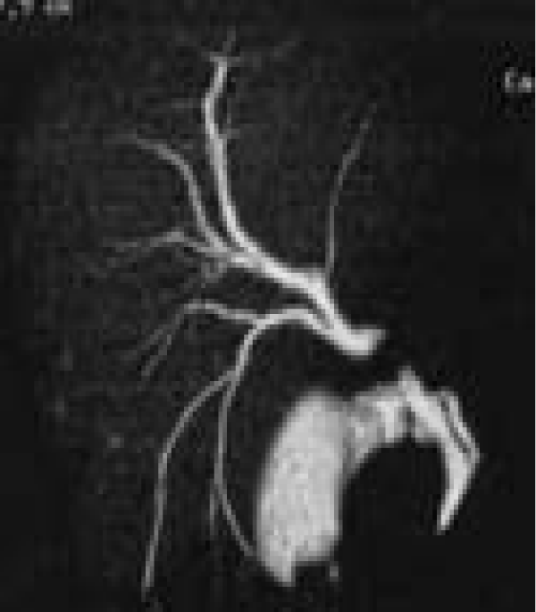
A 74–year–old woman was admitted because of a dilated left intrahepatic bile duct. Previously, she had undergone a right colectomy because of colonic carcinoma. However, minor leakage of the anastomosis occurred, and reoperation (drainage) was performed.After that, ileus developed and reoperation was done again. Computed tomography revealed dilatation of the left intrahepatic bile duct and mild dilatation of the right posterior intrahepatic duct (Figure 1). Drip infusion cholangiography revealed only the right anterior intrahepatic duct and stenosis at the hepatic hilum (Figure 2). Perihilarcholangiocarcinoma was diagnosed.
Figure 1 :Computed tomography revealed dilatation of the left intrahepatic bile duct and mild dilatation of the right posterior intrahepatic duct.
Figure 2 :Drip infusion cholangiography revealed only the right anterior intrahepatic duct and stenosis at the hepatic hilum.
Surgical procedures
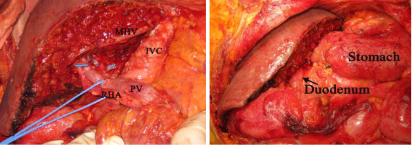
Laparotomy was performed using a Kent retractor. Severe adhesions of the jejunum were detected, precluding the use of a Rouxen– Y anastomosis fashioned from the jejunum. After kocherization of the duodenum to provide a tension–free anastomosis to the hepatic duct, an extended left hepatectomy with excision of the common bile duct and lymph–node dissection was performed. Intraoperative pathological examinations revealed that the stumps of the right intrahepatic bile ducts were negative for carcinoma. There were triple lumens of the right anterior bile ducts and double lumens of the right posterior bile ducts. Hepaticoplasty (i.e., the adjacent walls of the hepatic ducts were sutured together with single stitches using 6–0 polydioxane [PDS, Ethicon, NJ, USA] to obtain a single lumen for biliary anastomosis) was performed to make double lumens of the right anterior bile ducts and a single lumen of the right posterior bile duct. Unexpectedly, the duodenum was distant from the hepatic ducts. To provide a tension–free anastomosis, wide kocherization of the duodenum was performed again, and the right gastroepiploic artery and vein were cut. The Kent retractor was released transiently, and the anastomosis was confirmed to be tension–free. As an internal stent, a 10–Fr silicon drain with channels along the sides (BLAKE Silicone Drain, Ethicon, NJ, USA),approximately 20 mm in length, was used [21]. Before completing suture of the posterior row, the stent was inserted into each lumen. Five stents were placed within the hepatic duct and duodenal lumen to serve as the internal stents for the anastomosis. The stent was fixed to the anterior row of stitches with 5–0 polydioxane sutures. Three anastomoses of the intrahepaticbile ducts to the duodenum were established by means of a single–layer anastomosis with continuous sutures (5–0 polydioxane) (Figure 3). Fixation of the greater omentum to the peritoneum was not necessary to prevent delayed gastric emptying [22,23] because the stomach could not come in contact with the cut surface of the liver, a potential cause of adhesion.
Figure 3 :Before completing suture of the posterior row, the stent was inserted into each lumen. Five stents were placed within the hepatic duct and duodenal lumen to serve as the internal stents for the anastomosis. The stent was fixed to the anterior row of stitches with 5-0 polydioxane sutures. MHV: middle hepatic vein, IVC: inferior vena cava, PV: portal vein, RHA: right hepatic artery (a). Three anastomoses of the intrahepaticbile ducts to the duodenum were established by means of a single-layer anastomosis with continuous sutures(b).
The postoperative course was uneventful, and the patient was discharged on postoperative day 12. After discharge, upper gastrointestinal endoscopy revealed no duodeno gastric bile reflux.
Case 2
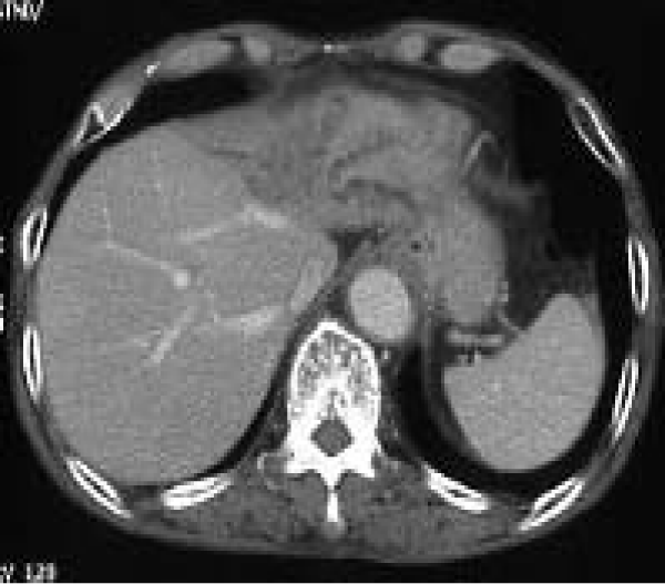
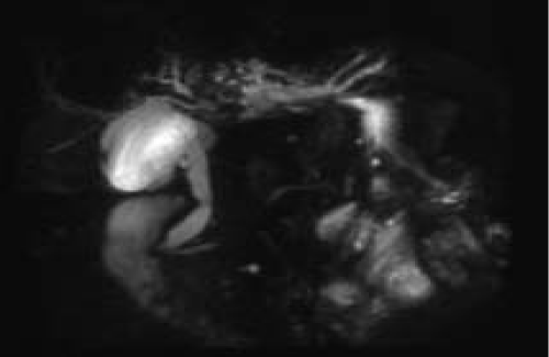
A 76–year–old man with dilatation of the left intrahepatic duct was admitted. Previously, he had undergone a distal gastrectomy (BillrothII reconstruction) because of gastric carcinoma. Computed tomography and magnetic resonance cholangiopancreatography revealed the dilated left intrahepatic bile duct (Figure 4,5). Perihilarcholangiocarcinoma was diagnosed.
Figure 4 :Computed tomography revealed the dilated left intrahepatic bile duct.
Figure 5 :Magnetic resonance cholangiopancreatography revealed the dilated left intrahepatic bile duct.
Surgical procedures
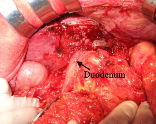
Laparotomy was performed using a Kent retractor. Adhesions of the jejunum were detected. Wide kocherization of the duodenal stump was performed to provide a tension–free anastomosis. The Kent retractor was released transiently, and the anastomosis was confirmed to be free of tension. An extended left hepatectomy with excision of the common bile duct and lymph–node dissection was performed. Intraoperative pathological examinations revealed that the stump of the right hepatic duct was negative for carcinoma. Because the right hepatic duct had a single lumen, hepaticoplasty was unnecessary. HD was performed using a single–layer anastomosis with continuous sutures (5–0 polydioxane). After completing suture of the posterior row, the stent was inserted into the hepatic duct. As internal stents, 10–Fr silicon drains with channels along the sides, approximately 20 mm in length, were used. The stent was fixed to the anterior row of stitches with 5–0 polydioxane sutures. Anastomosis of the right hepatic duct to the duodenal stump was established (Figure 6). Fixation of the greater omentum to the peritoneum was not necessary to prevent delayed gastric emptying [22,23] because the residual stomach did not come in contact with the cut surface of the liver, a potential cause of adhesion.
Figure 6 :Anastomosis of the right hepatic duct to the duodenal stump was established.
The postoperative course was uneventful, and the patient was discharged on postoperative day 10. After discharge, upper gastrointestinal endoscopy revealed no duodenogastric bile reflux.
Discussion
Biliary reconstruction using the duodenum has been successfully performed in liver transplantation and the treatment of biliary obstruction, common bile duct stones, biliary injury, choledochal cysts, and biliary atresia [2–14]. We decided to perform HD instead of HJ in the 2 patients described here, because it was technically difficult to perform HJ. No complication occurred after HD. One disadvantage of HD is that, if an anastomotic leak does occur, the leakage volume can be much greater than with HJ because the leak would contain both gastric and pancreatic juices rather than bile alone in contrast to HJ. A second advantage of HD is that it is unnecessary to createa Roux–en–Yjejunal limb, associated with a higher incidence of adhesive bowel obstruction than HD.A third advantage of HD is that the biliary system can be easily accessed by endoscopy, which is not feasible with HJ.
One drawback of HD is anastomotic tension, which is higher than that of choledochoduodenostomy, especially in patients with perihilarcholangiocarcinoma. We performed wide kocherization of the duodenum and confirmed that the anastomosis was tensionfree by releasing the Kent retractor transiently. Wide kocherization of the duodenum was an important factor in creating a tension–free anastomosis.
Shimotakahara [7] compared HD with HJ after excision of primary choledocal cysts in children. Complications after cyst excision occurred in 5 of the 12 patients in the HD group (42%). Four patients had bilious gastritis and 1 had temporary liver dysfunction. In the HJ group, 2 of the 28 patients (7.1%) had postoperative complications. Both patients had adhesive bowel obstruction, and 1additionally had cholangitis. Liem [14] reported that cholangitis occurred at a rate of 5.3% and bilious gastritis at a rate of 14.3% after laparoscopic cyst excision and HD for choledochal cysts in children. Moraca [5] compare dlong–term biliary function between HD and HJ in patients who underwent treatment of major bile–duct injuries during cholecystectomy. On long–term follow–up, no patient had cholangitis, jaundice, or liver failure in either group. Bennet [8] reported that preliminary experience suggested that choledochoduodenostomy is a safe technique for cadaveric liver transplantation. Campsen [2] documented the outcomes of 7 patients who underwent living donor transplantation with HD as the primary type of biliary anastomosis. There were no deaths or re–transplants during the follow–up period. One patient had cholangitis that responded to intravenous antibiotics and endoscopic removal of the stent. Various complications have occurred after HD or HJ, including bilious gastritis, temporary liver dysfunction, bowel obstruction, and cholangitis. Bilious gastritis occurred after HD in some children with choledocal cysts, but has not been reported after HD in adults. The severest complication is bowel obstruction or cholangitis. One advantage of HD is that the creation of a Roux–en Yjejunal limb and one anastomosis, associated with a higher incidence of adhesive bowel obstruction than HD, is unnecessary. After biliary reconstruction using the duodenum, cholangitis results from anastomotic stenosis and not reflux of the duodenal contents into the biliary tree [24]. The prevention of anastomotic stenosis is prerequisite to the prophylaxis of cholangitis.
In this study, we used a 10–Fr silicon drain with channels for internal biliary stenting of HD. We previously compared two types of stentsin patients who underwent surgery for perihilarcholangiocarcinoma. In one group, a 10–Fr silicon drain with channels along the sides was used as a stent for HJ (channel stent group), while in the other a 5–Fr silicon drain with an internal lumen and side holes was used (intraluminal stent group). Leakage developed in 4 patients (36.4%) in the intraluminal stent group versus 2 (20.0%) in the channel stent group. Cholangitis developed in 3 patients with leakage (27.3%) in the intraluminal stent group versus no patient in the channel stent group. Our results suggested that the use of a 10–Fr stent helps to maintain a 10–Fr intraluminal diameter of the anastomosis, even in the presence of biliary leakage. A 10–Fr stent may thus prevent cholangitis due to anastomotic stenosis [21]. No complication occurred after operation in the present study.
Hakamada [25] reported that cholangiocarcinoma developed in 7.4% of patients a mean interval of 18 years after transduodenalsphincteroplasty. Maeda [26] documented the development of bile duct cancer 21 years after choledochoduodenostomy. Tocchi [27] estimated that the incidences of cholangiocarcinomas after sphincteroplasty,choledochoduodenostomy, and hepaticojejunostomy were 4.8, 7.6, and 1.9%, respectively, occurring at intervals of 11 to 18 years. Therefore, cholangiocarcinoma as a delayed complication of transduodenalsphincteroplasty and choledochoenteric anastomosis has become a serious issue.
In conclusion, we reviewed our experience with HD as an alternative to Roux–en–Y biliary anastomosis in patients undergoing hepatectomy for perihilarcholangiocarcinoma, reported ourpreliminary findings in 2 patients, and speculated on future applications. No complication occurred after operation in this study. We emphasize that wide kocherization of the duodenum is necessary to provide a tension–free anastomosis, which should be confirmed after transiently releasing the Kent retractor. HD may be a reasonable alternative to Roux–en–Y biliary anastomosis in patients undergoing hepatectomy for perihilarcholangiocarcinoma. Additional studies and longer follow–up are needed, however, to confirm these findingsand to accurately assess the rates and types of complications associated with HD. Further studies in larger numbers of patients are needed before HD can be designated a standard of care.
References
- Sano T, Shimada K, Sakamoto Y, Esaki M, Kosuge T. Changing trends in surgical outcomes after major hepatobiliary resection for hilar cholangiocarcinoma: a single-center experience over 25 years. J Hepatobiliary Pancreat Surg. 2007; 14: 455-462.
- Campsen J, Zimmerman MA, Mandell MS, Wachs M, Bak T, Forman L, et al. Hepaticoduodenostomy is an alternative to Roux-en-Y hepaticojejunostomy for biliary reconstruction in live donor liver transplantation. Transplantation. 2009; 87: 1842-1845.
- Cole FR, Pisillo CJ. Choledochoduodenostomy: an evaluation of its indications. Int Surg. 1966; 46: 108-112.
- Todani T, Watanabe Y, Mizuguchi T, Fujii T, Toki A. Hepaticoduodenostomy at the hepatic hilum after excision of choledochal cyst. Am J Surg. 1981; 142: 584-587.
- Moraca RJ, Lee FT, Ryan JA Jr, Traverso LW. Long-term biliary function after reconstruction of major bile duct injuries with hepaticoduodenostomy or hepaticojejunostomy. Arch Surg. 2002; 137: 889-893.
- Srivengadesh G, Kate V, Ananthakrishnan N. Evaluation of long-term results of choledochoduodenostomy for benign biliary obstruction. Trop Gastroenterol 2003; 24: 205-207.
- Shimotakahara A, Yamataka A, Yanai T, Kobayashi H, Okazaki T, Lane GJ, et al. Roux-en-Y hepaticojejunostomy or hepaticoduodenostomy for biliary reconstruction during the surgical treatment of choledochal cyst: which is better? Pediatr Surg Int. 2005; 21: 5-7.
- Bennet W, Zimmerman MA, Campsen J, Mandell MS, Bak T, Wachs M, et al. Choledochoduodenostomy is a safe alternative to Roux-en-Y choledochojejunostomy for biliary reconstruction in liver transplantation. World J Surg. 2009; 33: 1022-1025.
- Degenshein GA. Choledochoduodenostomy: an 18 year study of 175 consecutive cases. Surgery. 1974; 76: 319-324.
- Bhandarkar DS, Shah RS. Laparoscopic choledochoduodenostomy for retained bile duct stone. J Postgrad Med. 2005; 51: 156-157.
- de Aretxabala X, Bahamondes JC. Choledochoduodenostomy for common bile duct stones. World J Surg. 1998; 22: 1171-1174.
- Onken JE, Pappas T, Baillie J. Long term results of choledochoduodenostomy in the treatment of choledocholithiasis. Gastrointest Endosc. 1992; 38: 403-405.
- Ramirez P, Parrilla P, Bueno FS, Abad JM, Muelas MS, Candel MF, et al. Choledochoduodenostomy and sphincterotomy in the treatment of choledocholithiasis. Br J Surg. 1994; 81: 121-123.
- Liem NT, Dung le A, Son TN. Laparoscopic complete cyst excision and hepaticoduodenostomy for choledochal cyst: early results in 74 cases. J Laparoendosc Adv Surg Tech A. 2009; 19: S87-90.
- Bourgeois N, Deviere J, Yeaton P, Bourgeois F, Adler M, Van De Stadt J, et al. Diagnostic and therapeutic endoscopic retrograde cholangiography after liver transplantation. Gastrointest Endosc. 1995; 42: 527-534.
- Sossenheimer M, Slivka A, Carr-Locke D. Management of extrahepatic biliary disease after orthotopic liver transplantation: review of the literature and results of a multicenter survey. Endoscopy. 1996; 28: 565-571.
- Colonna JO 2nd, Shaked A, Gomes AS, Colquhoun SD, Jurim O, McDiarmid SV, et al. Biliary strictures complicating liver transplantation. Incidence, pathogenesis, management, and outcome. Ann Surg. 1992; 216: 344-350.
- Greif F, Bronsther OL, Van Thiel DH, Casavilla A, Iwatsuki S, Tzakis A, et al. The incidence, timing, and management of biliary tract complications after orthotopic liver transplantation. Ann Surg. 1994; 219: 40-45.
- Lerut J, Gordon RD, Iwatsuki S, Esquivel CO, Todo S, Tzakis A, et al. Biliary tract complications in human orthotopic liver transplantation. Transplantation. 1987; 43: 47-51.
- Ryozawa S, Iwamoto S, Iwano H, Ishigaki N, Taba K, Sakaida I. ERCP using double-balloon endoscopes in patients with Roux-en-Y anastomosis. J Hepatobiliary Pancreat Surg. 2009; 16: 613-617.
- Yoshida H, Mamada Y, Taniai N, Mizuguchi Y, Kakinuma D, Ishikawa Y, et al. Silicon drain with channels along the sides for internal biliary stenting of hepaticojejunostomy in hepatic hilar malignancies. J Gastroenterol Hepatol 2009; 24: 752-756.
- Yoshida H, Mamada Y, Taniai N, Mizuguchi Y, Shimizu T, Kakinuma D, et al. Fixation of the greater omentum for prevention of delayed gastric emptying after left hepatectomy with lymphadenectomy for cholangiocarcinoma. J Hepatobiliary Pancreat Surg. 2007; 14: 392-396.
- Yoshida H, Mamada Y, Taniai N, Mizuguchi Y, Shimizu T, Takahashi T, et al. Fixation of the greater omentum for prevention of delayed gastric emptying after left-sided hepatectomy: a randomized controlled trial. Hepatogastroenterology. 2005; 52: 1334-1337.
- Escudero-Fabre A, Escallon A Jr, Sack J, Halpern NB, Aldrete JS. Choledochoduodenostomy. Analysis of 71 cases followed for 5 to 15 years. Ann Surg. 1991; 213: 635-642.
- Hakamada K, Sasaki M, Endoh M, Itoh T, Morita T, Konn M. Late development of bile duct cancer after sphincteroplasty: a ten- to twenty-two-year follow-up study. Surgery. 1997; 121: 488-492.
- Maeda A, Yokoi S, Kunou T, Saeki S, Murata T, Niinomi N, et al. Bile duct cancer developing 21 years after choledochoduodenostomy. Dig Surg. 2003; 20: 331-334.
- Tocchi A, Mazzoni G, Liotta G, Lepre L, Cassini D, Miccini M. Late development of bile duct cancer in patients who had biliary-enteric drainage for benign disease: a follow-up study of more than 1,000 patients. Ann Surg. 2001; 234: 210-214.
