Case Report
Austin J Surg. 2014;1(3): 1013.
Sclerosing Perivascular Epitheloid Cell Tumor of the Falciform Ligament in a 34-year Old Woman
Andreas Puntschart1*, Katharina Meszaros1,2, Peter Kornprat1, Bernadette Liegl-Atzwanger2 and Hans Joerg Mischinger1
1Department of General Surgery, Medical University of Graz, Austria
2Department of Pathology, Medical University of Graz, Austria
*Corresponding author: Andreas Puntschart, Department of General Surgery, Medical University of Graz, Auenbruggerplatz 29, A-8036 Graz, Austria
Received: May 09, 2014; Accepted: May 30, 2014; Published: June 02, 2014
Abstract
PEComas represent rare, heterogeneous mesenchymal neoplasms derived from perivascular epitheloid cells exprimating smooth muscle as well as melanocytic markers. Most of described cases showed a benign clinical course; nevertheless, rare cases with development of metastases were described. The extremely rare sclerosing PEComa subtype with strong hyalinization of stroma was mainly reported in the retroperitoneum.
We report a 34–year old female patient with a sclerosing PECOMA of the falciform ligament, who was primarily admitted with an intra–abdominal mass. Radiologic examination revealed a partially cystic tumor near to the hilum of the liver and led to the radiologic diagnosis of a GIST (gastrointestinal stromal tumor).
The tumor could be resected completely; definitive histopathologic examination showed a sclerosing subtype of PEComa. The further postoperative course was uneventful; the patient could be discharged home on postoperative day eight. One year postoperatively, the patient is under close clinical and radiological follow–up proving absence of tumor recurrence.
The extreme rarity and the uncertain malignant potential of sclerosing PEComa subtype occurring outside the retro peritoneum represents a clinical challenge because of lacking oncological guidelines for the ideal treatment and follow–up protocol. The presented case shows that in such cases, a tailored multi–disciplinary approach and close follow–up intervals are mandatory.
Keywords: Sclerosing PEComa; Mesenchymal tumor; Falciform ligament
Introduction
Perivascular Epitheloid Cell tumors (PEComas) represent a rare tumor entity deriving from perivascular epitheloid cells exprimating smooth muscle as well as melanocytic markers in immunohistochemical assay [1,2]. Most of described cases showed a benign clinical course; nevertheless, rare cases with development of metastases were described [3]. The extremely rare sclerosing PEComa subtype associated with strong stroma hyalinization was mainly reported in the retroperitoneum [4,5].
We present a case of a 34–year old female patient with a sclerosing PEComa of the falciform ligament.
Case Presentation
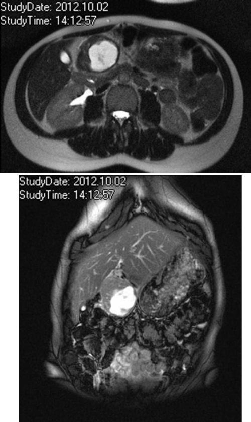
A 34–year old female primarily admitted with a palpable solid intra–abdominal mass. Magnetic resonance imaging of the abdomen revealed a partially cystic mass of 4.5 x 5.0 centimeters located near to the hilum of the liver, immediately ventrally of the pancreatic head without infiltration of the adjacent structures (Figure 1A,1B).Especially in the T1 weighted images a solitary, marginal component of the tumor with contrast enhancement became obvious. Becauseof the central cystic transformation of the tumor with radiomorphologic signs of gastrointestinal stromal tumors, a PET–scan(positron emission tomography) using FDG (Fluordesoxyglucose)was performed. The PET–scan showed a missing tracer uptake of the cystic tumor but proved absence of multifocal tumor occurrence and⁄or suspected metastatic lesions.
Figure 1 :T2-weighted axial (A) and coronal (B) magnetic resonance images: A partially cystic mass near the liver hilum, ventrally to the pancreatic head without infiltration of the adjacent structures.
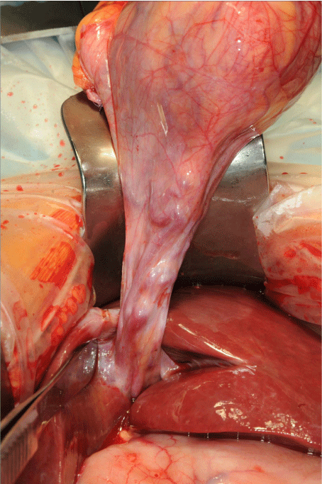
According to the above mentioned radio morphologic signs the patient was scheduled for surgical resection of the suspected GIST. The surgical approach was performed via upper median laparotomy. The peritoneal veins showed significant dilatation; a stalked tumor of a diameter of around seven centimeter appeared (Figure 2). The pedicle of the tumor containing large tumor–supplying vessels originated between the third and fourth liver segment; but without any adherence to the gastric wall as primarily suspected according to preoperative radiologic imaging. The tumor was resected from its pedicle; intraoperative histopathologic rapid section as well as the definitive section proved complete tumor resection. The postoperative course was uneventful. The patient could be discharged home on postoperative day eight. One year postoperatively, closed clinical and radiological follow–up proved freedom from tumor recurrence.
Figure 2 :Macroscopic view: Tumor with adherent pedicle originating between the third and fourth liver segment.
Results
Pathologic findings
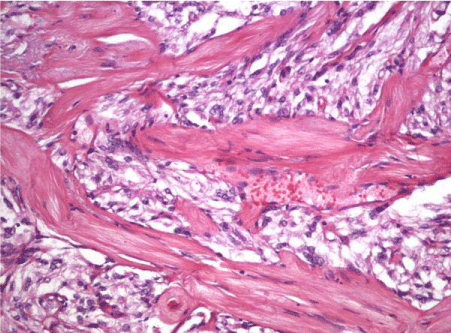
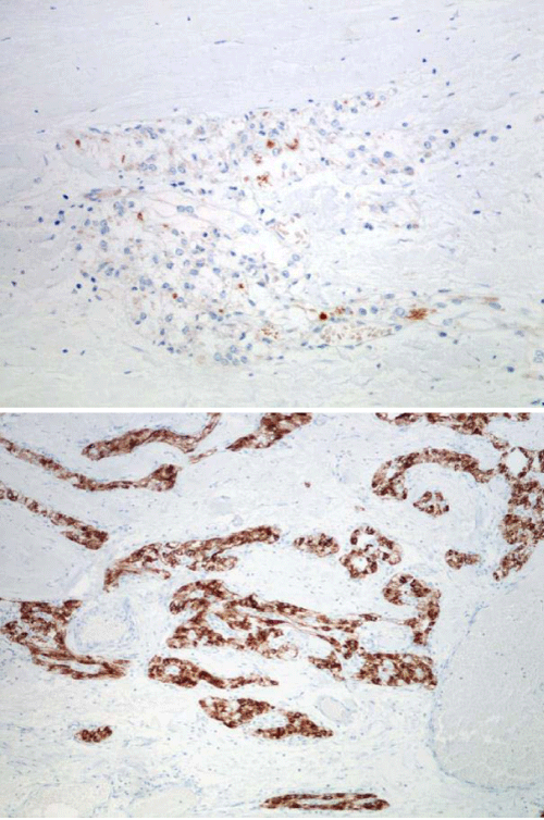
The tumor was located in the falciform ligament measuring 6 cm in diameter with a yellow–brown tan with cystic structure in the center and a 1 mm calcified capsula. The adjacent soft tissue contained numerous varicous partially thrombotic blood vessels; the cystic structures were filled with brown, gelatinous fluid. Detailled histopathologic examination showed mesenchymal tumorous tissue with vascular structures; the tumor cells partially located in nest forms. Most parts of the tumor exhibited significant sclerosis; singular giant cells and cytological atypias could be detected (Figure 3A, 3B). Immunohistochemic assays demonstrated a positive reaction for smooth muscle actin (SMA) as well as desmin (Figure 4A). The positive antibody reaction against CD 31 and CD 34 proved the tumor containing a significant number of vascular structures; a multifocal positive reaction could be obtained with HMB45 antibodies (Figure 4B), the reaction for S100 was negative. These findings finally confirmed the diagnosis of sclerosing PEComa.
Figure 3 :Microscopic hematoxylin and eosin appearance: Sclerosing Pecoma composed of cords and trabeculae of cytologically uniform bland epitheloid cells. The tumor contained abundant densely sclerotic stroma and tumor cells focally associated with blood vessel walls.
Figure 4 :Positive immunohistochemical reaction for HMB-45 (A) and desmin (B).
Discussion
Perivascular epitheloid cell tumors represent a rare tumor entity which was firstly described by Bonetti and coworkers in 1992 [1]. The extremely rare sclerosing PEComa subtype associated with strong stroma hyalinization was mainly reported in the retro peritoneum of middle–aged women [4,5], but can occur in various visceral and soft tissue sites [6].
Mostly, this tumor entity is considered to be benign; nevertheless, there has been evidence of rare cases with clinical and pathological characteristics of malignant disease [3,6,7]. Only one case out of thirteen described in the review of Hornick et al. exhibited unfavorable outcome with development of metastases of the liver, the lungs and the abdominal wall [4].The remaining patients of this case series showed freedom from tumor recurrence.
As described in our case, definitive histopathologic section followed by immunohistochemic assay is necessary for correct diagnosis of this very special and rare disease entity; no typical radio morphologic signs or clinical surrogates are available for correct preoperative diagnosis.
Complete surgical resection represents the therapy of choice in all cases of unclear tumor entity; no adjuvant treatment was described for sclerosing PEComa; only one patient described by Hornick et al. was experimentally treated with epirubicine and carboplatine [4].
Folpe et al. described a series of 7 patients over a 50–year period with a similar tumor entity in the falciform ligament [8]; nevertheless, none of these patients exhibited the sclerosing subtype which is described in our case. All the described tumors exprimated the same immunohistochemical characteristics; three cases were positive for smooth muscle actin and myosin; but none of tumors exprimated desmin. In another series of Folpe et al. describing 26 cases of perivascular epitheloid cell neoplasms of soft tissue and gynecologic origin [3] the authors classified PEComas according to their histopathological as well as clinical features into “benign”, “of uncertain malignant potential” and “malignant. According to this classification the patient described in our report was considered of uncertain malignant potential. Nevertheless, this classification cannot be considered as gold standard because of the very limited patient number of this rare, heterogenous tumor entity.
On our knowledge, the occurrence of sclerosing PEComa in the falciform ligament has not been described so far; so there is no evidence for either malignant or benign behavior of the tumor.
The extreme rarity and the uncertain malignant potential of sclerosing PEComa subtype represent a clinical challenge because of lacking oncological guidelines for the ideal treatment and followup protocol. The presented case demonstrates that in such cases, a tailored multi–disciplinary approach and close follow–up intervals are mandatory.
References
- Bonetti F, Pea M, Martignoni G, Zamboni G. PEC and sugar. Am J Surg Pathol. 1992; 16: 307-308.
- Zamboni G, Pea M, Martignoni G, Zancanaro C, Faccioli G, Gilioli E, et al. Clear cell "sugar" tumor of the pancreas. A novel member of the family of lesions characterized by the presence of perivascular epithelioid cells. Am J Surg Pathol. 1996; 20: 722-730.
- Folpe AL, Mentzel T, Lehr HA, Fisher C, Balzer BL, Weiss SW. Perivascular epithelioid cell neoplasms of soft tissue and gynecologic origin: a clinicopathologic study of 26 cases and review of the literature. Am J Surg Pathol. 2005; 29: 1558-1575.
- Hornick JL, Fletcher CD. Sclerosing PEComa: clinicopathologic analysis of a distinctive variant with a predilection for the retroperitoneum. Am J Surg Pathol. 2008; 32: 493-501.
- Santi R, Franchi A, Villari D, Paglierani M, Pepi M, Danielli D, et al. Sclerosing variant of PEComa: report of a case and review of the literature. Histol Histopathol. 2012; 27: 1175-1181.
- Hornick JL, Fletcher CD. PEComa: what do we know so far? Histopathology. 2006; 48: 75-82.
- Harris GC, McCulloch TA, Perks G, Fisher C. Malignant perivascular epithelioid cell tumour ("PEComa") of soft tissue: a unique case. Am J Surg Pathol. 2004; 28: 1655-1658.
- Folpe AL, Goodman ZD, Ishak KG, Paulino AF, Taboada EM, Meehan SA, et al. Clear cell myomelanocytic tumor of the falciform ligament/ligamentumteres: a novel member of the perivascular epithelioid clear cell family of tumors with a predilection for children and young adults. Am J SurgPathol. 2000: 24: 1239-1246.


