Editorial
Austin J Surg. 2014;1(4): 1017.
A Delayed Repair of an Iatrogenic Imperforate Anus
Deeba S and Khoury G*
Clinical Professor of Surgery, American University of Beirut, Lebanon
*Corresponding author: Ghattas KHOURY, MD, FRCS.FACS, Clinical Professor of Surgery, American University of Beirut, Lebanon
Received: July 09, 2014; Accepted: July 14, 2014; Published: July 18, 2014
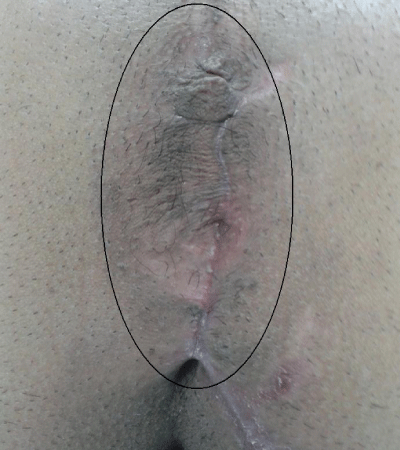
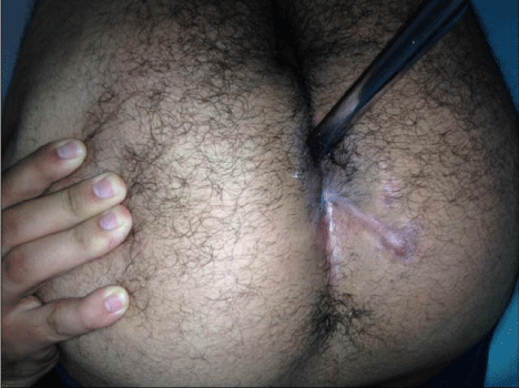
In this editorial we report a case of delayed anal reconstruction. This is a 30 year old male that was injured by shrapnel from a blast injury in very close proximity about six months prior to his presentation. The shrapnel entered in his buttock and avulsed his rectum and anus causing excessive hemorrhage. At that time, he was managed in a field hospital by a laparotomy and a colostomy for diversion along with complete primary closure of his anus for control of bleeding. He recovered and came to our service for reconstruction (Figure 1).
Figure 1 :Closed off and healed anal verge within black ellipse.
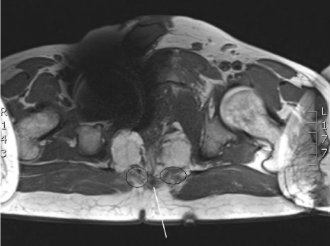
An MRI of pelvis was ordered to assess the musculature of the pelvic floor and anal sphincter apparatus. It visualized a distal rectal stump, but the anus was not well seen. The levator ani muscles were atrophic but present. The rectal lumen is well visualized till approximately 2 cm below the coccygeal tip distally. The external sphincter is most likely present but the internal sphincter integrity could not be determined on MRI due to artifact from in situ shrapnel that obscured the anus and anorectal junction (Figure 2). On exam when you ask the patient to constrict his anal muscles, a shadow of a moving sphincter can be seen in the perianal subcutaneous skin that is now closed off and scarred.
Figure 2 :MRI scan of pelvis showing atrophic levator muscles within ellipse and present sphincter at tip of white line.
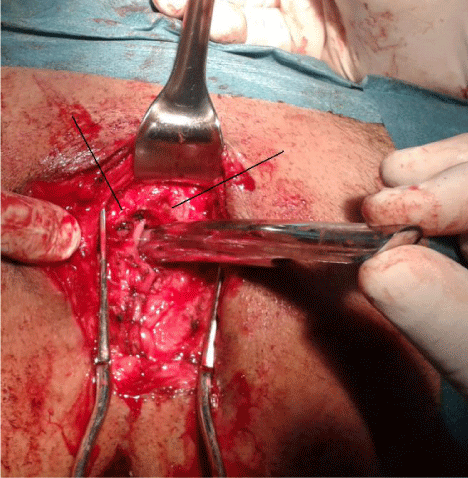
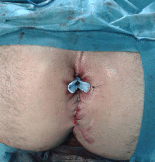
In the lithotomy position and under general anesthesia, he underwent exploration of his perineum. The healed closed anal verge was opened and dissection carried out until the distal rectal stump was identified along with the external sphincter and levator ani muscles. A pull down Duhamel type hand sewn rectoanal anastomosis was performed along with a sphincteroplasty to reconstruct the anus and achieve continuity. A rectal tube was left in the repair to avoid strictures (Figure 3 & 4).
Figure 3 :Anal reconstruction with Hagar dilator in distal rectum and the external sphincter muscle shown at tip of lines.
Figure 4 : Rectal tube left in situ at the end of reconstruction.
