Special Issue
Austin J Surg. 2014;1(5): 1021.
Recurrent Laryngeal Nerve Palsy Following Blunt Trauma of Lateral Neck
Tuzuner A1*, Demirci S1, Aydogan F1, Ceylan T2 and Karadas H1
1Department of Otolaryngology, Ankara Research and Training Hospital, Turkey
2Department of Otolaryngology, Diskapi Yildirim Beyazit Research and Training Hospital, Turkey
*Corresponding author: Tuzuner A, Department of Otolaryngology, Ministry of Health Ankara Training and Research Hospital, Cebeci, Ankara, Turkey
Received: July 29, 2014; Accepted: Aug 02, 2014; Published: Aug 06, 2014
Abstract
Objective: Laryngotracheal traumas are rare conditions and may cause wide spectrum of complications on the laryngeal airway. Impact of trauma may affect soft tissue, nerves and cartilage of larynx. For each injury, different treatment modalities from simple observation to urgent surgical intervention may be necessary.
Method: We would like to present a 24 year-old female patient who had a blunt trauma from lateral side of anterior neck that was resulted in hoarseness and breathiness of the voice in following days.
Results: On video-laryngostroboscopy, endolaryngeal soft tissue damage with restricted left vocal fold movement was observed. Partial recurrent laryngeal nerve damage showed on electromyography. Patient informed about the situation and injection laryngoplasty with hyaluronic acid was planned to be used to improve the voice quality during regeneration period.
Conclusion: Recurrent laryngeal nerve palsy due to blunt trauma is a very rare condition and laryngeal EMG is observed to be a very helpful tool for diagnosis and decision for treatment.
Keywords: Blunt neck trauma; Recurrent laryngeal nerve palsy; Laryngeal electromyography; Dysphonia
Introduction
Unilateral vocal fold paralysis (UVFP) causes breathiness, weakness of voice, swallowing problems due to restricted vocal fold motion which effects glottal closure. Most prominent causes of UVFP are idiopathic, iatrogenic injury (thyroidectomy, lung-mediastinum surgery, cervical spine esophageal, skull base, heart surgery etc.), radiation to the neck, oncologic diseases and blunt or sharp traumas to the neck region [1,2].
Blunt neck traumas involving larynx are very rare conditions and different type of injury to the laryngotracheal framework may result in from a simple soft tissue edema to life-threatening laryngotracheal separation. On clinical evaluation, patients may suffer from dyspnea, voice impairment, globus sensation or cough [3]. In the acute period of the injury airway evaluation is the priority for the patient. In the case of severe injury due to laryngeal collapse the emergent tracheotomy might be necessary. Complication rates are as high as 40% following neck traumas and blunt trauma seems to be less severe injury risk than sharp traumas [4,5]. Following trauma when acute period is overcome, the most presenting symptom is hoarseness that could be recurrent laryngeal nerve/superior laryngeal nerve paralysis, arytenoid subluxation or laryngeal cartilage fractures [6].
In the present report we would like to present a rare case that had isolated blunt trauma to the left side of her neck resulted in dysphonia and breathiness. After resolution of acute laryngeal findings, etiology is distinguished from two major causes of this condition.
Case Presentation
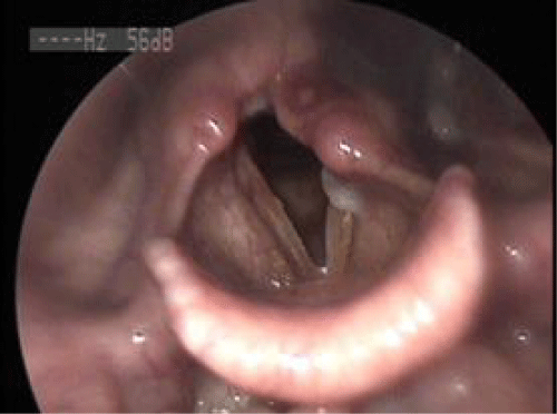
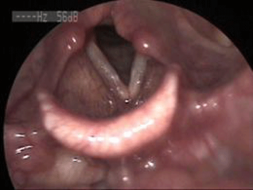
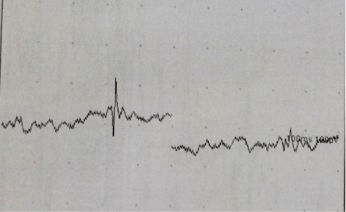
A twenty-four years old female who has dysphonia and globus sensation of throat following blunt injury to her left lateral side of anterior neck due to accidental car door crush. Patient applied our otolaryngology clinic two days ago. There was a mild soft tissue swelling anterior to the left sternocleidomastoid muscle with inspection and she had tenderness on the same region during palpation. The video-laryngostroboscopy showed endolaryngeal soft tissue swelling and ecchymosis on the left arytenoid mucosa, piriform sinus and restricted left vocal fold movement with 2 mm. opening between folds during phonation was observed (Picture 1). Considering edema and possible recurrent laryngeal nerve paralysis she was put on prednisolone treatment 1mg/kg for a week tapered by 20 mg per day and proton pomp inhibitor (30 mg lansoprazole daily). After one month follow-up, she still had similar symptoms and video-laryngostroboscopy (VLS) showed left vocal fold palsy with better closing defect less than 2 mm. glottal closures with phonation and scar tissue on the left piriform sinus mucosa (Picture 2). Laryngeal electromyography (EMG) planned to rule out arytenoid subluxation and design the proper treatment modality (Figure 1). Partial recurrent laryngeal nerve damage was observed showed on electromyography. Patient was informed about this temporary situation and injection laryngoplasty with hyaluronic acid was planned to improve the voice quality during regeneration period.
Picture 1: Acute period after trauma. Echymosis on the left arythenoid and piriform sinus mucosa with left laryngeal palsy.
Picture 2: Two months after trauma. Scar tissue on the left piriform sinus mucosa with restricted left VF movement.
Figure 1 : Left thyroarytenoid muscle EMG. Abnormal spontaneous activity with fibrillation potential which suggests denervation.
Discussion
Glottic incompetence due to UVFP is the major problem with these patients and full recovery on vocal fold motion during the first year of the paralysis is required for permanent interventions. Recovery rates following UVFP in patients reported range from 22% to 39% for different causes in the literature [7,8]. Even specific etiologic causes such as cardiovascular surgery have higher recovery rates [9].
Vocal fold paralysis Neck traumas related to larynx have high mortality rates and may be related to a blunt or penetrant injury. Gussack et al. reported over 109 head and neck injuries in 30,000 trauma victims over the past 5 years [4].
In the acute period of laryngeal trauma, voice associated dysfunctions are common recurring symptoms which would occur due to soft tissue edema, hemorrhage, cartilage fractures, cricoarytenoid subluxation and recurrent laryngeal nerve injury. Soft tissue edema, ecchymosis, vocal fold palsy, laryngeal cartilage dislocations could be observed using laryngostroboscopy. Following resorption of edema and hemorrhage if hoarseness and vocal fold restriction persists in clinical examination two main conditions are considered. These conditions are likely to be either recurrent nerve palsy or arytenoid subluxation and very rarely external brunch of superior laryngeal nerve palsy. Differentiating one of the three diagnoses is important for treatment choice. Laryngostroboscopy findings and laryngeal EMG are the most common clinical evaluation methods used to figure out the main underlying pathology [10]. Palpation under suspension laryngoscopy is mandatory for cricoarytenoid subluxation.
Temporary or permanent voice impairment might be observed due to involvement of laryngeal structures. Mostly soft tissue damage effecting vocal fold movements are resolve in weeks and a follow-up with or without a steroid treatment is sufficient [11]. When recurrent nerve palsy is the diagnosis, partial or complete paralysis of the nerve should be discriminated by laryngeal EMG. If regeneration potentials are present, follow-up or injection laryngoplasty could be offered to the patient to improve voice quality. Permanent damage to the nerve have similar intervention in the early stages of trauma, however patient should be aware of permanent voice disturbance which requires laryngeal framework surgery. Cricoarytenoid subluxation is the more rare type of hoarseness following injury and early surgical intervention relocate the joint is useful for resolution of symptoms [10].
Conclusion
UVFP accompanying blunt trauma is a rare condition and a careful examination of the neck and glottic region is important to differentiate the other etiologic factors. Laryngeal EMG is a useful method to reveal the improvement expectation with these patients as well as accuracy of diagnosis. If partial recovery has been detected patient may put on follow-up without and intervention for early period during three month or injection laryngoplasty could be performed.
References
- Fang TJ, Pei YC, Li HY, Wong AM, Chiang HC. Glottal gap as an early predictor for permanent laryngoplasty in unilateral vocal fold paralysis. Laryngoscope. 2014.
- Alghonaim Y, Roskies M, Kost K, Young J. Evaluating the timing of injection laryngoplasty for vocal fold paralysis in an attempt to avoid future type 1 thyroplasty. J Otolaryngol Head Neck Surg. 2013; 42: 24.
- Wu MH, Tsai YF, Lin MY, Hsu IL, Fong Y. Complete laryngotracheal disruption caused by blunt injury. Ann Thorac Surg. 2004; 77: 1211-1215.
- Gussack GS, Jurkovich GJ, Luterman A. Laryngotracheal trauma: a protocol approach to a rare injury. Laryngoscope. 1986; 96: 660-665.
- Schaefer SD. The acute management of external laryngeal trauma. A 27-year experience. Arch Otolaryngol Head Neck Surg. 1992; 118: 598-604.
- Sulica L. The natural history of idiopathic unilateral vocal fold paralysis: evidence and problems. Laryngoscope. 2008; 118: 1303-1307.
- Munin MC, Rosen CA, Zullo T. Utility of laryngeal electromyography in predicting recovery after vocal fold paralysis. Arch Phys Med Rehabil. 2003; 84: 1150-1153.
- Young VN, Smith LJ, Rosen C. Voice outcome following acute unilateral vocal fold paralysis. Ann Otol Rhinol Laryngol. 2013; 122: 197-204.
- Joo D, Duarte VM, Ghadiali MT, Chhetri DK. Recovery of vocal fold paralysis after cardiovascular surgery. Laryngoscope. 2009; 119: 1435-1438.
- Schroeder U, Motzko M, Wittekindt C, Eckel HE. Hoarseness after laryngeal blunt trauma: a differential diagnosis between an injury to the external branch of the superior laryngeal nerve and an arytenoid subluxation. A case report and literature review. Eur Arch Otorhinolaryngol. 2003; 260: 304-307.
- Kumral TL, Yildirim G, Uyar Y, Kuzdere M, Yurtseven C, Gümrükçü S, et al. Travmatik vokal kord paralizisi Turk Arch Otolaryngol. 2011; 49: 74-77.


