
Case Report
Austin J Surg. 2014;1(6): 1030.
Cytomegalovirus Colitis Associated with Ileocolic Intussusception in a Child Status Post Heart Transplant
Kemp CD1, Scholz S3, Subhawong AF2, Abdullah F1 and Rasmussen SK4*
1Division of Pediatric Surgery, Johns Hopkins University, USA
2Department of Pathology, Johns Hopkins University, USA
3Department of Surgery, Children’s Hospital of Pittsburgh, USA
4Division of Pediatric Surgery, University of Virginia, USA
*Corresponding author: Rasmussen SK, Division of Pediatric Surgery, University of Virginia, Charlottesville, VA, USA
Received: May 12, 2014; Accepted: August 30, 2014; Published: September 05, 2014
Abstract
A pediatric heart recipient presented with intermittent abdominal pain. Workup revealed intussusception with associated pneumatosis. A right hemicolectomy was performed. Pathologic examination revealed cytomegalovirus (CMV) colitis. This is the first case of CMV-colitis associated with intussusception in a pediatric transplant patient. We present the findings and review of the literature.
Keywords: Cytomegalovirus infection; Colitis; Pediatric heart transplant; Pediatric immunosuppression; Pneumatosis intestinalis
Abbreviations
CMV: Cytomegalovirus; CT: Computed Tomography; PCR: Polymerase Chain Reaction
Case Presentation
A 9 year old boy with a history of cardiac transplant for hypoplastic left heart syndrome (HLHS) presented to the Johns Hopkins Hospital with acute abdominal pain. His post-cardiac transplant course was significant for two episodes of biopsy-proven rejection in the intervening 6 months prior to presenting to the pediatric surgery service. On the day of presentation, his chief complaint was abdominal pain and diarrhea. His outpatient immunosuppression regimen consisted of tacrolimus, mycophenolate mofetil, and prednisolone. A plain film of the abdomen was obtained (Figure 1). This demonstrated an ileocolic intussusception with extensive pneumatosis of both the intussusceptum and the intussuscipiens. The patient’s clinical status (tachycardic with diffuse abdominal pain) and the findings of pneumatosis suggested significant bowel compromise. It was determined that an attempt at air contrast reduction in an immunosuppressed patient with such extensive pneumatosis was contraindicated and operative intervention was planned immediately A computed tomography (CT) scan of the abdomen and pelvis was performed to evaluate for pneumoperitoneum (Figure 2). This was negative and the child was taken directly to the operating room.
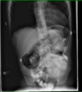
Figure 1: Plain film of the abdomen demonstrating an ileocolic intussusception with pneumatosis of the bowel wall indicated by an arrow.
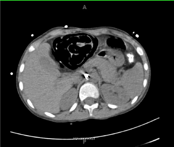
Figure 2: Representative axial section of CT obtained demonstrating pneumatosis (arrow).
A initial diagnostic laparoscopy confirmed the ileocolic intussusception. Though there was no sign of intestinal ischemia or necrosis, there was extensive pneumatosis (Figure 3) of the terminal ileum and the right colon. Both the intussuscipiens and intussusceptum were affected. The transition to normal-appearing bowel was abrupt. A right hemicolectomy was performed with a primary stapled side-to-side, functional end-to-end anastomosis. Final pathological analysis of the specimen revealed CMV colitis. Figure 4 demonstrates the CMV inclusion bodies seen on histologic analysis of the colectomy specimen. The child received treatment for his CMV infection and was discharged home on post-operative day 9 after an uneventful recovery. He did well following discharge. Titers of CMV, which had peaked at 995ng/mL, returned to undetectable levels.
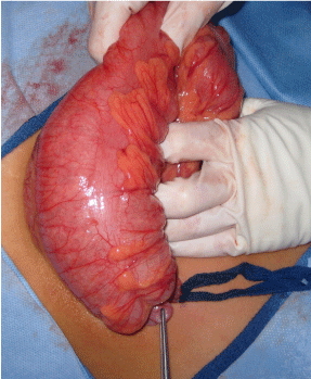
Figure 3: Intraoperative photograph of right colon. The tip of the intussuscepted appendix is evident in the forceps (star). The arrow indicates the pan-pneumatosis found in the bowel wall, despite no evidence of compromised perfusion.
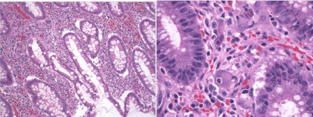
Figure 4: Immunohistochemistry of resected colon. A. Low magnification demonstrates hemorrhage and a mixed inflammatory infiltrate within the mucosa (hematoxylin and eosin, 40X). B. Higher magnification reveals scattered stromal cells (arrows) with the classic “owl’s eye” inclusions of cytomegalovirus (hematoxylin and eosin, 160X).
A thorough review of his post-cardiac transplant course revealed that our patient had recently been taken off of his CMV prophylaxis. Additionally, a recent episode of tacrolimus toxicity required adjustments to his immunosuppressant medications. He had also had two episodes of biopsy-proven rejection which were managed with high-dose steroid pulses. The most recent episode of rejection was 5 months after his heart transplant, approximately 7 weeks prior to his intussusception.
Unfortunately, the boy succumbed to his heart disease 18 months after his heart transplant. However, there were no recurrent episodes of CMV colitis, and no complications from his colectomy.
Discussion
This case represents a complex interplay of several important disease states and strategies in the management of complicated clinical issues in the pediatric transplant patient. Specifically, there are three clinical conditions in this presentation that impacted this patient’s clinical course: intussusception, CMV infection, and cardiac transplant status.
Intussusception is one of the most common causes of intra-abdominal emergencies in children. It is caused when an intestinal lead point telescopes into the distal bowel, causing a bowel obstruction. Intussusception manifests most commonly with episodes of intermittent abdominal pain in young toddlers. In the vast majority of pediatric cases, specific pathology is not present and enlarged mesenteric lymph nodes function as the lead point. Intussusception is most commonly managed initially with retrograde air-contrast enema reduction, which is diagnostic and therapeutic. In fact, most children altogether avoid an operation with this treatment. In cases where patients present with advanced peritonitis, or when bowel perforation is suspected, contrast reduction is contraindicated. In these cases, immediate operative reduction and/or resection are mandated.
CMV colitis is a common opportunistic infection in transplant recipients, and it can cause a variety of diseases, including colitis. It can have devastating effects on the patient. For this reason, organs and recipients are serotyped for CMV pre-operatively in order to minimize the chances of a CMV-negative recipient becoming infected. In patients who are CMV-positive, prophylaxis with valgancyclovir is maintained for several months post-transplant. In our patient, who stopped his valgancyclovir weeks prior to his intussusception, the possible role of CMV colitis in his intussusception cannot be overlooked.
Finally, all transplant organ recipients must maintain life-long immunosuppression to prevent rejection of the donor organ. There is the constant dilemma of the goal to eliminate the risk of rejection and still have a host with a competent immune system. As a result, transplant recipients are at life-long risk of opportunistic infections, of which CMV is one.
The clinician must also avoid toxicity related to overdoses of the immunosuppressant medications. Supra-therapeutic levels of different immunosuppressants can require temporary cessation of the medications in order to avoid or minimize organ impairment. These temporary cessations can make the patient vulnerable to further episodes of rejection.
In the two months prior to his presentation to the pediatric surgery service, our patient had several episodes of intermittent abdominal pain and diarrhea. Review of his chart revealed that he recently had an episode of tacrolimus toxicity, prompting alteration of his immunosuppressant regimen. During the time that his serum tacrolimus level was too high, he may have been more susceptible to opportunistic infections. The patient was on valgancyclovir during his first five months post-transplant. The valgancyclovir was given during the patient’s first episode of rejection in the immediate postoperative period after his heart transplant. However, valgancyclovir was not utilized during the second episode five months post-heart transplant. Interestingly, when the patient originally presented with vague complaints of diarrhea and abdominal pain, CMV titers had been undetectable by polymerase chain reaction (PCR). During his admission for the intussusception, the CMV titer was 995copies/mL. The timing of these two events associated with his presentation of intussusception is suggestive that the CMV colitis caused the ileocecal pneumatosis and mesenteric lymphadenitis which functioned as a lead point for the intussusception, or that the hyperperistalsis causing diarrhea from CMV enterocolitis triggered the intussusception. It is unlikely that pneumatosis occurred as a result of the intussusception. Bowel which is obstructed in an intussusception can develop pneumatosis secondary to effects of local tissue compression and resulting vascular compromise. This would certainly be a consequence of ischemia, and would result in decreased bowel viability. However, during our exploration, the bowel was well perfused with no evidence of ischemia, implying that the pneumatosis developed before the intussusception, and not as a result of it.
Once confronted in the OR with viable-appearing colon that had extensive pneumatosis, we decided to undertake a right hemicolectomy. The intussusception reduced manually quite easily, and there were no areas of questionable perfusion of the intestinal wall. However, given the child’s physiologic status on presentation, his immunosuppressed status, and the fact that we did not yet know what was causing the pneumatosis, we felt it most prudent to remove the affected colon and perform a primary anastomosis. In particular, the chronic use of steroids contributed to the decision to perform a resection.
A brief discussion concerning temporary diversion was undertaken. We decided against this because the child was nutritionally replete and therefore we could expect him to heal an anastomosis. Additionally, the unaffected ileum and transverse colon were of normal caliber and appearance. Primary anastomosis in emergent hemicolectomies is widely reported in the literature. For example, a recent review of 207 consecutive emergency right hemicolectomies at a single institution revealed that stoma creation and ASA score were the only significant predictors of poor outcome [1]. Other studies have demonstrated that stoma complications are significant after right hemicolectomy and that they may be riskier than primary anastomosis [2-4].
There is one prior case reported in the literature of CMV-colitis being associated with intussusception [5]. Interestingly, this report is of recurrent intussusceptions occurring in an adult liver transplant patient. It has been reported that liver transplantation is a risk factor for intestinal intussusception [6]. This patient described in this case report had recurrent intussusceptions diagnosed by ultrasound and computed tomography. A colonoscopy done to elucidate the cause of the recurrent intussusceptions revealed CMV colitis on biopsy. This patient’s symptoms resolved with treatment of her CMV colitis and follow-up biopsies were negative for CMV disease. Similarly in our patient, follow-up CMV levels showed a decline and eventually were undetectable postoperatively.
There is another report of a colo-colonic intussusception in a heart transplant patient [7]. However, it is not described whether this case was associated with CMV infection. Similar to our case, initial radiographic findings were quite impressive, and this child underwent primary resection with anastomosis at operation as well. This patient had no complications associated with anastomosis, supporting the use of anastomosis in emergency colonic procedures in pediatric transplant patients.
CMV infection is known to cause a broad spectrum of morbidity and mortality in the pediatric population. In particular, infants who are congenitally infected with the virus, as well as organ transplant patients are affected. CMV colitis can be detected on plain abdominal radiograph in neonates with CMV colitis [8]. This is the first case report of a pediatric transplant patient experiencing intussusception associated with CMV colitis. The possibility of this complication arising should add caution to undertake careful consideration about whether to withdraw CMV prophylaxis in pediatric transplant patients.
Acknowledgement
The authors wish to express gratitude to Dr. Paul M. Colombani, for his comments in the preparation of this manuscript.
References
- Tan KK, Liu JZ, Yeow Y, Gunasekaran S, Tan JJ. Is emergency right hemicolectomy still associated with significant morbidity and mortality rates? An institution's experience of 207 cases over 6 years. Int j colorectal dis. 2011; 26: 1157-1161.
- Miller FB, Nikolov NR, Garrison RN. Emergency right colon resection. Arch Surg. 1987; 122: 339-343.
- Mealy K, Salman A, Arthur G. Definitive one-stage emergency large bowel surgery. Br J Surg. 1988; 75: 1216-1219.
- Wyrzykowski AD, Feliciano DV, George TA, Tremblay LN, Rozycki GS, Murphy TW, et al. Emergent right hemicolectomies. Am Surg. 2005; 71: 653-656.
- Pischke S, Tutarel O, Greten TF, Heim A, Wedemeyer J, Herzog P, et al. [CMV-enterocolitis as a cause for repeated intestinal intussusceptions in an adult patient after liver transplantation?]. Zeitschrift fur Gastroenterologie. 2010; 48: 688-692.
- Pischke S, Karsten W, Hadem J, Schmidt S, Heiringhoff Heinz K, Helfritz F, et al. Liver transplantation: A new risk factor for intestinal intussusceptions. Ann Hepatol. 2011; 10: 38-42.
- Sanchez S, Javid P, Ricca R, Avansino J. Colocolonic intussusception in a four-yr-old with a heart transplant: a case report and review of the literature. Pediatr Transplant. 2012; 16: E225-228.
- Van der Jagt EJ, van Son WJ, van der Woude FJ, Meijer S, Slooff MJ, Tegzess AM, et al. Pneumatosis intestinalis related to cytomegalo-virus infection--a new etiology? Eur J Radiol. 1987; 7: 28-29.