Research Article
Austin J Surg. 2014;1(7): 1031.
Soft Tissue Reconstruction of Foot and Ankle Defects: Free Vs Pedicled Flaps with the Use of 6 Different Flaps in 50 Cases of Road Traffic Accidents
Khurram MF*, Ahmad I and Nanda M
Department of Burns, Plastic and Reconstructive Surgery, JN Medical College, India
*Corresponding author: Mohammed Fahud Khurram, Department of Burns, Plastic and Reconstructive Surgery, JN Medical College, AMU, G-2, Azim Green Home, New Sir Syed Nagar, Aligarh, UP-202002, India
Received: July 14, 2014; Accepted: September 06, 2014; Published: September 11, 2014
Abstract
Background: Soft tissue reconstruction of the foot and ankle still remains a complex and challenging process despite advances in the transfer of fasciocutaneous, musculocutaneous, and composite flaps. The development of microsurgery and the expansion of plastic surgery techniques have led to an increase in number of reconstructive options for the salvage of lower extremities and thereby causing dilemma regarding use of pedicled flaps for foot reconstruction. We present our comparative study with the use of pedicled as well as free flaps for soft tissue reconstruction of the foot and ankle.
Materials and Methods: From 2008 to 2013, the soft tissue defects of traumatic injuries of the foot & ankle were reconstructed using 6 different flaps in 50 cases (32 male and 18 female). There were 22 pedicled flaps and 28 free flaps used in reconstruction. The pedicled flaps included reverse sural flap, pedicled peroneal artery perforator flap and pedicled tibial artery perforator flap. The free flaps were LD musculocutaneous flap, ALT musculocutaneous flap and Vastuslateralis muscle flap. The sensory nerve coaptation was performed in 8 patients only.
Results: Among 28 free flaps, 3 flaps were completely lost as against 1 flap in pedicled flap group, in which the defects were managed by the secondary procedures. The donor site complications were seen in 1 case with the free flaps and 3 cases with pedicled flap. All limbs were preserved and the patients regained walking and daily activities. Out of 10 pedicled flaps and 8 free flaps used to reconstruct hind foot, ulcers developed in 2 pedicled flaps and 3 free flaps (muscle with STSG) after weight bearing. All the patients in whom nerve co-optation was done regained protective sensation from 6 to 12 months postoperatively.
Conclusion: A careful pre & per-operative planning with special emphasis to be given on size and location of the defect and correct contour match and insetting should allow for maximal functional and aesthetic result with minimal post-operative morbidity.
The loco-regional flaps are good options for the coverage of defects around ankle & dorsal hind foot. Plantar foot, forefoot and large size defects could be reconstructed with free flaps.
Large defects with exposed bone/ implant with or without infection are best handled with a free flap.
Keywords: Foot and ankle defect; Free flap; Loco-regional flap; Road traffic accidents
Abbreviations
RTA: Road Traffic Accidents; RSA: Reverse Sural Artery Flap; PTAF: Posterior Tibial Artery Based Flap; PAP: Peroneal Artery Perforator Based Flap; LD: Latissimusdorsi Flap; ALT: Anterolateral Thigh Flap; VL: Vastuslateralis Muscle
Introduction
The foot is an important part of the body which supports the body in standing posture and provides a stable interface between the ground and the body during walking. Its function depends on many factors which can be affected by variety of pathological processes and trauma.
Due to ever increasing number of vehicles, the road accidents have increased. Road traffic accidents (RTA) is the most common cause of injury to the foot (73.88%) [1], and out of that more than 70% has open fractures [1].
Soft tissue reconstruction of the foot and ankle still remains a complex and challenging process despite advances in the transfer of fasciocutaneous, musculocutaneous, and composite flaps [2]. The development of microsurgery and the expansion of plastic surgery techniques have led to an increase in number of reconstructive options for the salvage of lower extremities. Free flaps offer a great variety of available tissues to cover larger, multifocal or multi-structural defects. They also improve the perfusion of the infected and poorly perfused areas [3].
Although the plastic surgeons is influenced by the success and failure of past endeavours, it is imperative to perform ongoing re-evaluation of the one`s results and to modify one`s approach accordingly [2].
We present our experience with the use of pedicled as well as free flaps for soft tissue reconstruction of the foot and ankle sustained during or after road traffic accidents.
Material and Methods
This study was conducted in prospective manner from 2008 to 2013. Fifty patients (32 male and 18 female) with history of road traffic accidents and the soft tissue defects of the foot & ankle were included. They either had compound injury at the time of accident, or after debridement or after orthopedic intervention with exposed bone or implant. These were reconstructed using 6 different flaps. The reconstructive option depended on the site, size and cause of the defect. There were 22 pedicled flaps and 28 free flaps used in reconstruction. The patient details were recorded in a Performa which included history, examination (local as well as general), lab tests, radiographs, Doppler/ angiography as and when required. The procedures were explained to the patient and informed consent taken and recorded. Standard techniques were used to raise the flaps. All the patients with free flaps were admitted in the ICU for at least 2 days with proper monitoring of the flaps. The duration of hospital stay and other outcome analysis was also recorded.
The pedicled flaps which were used are:-
- Reverse sural flap,
- Pedicled peroneal artery perforator flap, and
- Pedicled tibial artery perforator flap.
- LD musculocutaneous flap,
- ALT musculocutaneous flap, and
- Vastuslateralis muscle only flap.
- Postoperative complications including donor site morbidity,
- Hospital stay,
- Time to heal,
- Length of time the wound stayed healed,
- Limb salvage, and
- Patient survival.
- Patient satisfaction and ambulatory status were also assessed.
- Any limb that was amputated at or proximal to the ankle was regarded as a failure.
The following free flaps were used
The sensory nerve coaptation was performed in 08 patients only.
Outcome analysis consisted of
Statistical analysis was performed using Fisher’s exact test and chi-square test, depending on the data being analyzed.
Standard follow up protocol was followed which included regular visit after discharge and long term problems were noted (especially after full weight bearing and walking) and rectified accordingly.
Results
- The patient’s demographic properties are shown in Table 1.
- The mean age was 39 years ± 7.8, with a 1.7:1 male-to-female ratio.
- All wounds after debridement had exposed bone, tendon or osteosynthetic implant at their base.
- Among 28 free flaps, 3 flaps were completely lost, whereas in pedicled flap group only 1 flap failed completely, and resultant defects were managed by the secondary procedures.
- The donor site complications were seen in 1 case with the free flaps and 3 cases with pedicled flap.
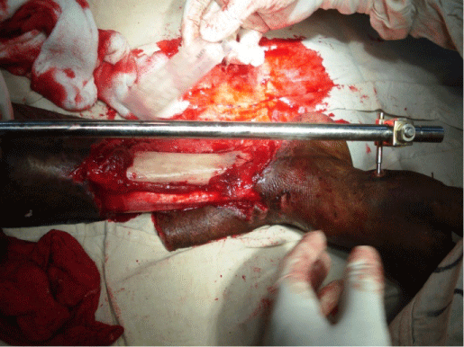
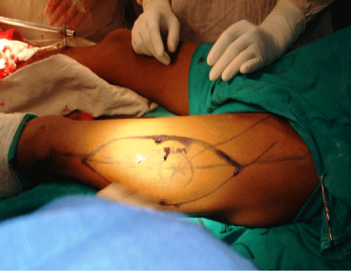
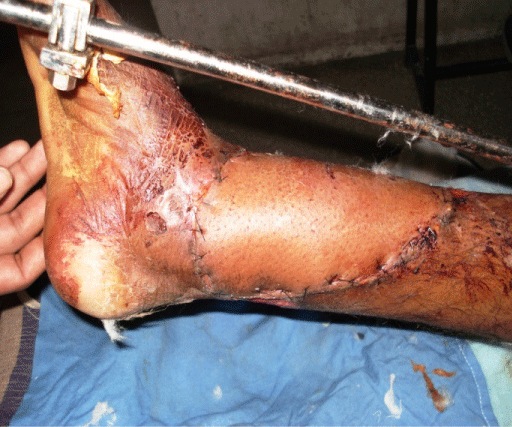
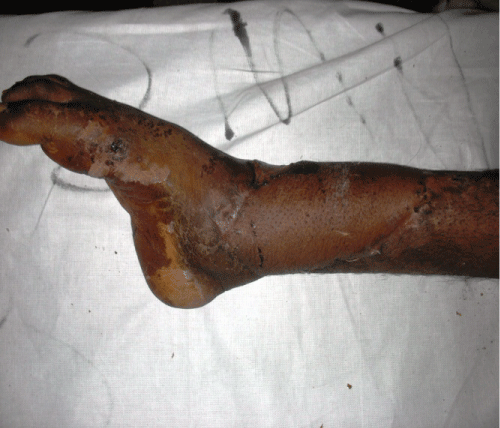
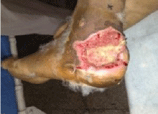
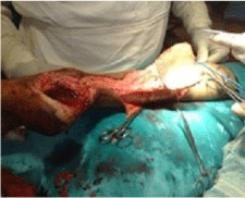
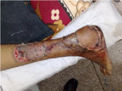
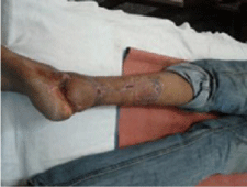
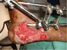
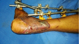
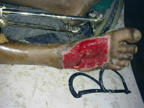
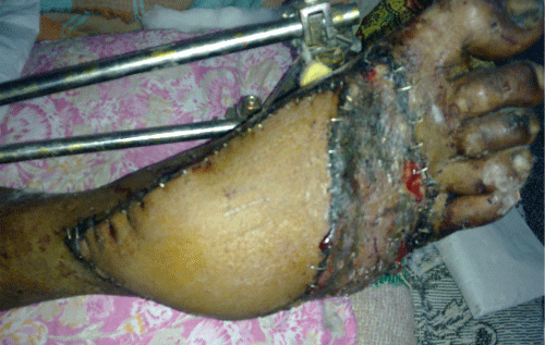
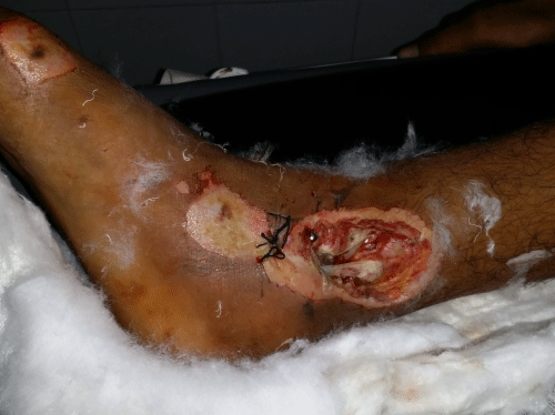
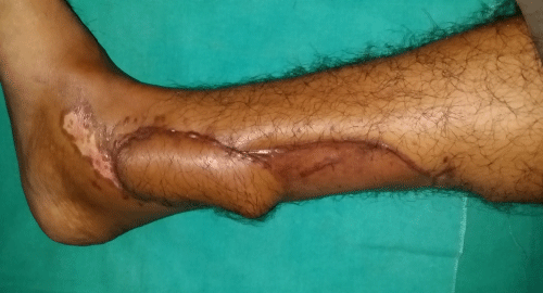
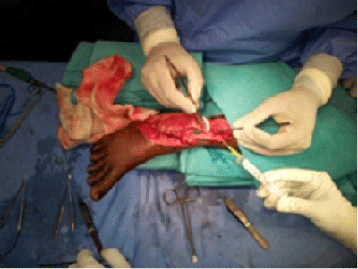
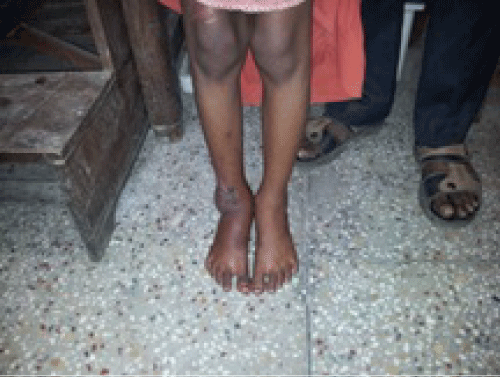
- All limbs were preserved and the patients regained walking and daily activities (80 & 85% cases) [Table 2 & 3] [Figures 1-6].
Figure 1a: Pre operative photograph of a patient with exposed lower tibia and ankle joint.
Figure 1b: Intra operative picture showing marking of free anterolateral thigh flap.
Figure 1c: Post operative picture (after 7 days).
Figure 1d: Post operative picture (after 6 weeks).
Figure 2a: Preoperative picture with post road traffic accident defect over posterior heel.
Figure 2b: Reverse sural flap dissected and raised.
Figure 2c: Postoperative day 7th photograph showing reverse sural flap covering the heel defect.
Figure 2d: Postoperative day 28th photograph showing excellent result.
Figure 3a: Pre-operative photograph of a patient with injury to posterior part of foot with exposed underlying bone and tendons after debridement.
Figure 3b: Post operative photograph showing coverage with free latissimus dorsi flap with STSG.
Figure 4a: Defect over dorsum of foot.
Figure 4b: Post operative photograph showing coverage with free anterolateral thigh flap.
Figure 5a: Post RTA defect over Right leg with exposed tibia.
Figure 5b: Post operative photograph showing excellent result with posterior tibial artery perforator based flap.
Figure 6a: Defect over dorsum and ankle of right foot being covered with free vastuslateralis muscle flap.
Figure 6b: Post operative photograph after 8 weeks with excellent weight bearing and walking ability.
Out of 10 pedicled flaps and 8 free flaps used to reconstruct hind foot, ulcers developed in 2 pedicled flaps and 3 free flaps (muscle with STSG) after weight bearing (Table 4).
All the patients in whom nerve co-optation (n=8) was done regained protective sensation from 6 to 12 months postoperatively.
Discussion
Ever increasing number of vehicles has led to increase in road traffic accidents (RTA) and that too in younger age group. RTA is the most common cause of injury to the foot especially in young people. Road accidents have emerged as a major global public health problem of this century and now recognized as veritable neglected pandemic [4]. The problem is so severe that by 2020, it is projected that road traffic disability adjusted life years (DALYs) lost will move from being the 9th leading cause of DALYs lost to the 3rd leading cause in the world and will be 2nd leading cause in the developing countries [5].
Some of the factors that increase the risk of road accidents in India are unsafe traffic environment, poor road infrastructure and encroachment, traffic mix and unsafe driving behavior and training [6,7].
Foot is involved in almost 70% of RTA cases and compounding is seen in 43% cases [8,9]. Foot and ankle poses a difficult reconstructive challenge for plastic surgeons. There are various options available for reconstruction of soft tissue defect of foot and ankle viz. local fasciocutaneous flap (PTAF, PAP. RSA, DPA etc.), muscle and musculocutaneous flaps (peroneus brevis, reverse soleus) and free flaps.
There are numerous studies that confirm the advantage of immediate soft tissue coverage of exposed bone, tendon, nerve, vessels or osteointegrated implants [10-13]. The choice of coverage depends on the size, site, shape and type of defect. Nowadays, the free flaps are being increasingly used to cover the defects of foot and ankle, but it requires special surgical expertise, longer operating time and aggressive monitoring than the pedicled flaps. The complications rate of free flaps ranges from 10% [14] to 38% [15] and from 21% [16-18] to 46% [19] for local flaps.
In our experience, complications were 6/22 and 5/28 in pedicled and free flap group respectively. The free flaps took longer average operating time (200 min) as compared to 90 minutes in pedicled group in this study.
As far as wound location is concerned we have used free as well as pedicled flap in almost equal frequency at all sites with comparable results (P value not significant). The local fasciocutaneous flaps can provide excellent alternative option to free flap but they have limited reach and unreliable especially if the surrounding area is also traumatized. They also leave ugly donor site scars because of skin grafting. Among the pedicled fasciocutaneous flaps, we used reverse sural flap in 08 cases, posterior tibial artery based flap and peroneal artery perforator based flap in 07 cases. The advantages of these flaps are less operating time, no sacrifice of major vessel, easy dissection, comparable success rates, and easy reach to the weight bearing areas of foot, good color match and less aggressive monitoring. The disadvantages include higher rate of venous congestion, single team approach, donor site morbidity, difficulty in tailoring/ customization because of unclear blood supply source (both arterial and venous drainage).
The free flaps have the advantages of good contour match, composite flap technique can be used, less donor site morbidity, two team approach and these can be neurotized. Disadvantages of free flap are longer operating time and hospital stay, aggressive monitoring, microsurgery availability & expertise, neuropathic ulcer in case of muscle only flaps, sacrifice of major muscle (latissimusdorsi) & vessel (radial artery in radial forearm flap), and tedious dissection in case of perforator free flap.
El-Shazly et al. [20] described free tissue transfer as a valuable reconstructive procedure in foot reconstruction in their study of 11 patients.
Yue-Liang et al. [21] have used 14 different flaps in 226 cases and suggested that the sural flap and saphenous flap could be good options for the coverage of the defects at malleolus, dorsal hind foot and mid-foot. Plantar foot, forefoot and large size defects could be reconstructed with free ALT flap and for infected cases they have used LD muscle/ muculocutaneous flap. They suggest microsurgical free flaps have a high success rate, are relatively straightforward, and are still the reconstructive option of choice when covering foot and ankle defects. Free flaps do require general anesthesia, cardiac clearance, good recipient vessels, microsurgical skills, longer operative time, and longer hospital stay, and may require subsequent recontouring when healed. Free flaps have the advantage of abundant, healthy muscle and soft tissue in various thicknesses, depending on the location and size of the defect. The recipient site can be debrided aggressively to healthy tissue to yield a superior result. In addition, free flaps can be innervated to provide some sensory feedback.
A. Bhatti et al. [9] showed comparable results between free and pedicled flaps with higher medical complications, longer operating time, and increase hospital stay in free flap group.
Wyble EJ et al. [22] described their experience with 17 free flaps to reconstruct severe foot injuries of the foot.
Rainer et al. [23] published their study with the use of 77 free flaps for foot reconstruction and showed lower ulceration rate in muscle flaps with skin grafts than the fasciocutaneous flap in weight bearing areas and in non-weight bearing areas, they performed fasciocutaneous flap.
Similar to these above studies, our experience has comparable result between the 2 groups except in few conditions like donor site morbidity, duration of hospital stay, operating time and patient satisfaction (only aesthetic and not functional). Among 28 free flaps, 3 flaps were completely lost, whereas in pedicled flap group only 1 flap failed completely, and resultant defects were managed by the secondary procedures. The donor site complications were seen in 1 case with the free flaps and 3 cases with pedicled flap. All limbs were preserved and the patients regained walking and daily activities (80 & 85% cases). Out of 10 pedicled flaps and 8 free flaps used to reconstruct hind foot, ulcers developed in 2 pedicled flaps and 3 free flaps (muscle with STSG) after weight bearing. All the patients in whom nerve co-optation (n=8) was done regained protective sensation from 6 to 12 months postoperatively with no evidence of trophic ulcer upto 2 years of follow-up.
Conclusion
- A careful pre & per-operative planning with special emphasis to be given on size and location of the defect and correct contour match and insetting should allow for maximal functional and aesthetic result with minimal post-operative morbidity.
- Negative pressure wound therapy (NPWT) can be useful in decreasing the size of the defect by promoting the formation of granulation tissue.
- A backup plan should exist in both the cases because the dissected pedicled flap may have less bulk than estimated or may not have the necessary reach or free flap might fail.
- If an external fixator is to be placed, the pins should not compromise the potential harvest site of pedicled muscle flap or recipient vessels.
- The loco-regional flaps are good options for the coverage of defects around ankle & dorsal hindfoot. Plantar foot, forefoot and large size defects should be reconstructed with free flaps.
- For the infected wounds with dead space and large size, the free musculocutaneous flap (ALT with VL and LD) is the best choice.
- Large defects with exposed bone/ implant with or without infection are best handled with a free flap.
- Dhillon MS, Aggarwal S, Dhatt S, Jain M. Epidemiological Pattern of foot injuries: Preliminary assessment data from a tertiary hospital. J Postgrad Med Edu Res. 2012; 46: 144-147.
- Lawrence BC, Theodore UJ. Foot Reconstruction. In: Mathes SJ, editor. Plastic Surgery. IInd Edn. Philadelphia: Elsevier. 2006: 1403.
- Baumeister S, Germann G. Soft tissue coverage of the extremely traumatized foot and ankle. Foot Ankle Clin. 2001; 6: 867-903, ix.
- Dandona R, Mishra A. Deaths due to road traffic crashed in Hyderabad city in India: need for strengthening surveillance. Natl Med J India. 2004; 17: 74-79.
- Murray CJ, Lopez AD. Alternative projections of mortality and disability by cause 1990-2020: Global Burden of Disease Study. Lancet. 1997; 349: 1498-1504.
- Rahman SA, Chandrasala S. When to suspect head injury or cervical spine injury in maxillofacial trauma? Dent Res J (Isfahan). 2014; 11: 336-344.
- Vijayamahantesh SN, Vijayanath V. A cross-sectional study: pattern of injuries in non-fatal road traffic accidents in Bagalkot city of Karnataka. Intl J Medical toxicology and forensic medicine. 2012; 2: 27-32.
- Ganveer GB, Tiwari RR. Injury pattern among non-fatal road traffic accident cases: a cross-sectional study in Central India. Indian J Med Sci. 2005; 59: 9-12.
- Bhatti AZ, Adeshola A, Ismael T, Harris N. Lower leg flaps comparison between free versus local flaps. Int J Plast Surg. 2007; 3.
- Byrd HS, Spicer TE, Cierney G 3rd. Management of open tibial fractures. Plast Reconstr Surg. 1985; 76: 719-730.
- Small JO, Mollan RA. Management of the soft tissues in open tibial fractures. Br J Plast Surg. 1992; 45: 571-577.
- Yaremchuk MJ, Brumback RJ, Manson PN, Burgess AR, Poka A, Weiland AJ, et al. Acute and definitive management of traumatic osteocutaneous defects of the lower extremity. Plast Reconstr Surg. 1987; 80: 1-14.
- Tropet Y, Garbuio P, Obert L, Ridoux PE. Emergency management of type IIIB open tibial fractures. Br J Plast Surg. 1999; 52: 462-470.
- Lin SD, Lai CS, Tsai CC, Chou CK, Tsai CW. Clinical application of the distally based medial adipofascial flap for soft tissue defects on the lower half of the leg. J Trauma. 1995; 38: 623-629.
- Lin SD, Lai CS, Chiu YT, Lin TM, Chou CK. Adipofascial flap of the lower leg based on the saphenous artery. Br J Plast Surg. 1996; 49: 390-395.
- Attinger CE, Ducic I, Cooper P, Zelen CM. The role of intrinsic muscle flaps of the foot for bone coverage in foot and ankle defects in diabetic and nondiabetic patients. Plast Reconstr Surg. 2002; 110: 1047-1054.
- Heymans O, Verhelle N, Peters S. The medial adiposofascial flap of the leg: anatomical basis and clinical applications. Plast Reconstr Surg. 2005; 115: 793-801.
- Steed DL, Donohoe D, Webster MW, Lindsley L. Effect of extensive debridement and treatment on the healing of diabetic foot ulcers. Diabetic Ulcer Study Group. J Am Coll Surg. 1996; 183: 61-64.
- Arnold PG, Yugueros P, Hanssen AD. Muscle flaps in osteomyelitis of the lower extremity: a 20-year account. Plast Reconstr Surg. 1999; 104: 107-110.
- El-Shazly M, Makboul M. Microsurgical free tissue transfer as a valuable reconstructive procedure in foot reconstruction. Indian J Plast Surg. 2007; 40:141-146.
- Zhu YL, Wang Y, He XQ, Zhu M, Li FB, Xu YQ, et al. Foot and ankle reconstruction: An experience on the use of 14 different flaps in 226 cases. Microsurgery. 2013.
- Wyble EJ, Yakuboff KP, Clark RG, Neale HW. Use of free fasciocutaneous and muscle flaps for reconstruction of the foot. Ann Plast Surg. 1990; 24: 101-108.
- Rainer C, Schwabegger AH, Bauer T, NinkoviÄ M, Klestil T, Harpf C, et al. Free flap reconstruction of the foot. Ann Plast Surg. 1999; 42: 595-606.
References
