Case Report
Austin J Surg. 2014;1(7): 1033.
Rapid Recurrence of a Colloid Cyst after Endoscopic Resection
Fontenot P1 and Sandberg DI2*
1Department of Urology, University of Kansas, USA
2Department of Pediatric Surgery, University of Texas Health Science Center at Houston, USA
*Corresponding author: Sandberg DI, Department of Pediatric Surgery, Division of Neurosurgery, University of Texas Health Science Center at Houston, USA, 6431 Fannin Street MSB 5.144, Houston, TX 77005, USA
Received: August 21, 2014; Accepted: September 15, 2014; Published: September 24, 2014
Abstract
Background: Endoscopic resection of colloid cysts has gained popularity as a less invasive alternative to removal via open craniotomy. Previous publications have emphasized that recurrence rates are low after endoscopic removal of colloid cysts even when the resection is subtotal.
Case Description: We review the case of a 36 year-old man who underwent endoscopic resection of a large colloid cyst. All of the cyst contents and the majority of the cyst wall were removed, with a small remnant of the cyst wall left behind. Postoperative imaging studies immediately after surgery and six weeks later did not definitively demonstrate residual colloid cyst. A subsequent MRI scan nearly seven months post-operatively demonstrated a definitive recurrence of the colloid cyst, which was ultimately removed via an open craniotomy.
Conclusions: This unusually rapid recurrence of a colloid cyst following endoscopic resection has not been well-documented in previously published literature. This case is reported to alert neurosurgeons that close radiographic follow-up is important after endoscopic resection of colloid cysts and to add to the ongoing debate regarding the optimal means of managing these lesions.
Keywords: Colloid cyst; Endoscopic resection; Neuro-endoscopy
Introduction
Colloid cysts are benign tumors which are located in the anterior portion of the third ventricle [1-4]. They comprise between 0.5 to 2 percent of all intracranial tumors and 15 to 20% of all intraventricular tumors [5,6]. Colloid cysts can be incidental findings on imaging studies or they can present with headaches when they obstruct the foramen of Monro and cause hydrocephalus [1-4]. A rare but feared presentation of colloid cysts is sudden death, presumably from acute obstructive hydrocephalus but possibly related to other mechanisms such as hypothalamic compression [7-9,4]. Visual loss including blindness is also a rare presentation of colloid cysts [10].
Colloid cysts are typically easily identifiable on magnetic resonance imaging (MRI) scans based upon their round symmetry, location in the anterior third ventricle obstructing the foramen of Monro, and gelatinous contents [11]. Small, asymptomatic colloid cysts may be observed with serial imaging studies [12]. When colloid cysts are large, growing, cause obstructive hydrocephalus, or are clearly symptomatic, treatment is warranted. Cerebrospinal fluid (CSF) diversion via shunting is an option, but most neurosurgeons advocate surgical resection of the colloid cyst. Traditionally, surgical resection has been performed by craniotomy, most commonly via an interhemispheric transcallosal approach. With the advent of neuro-endoscopy, endoscopic removal has emerged as an alternative to craniotomy, and considerable debate exists regarding the overall advantages and disadvantages of open versus endoscopic approaches [2,8,13,14].
The main advantages of endoscopic colloid cyst removal include smaller, more cosmetically-appealing incisions, less post-operative pain, and shorter hospital stays [1,15,16,7,17,8,5,18,9,19,13,10,4,2 0,21]. However, complete endoscopic removal is technically more challenging in some cases than removal via open craniotomy, and recurrence rates may be higher [16,7,8,5,18,9,13,14]. Because open surgical procedures have low recurrence rates and acceptably low morbidity, it is the burden of endoscopic neurosurgeons to demonstrate that the endoscopic approach can yield similar success with long-term follow-up.
In previous studies, many colloid cysts do not recur at all after near total endoscopic resection [7,5,9,12,22,4]. When colloid cysts do recur after endoscopic removal, recurrence typically is noted between two and six years after surgery [1,2,17,19,13,14,20]. The earliest reported recurrence after endoscopic resection to date was eight months after surgery [19].
This case is noteworthy because of an unusually rapid recurrence after near-complete endoscopic resection of a large colloid cyst. This case is reported to alert neurosurgeons that recurrence can occur after near-total endoscopic resection of colloid cysts earlier than previous reports suggest and to add to the ongoing debate regarding the optimal means of managing these lesions.
Case Presentation
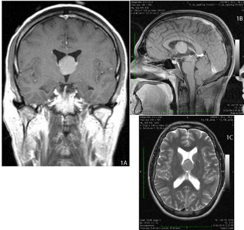
A 36 year-old man presented with an 18 month history of headaches occurring with increasing frequency and severity. A computed tomography (CT) scan demonstrated a lesion in the anterior third ventricle measuring greater than 2 centimeters in diameter. Magnetic resonance imaging (MRI) showed that the lesion had signal characteristics consistent with a colloid cyst (Figures 1a, 1b, 1c). The ventricles were not appreciably enlarged, and there was no trans-ependymal absorption of CSF.
Figure 1:Pre-operative MRI scan demonstrating colloid cyst in the anterior third ventricle measuring greater than 2 centimeters in diameter.
Figure 1a:Coronal T1-weighted MRI with gadolinium.
Figure 1b:Sagittal T1-weighted MRI with gadolinium.
Figure 1c:Axial T2-weighted MRI.
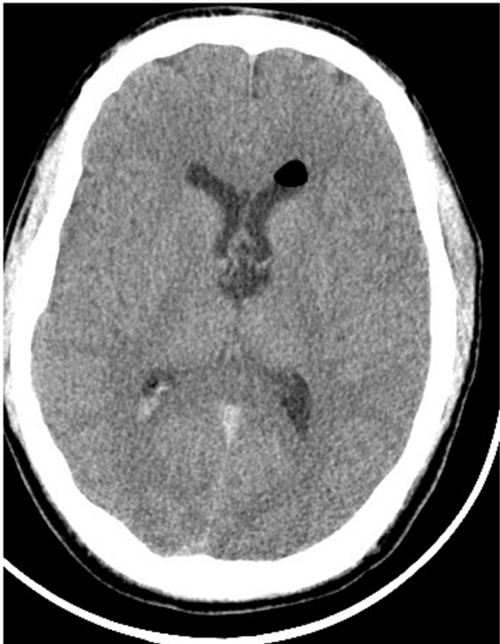
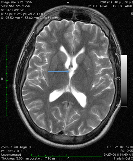
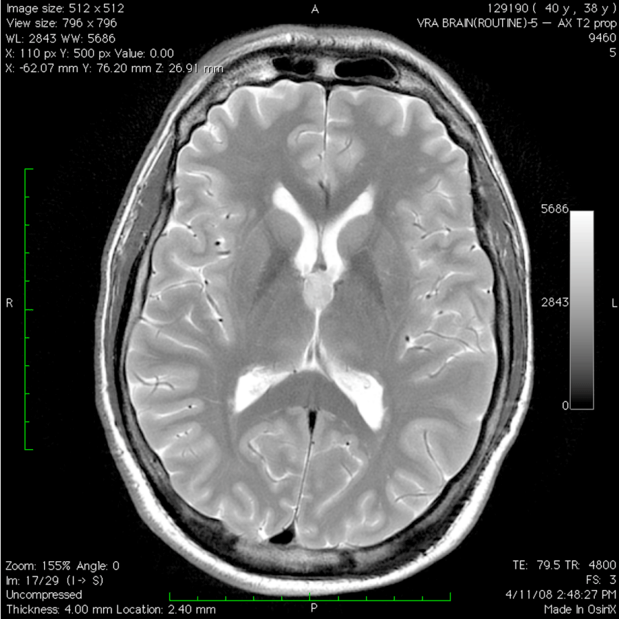
The patient underwent endoscopic removal of the colloid cyst via an anterior right frontal burr hole assisted by frameless stereotaxy. The surgery was performed with both a zero degree and a thirty degree rigid endoscope. All of the mucoid contents of the cyst were removed, and the majority of the capsule was also removed. A small portion of the capsule was not removed because it was technically difficult to do so without risking injury to the fornix. Pathological analysis confirmed the diagnosis of colloid cyst. No atypical pathological features were noted. Postoperatively, the patient was neurologically intact, and he was discharged from the hospital three days after surgery. A CT scan prior to discharge demonstrated no radiographic evidence of residual colloid cyst (Figure 2). An MRI scan performed six weeks postoperatively was reported by the radiologist to have no evidence of residual or recurrent colloid cyst. On our inspection of the T2-weighted images only, however, there was a suggestion of a small remnant at the right foramen of Monro (Figure 3). Interestingly, despite the excellent radiographic results, the patient had only minimal, if any, improvement in his headaches.
Figure 2: Non-contrast CT scan post-operatively demonstrating no radiographic evidence of colloid cyst.
Figure 3: Axial T2-weighted MRI scan 6 weeks postoperatively. There is a suggestion of cyst remnant at the right Foramen of Monro (arrow).
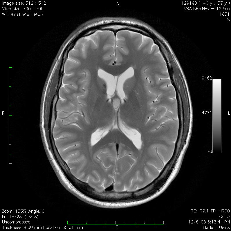
A follow-up MRI scan nearly seven months after surgery demonstrated a definitive recurrence of the colloid cyst (Figure 4). Because the cyst was considerably smaller than prior to surgery and the patient had no hydrocephalus, the recurrence was followed with serial imaging studies. Over the next year, the patient had four MRI scans and one CT scan, all of which showed no gross change in the size of the cyst. He continued to have headaches, which were unchanged from preoperatively. Seventeen months after the recurrence was first detected, a fifth MRI scan demonstrated growth of the cyst (Figure 5). At this point, the patient underwent craniotomy and complete resection of the cyst via an interhemispheric transcallosal approach. He has remained neurologically intact with no recurrence to date, two years after this second operation. He has no hydrocephalus, but he continues to have debilitating headaches, which have never improved.
Figure 4: Axial T2-weighted MRI nearly seven months after surgery demonstrating recurrent colloid cyst.
Figure 5: Axial T2-weighted MRI scan obtained seventeen months after MRI shown in Figure 4. An increase in the size of the recurrent colloid cyst is noted.
Discussion
In 1983, Powell et al. performed the first successful removal of a colloid cyst with an endoscope [6]. Over the past few decades, endoscopic removal has become the treatment modality of choice for many neurosurgeons in the setting of colloid cysts which are large, growing, or causing symptoms [15,21]. Potential advantages of endoscopic colloid cyst removal include smaller incisions with avoidance of a craniotomy, less post-operative pain, and shorter hospital stays [1,15,16,7,17,8,5,18,9,19,13,14,4,20,21].
One potential disadvantage of endoscopic colloid cyst removal is that it may be more technically challenging to remove the entire cyst wall with an endoscope than with open craniotomy. Moreover, several authors have noted higher rates of residual colloid cyst after endoscopic resection than after craniotomy [8,5,9,19,4]. Working unilaterally through the foramen of Monro, even with angled endoscopes, at times does not provide complete access to remove a cyst wall which extends medially or posteriorly around the foramen of Monro beyond the view of the endoscope [5,9,19].
In the current case, a remnant of cyst wall was left behind because removing it would have risked damage to the fornix or required significant torque on the brain. Previous publications do not adequately quantify the increased probability of recurrence if resection of a colloid cyst is nearly complete versus gross total. Some authors contend that leaving a remnant of the cyst does not necessarily increase the rate of recurrence [1,15,8,5,19,13]. Longatti et al., for example, reported a series of 61 patients who underwent endoscopic colloid cyst resection. 62.3% of these patients had evidence of residual cyst postoperatively. Over a mean follow-up period of 32 months, seven recurrences (11.4%) were detected, but only three recurrences (4.9%) were attributed to growth of residual colloid cyst [19]. It is important to note that the follow-up period in this and most other previous studies is relatively short, and colloid cysts typically recur very slowly. With craniotomy for open surgical resection of colloid cysts, it is technically easier to assure complete removal of the entire cyst wall and contents. In a recent meta-analysis reviewing contemporary literature, microsurgical resection of colloid cysts was associated with a higher rate of complete resection and a lower rate of recurrence [23]. We hypothesize that, in the current case, recurrence is most likely related to a remnant of cyst wall left behind that was not appreciated at the time of endoscopic surgery. Because endoscopic removal of colloid cysts may be associated with decreased morbidity than open craniotomy [23], there is much debate among neurosurgeons whether open craniotomy or endoscopic resection of colloid cysts is preferable.
When only stereotactic aspiration of the contents of colloid cysts is performed, early recurrence is common. In a series by Mathieson et al., 80% of colloid cysts recur after stereotactic aspiration alone, and 61% of these recurrences are within the first two months after surgery [24]. For this reason, most centers have abandoned stereotactic aspiration as a treatment option. However, early recurrence after near total removal of a colloid cyst is rarely noted in previous publications. The seven recurrences in the study by Longatti et al. occurred at 8,15,25,42,48,72 and 79 months after surgery [19]. Similarly, Horn et al., Kehler et al. and Greenlee et al. all found recurrences in their patients around 2 years after surgery [16,8,5]. Levine et al., Grondin et al. and Hellwig et al. reported recurrences between 4-6 years after surgery in their cases [7,17,18]. A literature search conducted by Grondin et al. noted no recurrence earlier than 1.8 years after surgery among 157 patients who underwent endoscopic colloid cyst resection [7,17,18]. The earliest reported recurrence after endoscopic resection was eight months after surgery in one of the seven recurrences noted by Longetti et al [19]. Consequently, the time to recurrence reported in this manuscript represents the earliest reported recurrence of a colloid cyst after near-total endoscopic resection.
In conclusion, we report an unusually rapid recurrence of a colloid cyst after near-total endoscopic resection. Close clinical and radiographic follow-up is necessary after resection of a colloid cyst, especially if resection of the cyst is not complete. This report will add to the ongoing debate regarding the relative merits of endoscopic versus open resection of colloid cysts.
References
- Acerbi F, Rampini P, Egidi M, Locatelli M, Borsa S, Gaini S. Endoscopic treatment of colloid cysts of the third ventricle: long-term results in a series of 6 consecutive cases. Journal of Neurological Sciences, 2007; 51: 53-60.
- Charalampaki P, Filippi R, Welschehold S, Perneczky A. Endoscope-assisted removal of colloid cysts of the third ventricle. Neurosurg Rev. 2006; 29: 72-79.
- Kumar V, Behari S, Kumar Singh R, Jain M, Jaiswal AK, Jain VK, et al. Pediatric colloid cysts of the third ventricle: management considerations. Acta Neurochir (Wien). 2010; 152: 451-461.
- Stachura K, Libionka W, Moskala M, Krupa M, Polak J. Colloid cysts of the third ventricle. Endoscopic and open microsurgical management. Neurol Neurochir Pol. 2009; 43: 251-257.
- Kehler U, Brunori A, Gliemroth J, Nowak G, Delitala A, Chiappetta F, et al. Twenty colloid cysts--comparison of endoscopic and microsurgical management. Minim Invasive Neurosurg. 2001; 44: 121-127.
- Powell MP, Torrens MJ, Thomson JL, Horgan JG. Isodense colloid cysts of the third ventricle: a diagnostic and therapeutic problem resolved by ventriculoscopy. Neurosurgery. 1983; 13: 234-237.
- Grondin RT, Hader W, MacRae ME, Hamilton MG. Endoscopic versus microsurgical resection of third ventricle colloid cysts. Can J Neurol Sci. 2007; 34: 197-207.
- Horn E, Feiz-Erfan I, Bristol R, Lekovic G, Goslar P, Smith K, et al. Treatment options for third ventricular colloid cysts: comparison of open microsurgical versus endoscopic resection. J Neurosurgery. 2007; 60: 613-620.
- Lewis A, Crone K, Taha J, Van Loveren H, Yeh Hwa-Shain, Tew J. Surgical resection of third ventricle colloid cysts: preliminary results comparing transcallosal microsurgery with endoscopy. J Neurosurgery. 1994; 81: 174-178.
- Ozmen E, Algin O. Blindness: An uncommon presentation of colloid cysts. Sultan Qaboos Univ Med J. 2014; 14: e128-129.
- Algin O, Ozmen E, Arslan H. Radiologic manifestations of colloid cysts: a pictorial essay. Can Assoc Radiol J. 2013; 64: 56-60.
- Pollock BE, Huston J 3rd. Natural history of asymptomatic colloid cysts of the third ventricle. J Neurosurg. 1999; 91: 364-369.
- Longatti P, Martinuzzi A, Moro M, Fiorindi A, Carteri A. Endoscopic treatment of colloid cysts of the third ventricle: 9 consecutive cases. Minim Invasive Neurosurg. 2000; 43: 118-123.
- Pinto FC, Chavantes MC, Fonoff ET, Teixeira MJ. Treatment of colloid cysts of the third ventricle through neuroendoscopic Nd: YAG laser stereotaxis. Arq Neuropsiquiatr. 2009; 67: 1082-1087.
- El-Ghandour NM. Endoscopic treatment of third ventricular colloid cysts: a review including ten personal cases. Neurosurg Rev. 2009; 32: 395-402.
- Greenlee JD, Teo C, Ghahreman A, Kwok B. Purely endoscopic resection of colloid cysts. Neurosurgery. 2008; 62: 51-55.
- Hellwig D, Bauer BL, Schulte M, Gatscher S, Riegel T, Bertalanffy H, et al. Neuroendoscopic treatment for colloid cysts of the third ventricle: the experience of a decade. Neurosurgery. 2003; 52: 525-533.
- Levine NB, Miller MN, Crone KR. Endoscopic resection of colloid cysts: indications, technique, and results during a 13-year period. Minim Invasive Neurosurg. 2007; 50: 313-317.
- Longatti P, Godano U, Gangemi M, Delitala A, Morace E, Genitori L, et al. Cooperative study by the Italian neuroendoscopy group on the treatment of 61 colloid cysts. Childs Nerv Syst. 2006; 22: 1263-1267.
- Tirakotai W, Schulte DM, Bauer BL, Bertalanffy H, Hellwig D. Neuroendoscopic surgery of intracranial cysts in adults. Childs Nerv Syst. 2004; 20: 842-851.
- Zohdi A, El Kheshin S. Endoscopic approach to colloid cysts. Minim Invasive Neurosurg. 2006; 49: 263-268.
- Sampath R, Vannemreddy P, Nanda A. Microsurgical excision of colloid cyst with favorable cognitive outcomes and short operative time and hospital stay: operative techniques and analyses of outcomes with a review of pervious studies. J Neurosurgery. 2010; 66: 368-376.
- Sheikh Ab, Mendelson ZS, Liu JK. Endoscopic versus microsurgical resection of colloid cysts: a systematic review and meta-analysis of 1,278 cases. World Neurosurg. 2014. [Epub ahead of print].
- Mathiesen T, Grane P, Lindquist C, von Holst H. High recurrence rate following aspiration of colloid cysts in the third ventricle. J Neurosurg. 1993; 78: 748-752.
