
1Department of Surgery, Montefiore Medical Center, USA
*Corresponding author: Grucela A, Department of Surgery, Montefiore Medical Center, Green Medical Arts Pavilion, 3400 Bainbridge Avenue, Bronx, NY, USA 10467
Received: September 26, 2014; Accepted: November 05, 2014; Published: November 05, 2014
Citation: Bellistri JP and Grucela A. Chagas Disease: An Interesting Presentation and Diagnosis. Austin J Surg. 2014;1(8): 1038. ISSN: 2381-9030.
Introduction: Chagasdisease is a vector-borne zoonosis that is caused by the protozoan Trypanosomacruzi (T. cruzi) and affects over 8 million people worldwide. Chronic Chagas disease is characterized predominantly by cardiac manifestations, gastrointestinal (digestive) manifestations or a combination of both. The digestive form develops in 10-15% of chronically infected individuals with the two most common manifestations being megaesophagus or megacolon, both of which are resultant from loss of myenteric neurons.
Case Report: We report an interesting case of Chagas disease in a patient who had many confounding signs and symptoms of disease resulting in a delay in diagnosis. A 61 year-old male underwent a sigmoid resection for a volvulus. He subsequently underwent multiple admissions for left sided chest pain, abdominal distention and constipation with failed ability to elucidate etiology. He then re-presented 4 years after his original surgery with worsening symptoms. A thorough history revealed he spent time in Mexico. Chagas disease was suspected, and serology was positive. He was then treated conservatively.
Discussion: In this case, the patient went misdiagnosed for many years. His confusing presentation with chest pain, along with his diaphragmatic eventration, and ultimate missed diagnosis of Chagas disease led to multiple admissions and limited quality of life. It is important for clinicians to be aware of this clinical entity and to consider testing when dealing with patients with sigmoid volvulus, colonic dilation and/or intestinal dysmotility. Proper diagnosis and direct treatment can avoid unwarranted admissions, unnecessary tests and procedures, and improve patient quality of life.
Keywords: Trypanosomacruzi; Chagas disease; Sigmoid volvulus; Megacolon
Trypanosomacruzi: T.Cruzi; CT: Computerized Tomograph
Chagas disease, also referred to as American trypanosomiasis, is a vector-borne zoonosis that affects over 8 million people worldwide [1,2]. Chagas disease is most prevalent in Mexico, Central America, and South America. It is estimated that about 300,000 people are infected in the United States with most of these people having acquired the illness in endemic countries [2]. Trypanosomacruzi (T. cruzi) is the protozoan parasite responsible for Chagas disease. Spread is predominantly through triatomine insects, although it may also be transmitted through blood transfusion or vertical transmission (mother to infant). It is clinically manifested as acute, latent (indeterminate), and chronic phases of illness [3]. Acute illness is characterized by self-limiting fever. This phase usually lasts 4-8 weeks. If identified during the acute phase, anti-trypanosome drugs are efficacious in achieving cure. For those patients not treated, 70- 60% never develop long-term complications of disease, but remain seropositive. These patients have the indeterminate form of Chagas disease [3].
Chronic Chagas disease is characterized predominantly by cardiac manifestations, gastrointestinal (digestive) manifestations, or a combination of both. The cardiac form is the most common manifestation of chronic disease, developing in 20-30% of individuals. This form is mostly characterized by conduction abnormalities, dilated cardiomyopathy, heart failure, and thromboembolic events [3,4].
The digestive form develops in 10-15% of chronically infected individuals with the two most common manifestations being megaesophagus or megacolon, both of which are resultant from loss of myenteric neurons [5]. Symptoms of megaesophagus include dysphagia, odynophagia and epigastric pain. Megacolon may present with obstipation, abdominal distention, or large bowel obstruction [6]. Sigmoid volvulus can also be a first presentation in patients with the digestive form of Chagas disease [7].
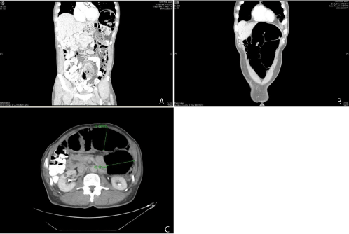
A 61 year-old man with a history of hypertension, hyperlipidemia, and coronary artery disease originally presented to the emergency room in 2010 with complaints of progressively worsening abdominal distention, nausea and vomiting for duration of one month. He originally presented to an outside hospital three days prior with similar complaints and was diagnosed with sigmoid volvulus. At that time, colonoscopic decompression was performed. Surgical intervention was advised, but the patient originally refused. He then presented with recurrent symptoms, and Computerized Tomography (CT) imaging was concordant with large bowel obstruction secondary to sigmoid volvulus (Figure 1). The patient was then agreeable to surgery, and he underwent repeat colonoscopic decompression followed by sigmoid resection with primary anastomosis. The patient tolerated surgery well, and the post-operative course was unremarkable.
The patient was followed by his gastroenterologist postoperatively with repeated complaints of constipation over the next few years. In 2013, CT imaging revealed a dilated colon through and including his anastomosis. He also was found to have a large left sided diaphragmatic eventration. Colonoscopic investigation revealed diverticulitis and a transverse colon polyp that was removed and found to be a tubulovillous adenoma on histopathologic analysis.
He also had 10 admissions between 2010 and 2013for chest pain. This was worked up multiple times and determined to be noncardiac in nature. Electrocardiogram exhibited non-specific T wave abnormalities, but sinus rhythm. Cardiac catheterization revealed a 30% mid right coronary artery stenosis and a 70% distal left anterior descending artery stenosis. However, dobutamine and treadmill stress echocardiography did not elicit any symptomatology or reveal any cardiac dysfunction. The left sided chest pain was determined to be attributable to the left diaphragmatic eventration. This was hen repaired with a laparoscopic diaphragmatic plication, which failed. Repeat imaging at one year postoperatively showed no change in diaphragm appearance. The patient reported feeling worse after this repair and continued to have chest pain.
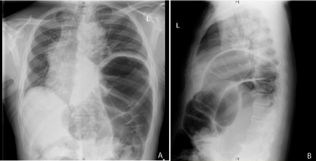
Four years after the initial sigmoid resection, the patient presented back to the emergency room complaining of recurrent chest pain, constipation, and increasing abdominal distention. Chest x-ray exhibited eventration of the left hemi diaphragmun changed from prior to repair with increased colonic distention from prior studies (Figure 2). The colorectal surgery service was consulted. Physical exam revealed a soft, mildly distended abdomen without any tenderness. On digital rectal exam, he had appropriate sphincter tone. He denied issues with constipation as a child and remarked that the constipation began worsening after his sigmoid resection despite the fact that there was no anastomotic stricture.
At this point in his workup, it appeared that his chest pain was more likely due to his giant dilated splenic flexure with eventration into his chest, rather than a cardiac etiology. We wished to determine the etiology of his colonic dilation and dysmotility. As his sphincter tone was normal, we did not favor a previously determined diagnosis of “tight anus.” A thorough history revealed that he had lived in Mexico for an extended period of time. About 15 years prior to initial presentation, he had moved from South Texas to New York. Decision was then made to test for T. cruzi antibody, and he was found to be seropositive. With little evidence to support use of antiparasitic agents in the latent chronic phase of Chagas disease, treatment was focused on supportive care while maintaining appropriate dietary and bowel hygiene. Should symptomatology progress, further surgical planning will be discussed.
Chagas disease is a parasitic infection with approximately 300,000 individuals infected within the United States [2]. As is the case with our patient, most of these patients acquired the infection while in an endemic area. Curative therapy relies on early detection and treatment within the acute phase of illness3. Effective anti-parasitic therapy for those patients in the chronic phase of illness has yet to be established. These patients rely on supportive therapy as the mainstay of treatment.
As in this case, the digestive form of chronic Chagas disease commonly presents with sigmoid volvulus. Colonic decompression followed by sigmoid resection with colorectal anastomosis is the recommended treatment. Institutions in endemic areas tend to perform more extensive resections with removal of aganglionic and dyskinetic segments of bowel with improved functional outcomes. Abdominoperineal pull-through operations remove all dysfunctional colon and rectum and avoid complications that ensue secondary to remaining diseased bowel [8]. In the United States, Chagas disease itself is not a common clinical entity. However, sigmoid volvulus secondary to Chagas disease should be suspected in those with a history of travel to endemic areas and testing prior to surgical intervention should be performed [7,9,10].
In this case, the patient went misdiagnosed for many years. His confusing presentation with chest pain, along with diaphragmatic eventration, and repeatedly missed diagnosis led to multiple admissions and limited quality of life.
Chagas disease affects over 8 million people worldwide and over 300,000 people in the United States [1,2]. It is important for clinicians in dealing with patients with sigmoid volvulus and/or colonic dilation or dysmotility to be aware and consider and test for this entity [11]. Proper diagnosis and direct treatment can avoid unwarranted admissions, unnecessary tests and procedures, and improve patient quality of life [9].
CT scan demonstrating sigmoid volvulus A) coronal section demonstrating mesenteric swirling B) Coronal section demonstrating dilated sigmoid colon in a closed loop configuration C) Axial section demonstrating transverse and descending colon of 7cm and 8.8cm, respectively.
(A) Posterior-Anterior (PA) and (B) Lateral chest x-ray demonstrating colonic distention and elevated left hemidiaphragm.
Austin Publishing Group is an emerging open access publisher specialising in Science, Technology and Medicine is dedicated to serve the biomedical community through its initiatives. Austin Publishing Group is an academic publisher with 100+ peer reviewed open access journals in various subjects such as biomedical, Pharma, Life Sciences, Environmental, Engineering and Management. Austin Publishing Group publishes Open Access eBooks providing free access to vast scientific literature.