Department of Cardiology and Cardiothoracic surgery, St Georges Hospital NHS Trust, United Kingdom
*Corresponding author: Smriti Saraf, Department of Cardiology and Cardiothoracic surgery, St Georges Hospital NHS Trust, London, United Kingdom
Received: November 20, 2014; Accepted: November 24, 2014; Published: November 28, 2014
Citation: Saraf S, Chandrasekaran V and Firoozi S. Leakage from an Internal Mammary Graft Anastomosis after Endoscopic Atraumatic Coronary Artery Bypass (Endo-Acab) Graft Surgery. Austin J Surg. 2014;1(9): 1042. ISSN: 2381-9030.
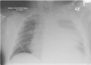
A 70 year old gentleman presented to the Emergency department with chest and abdominal pain. He had recently had an endoscopic atraumatic coronary artery bypass (ENDO-ACAB) 3 weeks ago with a LIMA graft to the LAD. While in emergency, he became haemodynamically compromised and suffered a pulse less electrical alternans arrest. Cardiopulmonary resuscitation was carried out promptly for 6 minutes with return of spontaneous circulation. A portable chest x-ray demonstrated complete white out of the left lung (Figure 1), and an electrocardiogram showed ST elevation in the anterior leads. A bedside echocardiogram demonstrated good overall left ventricular systolic function with hypokinesia of the anterior wall and apex, and the patient was taken to the cardiac catheterization laboratory for emergency coronary angiography.
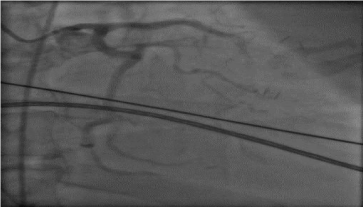
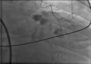
Coronary angiography was performed via the right femoral artery. The Left Main Stem (LMS), Left Circumflex (LCX) and Right Coronary Artery (RCA) were unobstructed. The Left Anterior Descending (LAD) artery was heavily calcified with sub-total occlusion in the mid-vessel (Figure 2). The Left Internal Mammary Artery (LIMA) graft was patent, but leakage of contrast was noted at the anastomosis site (Figure 3) with very sluggish flow in the LAD distal to the anastomosis. He was taken to theatre for emergency coronary artery bypass surgery and subsequently made good recovery.
ENDO-ACAB is currently the most minimally invasive bypass surgery available, and is used as a hybrid treatment option in combination with catheter based therapy [1].
CXR demonstrating white out of the left lung.

Subtotal occlusion of calcified LAD.

Leakage of contrast at anastomotic site of LIMA to LAD.
