Special Article: Laparoscopic Surgery
Austin J Surg. 2023; 10(1): 1294.
Abdominal Cocoon as a Rare Cause of Intestinal Obstruction: A Case Report
Hussein MA1, Abouelgreed TA2*, Saafan T3 and Diab EA4
1Department of General Surgery, Saudi German Hospital, Ajman, UAE
2Department of Urology, Faculty of medicine, Al-Azhar University, Cairo, Egypt
3Department of General Surgery, NMC Royal hospital, Sharjah, UAE
4Department of Radiology, AlQasemi Hospital, Sharjah, UAE
*Corresponding author: Abouelgreed TA Department of Urology, Faculty of medicine, Al-Azhar University, Cairo, Egypt
Received: January 17, 2023; Accepted: March 03, 2023; Published: March 10, 2023
Abstract
Abdominal cocoon is one of the rare causes of intestinal obstruction. It is referred as complete or partial small bowel encapsulation caused by the thick fibrocollagenous membrane. It is most common in young adolescent girls. We present a 34-year-old male patient with idiopathic abdominal cocoon causing intestinal obstruction. Few cases of male patients suffering from idiopathic abdominal cocoon have been reported in literature.
Introduction
Abdominal cocoon is referred as a complete or partial small bowel encapsulation caused by dense fibrocollagen membranes leading to acute or chronic small bowel obstruction. It was first termed as peritonitis chronic fibrosain capsulata by Owtschinnikow in 1907 and finally abdominal cocoon by Foo in 1978 [1]. It is most commonly seen in adolescent girls of tropical and subtropical region though few cases of male have also been reported in literature [1,2].
Case Presentation
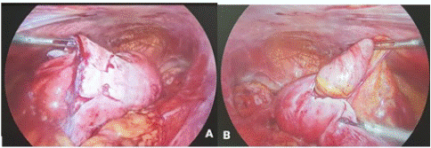
A 34-year-old male patient presented with acute abdominal pain, distension and recurrent vomiting for the last 2 days with signs of intestinal obstruction. The patient had a past history of recurrent abdominal pain during the last 2 months. There was no associated fever. On physical examination, there was tenderness over the right iliac fossa. The patient underwent Computed Tomography (CT) abdomen and pelvis (plain and contrasted) which showed evidence of dilated small bowel loops involving jejunal and ileal loops apart from the distal few centimeters of ilium (Figure 1). The distal ileal loops is seen clustered within thin wall sac like structure in the lower abdomen with convergent, crowded and congested mesenteric vessels associated with minimal streaks of edema. The sigmoid colon is displaced medially and right supero-lateral surface of urinary bladder is compressed. Normal enhancement pattern of the bowel walls. The above finding is suggesting small bowel obstruction due to internal hernia likely of trans mesenteric type (Figure 2). Laparoscopic exploration of the whole abdomen was done; the internal herniated ileum was found encased in a cocoon-like fibrotic tissue with a diameter of nearly 15 cm. We cut the fibrous membrane and the small bowel loosened. Circulation of the bowel segment was intact. On the 3rd post-operative day the patient was discharged.

Figure 1: (A-B), Sagittal and transverse sections of CT showing evidence of dilated small bowel loops involving jejunal and ileal loops.

Figure 2: A-B, Intraoperative view during exploration and excision of the Coccon.
Histopathology
Gross description: The specimen is identified as hernia sac and consists of 4 cystic like fragments together measuring 4.5 × 4.5 × 0.5 cm totally submitted in four cassettes.
Microscopic description: Sections examined show fibrous tissue infiltrated by plasma cells, multinucleated giant cells, plump activated fibroblasts with pale staining nuclei, histiocytes with small nuclei and abundant vacuolated cytoplasm, haemorrhage and granulation tissue with chronic inflammatory infiltrate.
Discussion
Abdominal cocoon is one of the rare diseases causing acute to sub acute intestinal obstruction. This is a rare peritoneal disorder characterized by a dense, off-white, membranous layer of fibrous connective tissue that surrounds some or all of the abdominal organs, resembling a silkworm cocoon. Due to the lack of specificity of clinical manifestations, it is difficult to differentiate from intestinal obstruction, perforation, or abdominal mass caused by other causes, so preoperative diagnosis is difficult, and the diagnosis mainly depends on intraoperative findings [3]. The prevalence of Abdominal Cocoon Syndrome (ACS) is unknown, but it occurs in 1.4–7.3% of peritoneal dialysis patients [4]. Abdominal cocoons are more common in males, and the male to female ratio is about 1.2 to 2:1 [5]. The role of laparoscopy is limited to elective investigation and auxiliary diagnosis, but due to the nature of adhesions and bowel entrapment, open exploration is unintentionally required for treatment [6]. In our case, we performed diagnostic laparoscopy and completed the procedure without conversion to laparotomy. The patient underwent emergency surgery for intestinal obstruction, during which an abdominal callus was found. Abdominal cocoons are characterized by small bowel obstruction caused by all or part of the small intestine being covered by a dense fibrous membrane, also known as idiopathic sclerosing peritonitis, small bowel incarceration, and small intestinal fibrous membrane herniation [7]. The etiology of abdominal cocoon can be divided into primary abdominal cocoon and secondary abdominal cocoon. Primary abdominal cocoon disease is primarily caused by embryonic body curling, abnormal mesoderm differentiation, and intestinal dorsal mesenteric dysplasia. It is frequently associated with the absence of the omentum, the absence of the gastrocolic ligament, intestinal or colonic malrotation, visceral transposition, cryptorchidism, hernia, and other diseases [8]. The following are the causes of the secondary abdominal cocoon: (1) Changes in the abdominal microenvironment: an abdominal and pelvic infection caused by a variety of factors, a history of abdominal surgery, long-term peritoneal dialysis, and a malignant tumor, among others. In addition to abdominal tuberculosis, autoimmune diseases, liver transplantation, and other immune factors can cause abdominal cocoon [9]. Some clinical workers have also observed that abdominal injury and hepatitis C are the causes in recent years [10]. There have also been reports of abdominal cocoons with abdominal free gas [11]. (2) Drug effects: Chemotherapy drugs, B receptor blockers, and mercury can cause peritonitis, which leads to the formation of the abdominal cocoon, which is associated with excessive collagen production and abdominal fibrosis [12]. Because the patient had a history of anemia and no other disease was discovered, it was diagnosed as a primary abdominal cocoon. Abdominal pain, distension, vomiting, and mass are all symptoms of the abdominal cocoon. The disease's duration varies. Preoperative diagnosis is challenging, and the majority is diagnosed based on intraoperative findings. According to Wei et al., the main clinical manifestations of the abdominal cocoon in 24 cases were incomplete or complete intestinal obstruction (88%), and abdominal mass (54%) [13]. According to Machado et al., the most common clinical manifestations of the abdominal cocoon were abdominal pain (72%), abdominal distension (44.9%), and abdominal mass (30.5%) [14]. There are currently few reports of abdominal cocoon with intestinal perforation. Preoperative gastrointestinal radiography and high-resolution CT examination are useful in determining the presence of an abdominal cocoon [15]. However, in patients with acute intestinal obstruction or perforation, gastroenterography may aggravate abdominal symptoms, so it should be used with caution [16]. CT diagnosis has high technical requirements for doctors; this patient had an abdominal CT examination, and an abdominal cocoon was discovered; therefore, the diagnosis of abdominal cocoon was based primarily on the CT examination findings. The operation's principle is to remove the capsule, release adhesion, and relieve the obstruction [17]. The following are the operation methods: (1) Intestinal resection: this procedure is appropriate for cases of intestinal ischemia and necrosis. We removed a portion of the necrotic small intestine in this case due to intestinal necrosis. (2) Intestinal arrangement surgery: it is appropriate for cases with severe adhesion that cannot be separated in order to relieve obstruction. To avoid causing too many other injuries, such as postoperative intestinal fistula, necrosis, and other complications, it should not be widely separated in order to completely remove the capsule. (3) Appendectomy: it is appropriate for patients who have an obvious appendiceal fecal stone discovered during the operation and the possibility of developing acute appendicitis later on.
Conclusions
Bowel obstruction caused by abdominal cocoon is rare. In clinical work, when encountering patients with intestinal obstruction, we must carefully inquire about their medical history. If the patient has no history of digestive system diseases, the possibility of abdominal cocoon obstruction should be considered.
Data Availability Statement
The original contributions presented in the study are included in the article/supplementary material; further inquiries can be directed to the corresponding author/s.
Ethics Statement
The studies involving human participants were reviewed and approved by first author institute. The patients/participants provided their written informed consent to participate in this study. Written informed consent was obtained from the individual(s) for the publication of any potentially identifiable images or data included in this article.
Author Contributions
TA and MH designed this study, collected the information, images, and wrote the manuscript. ED and TS, reviewed the manuscript. All the authors read and approved the final manuscript.
Conflicts of Interest
There are no conflicts of interest.
References
- Solak A, Solak I. Abdominal cocoon syndrome: Preoperative diagnostic criteria, good clinical outcome with medical treatment and review of the literature. Turk J Gastroenterol. 2012; 23: 776–779.
- Oran E, Seyit H, Besleyici C, Ünsal A, Alis H. Encapsulating peritoneal sclerosis as a late complication of peritoneal dialysis. Ann Med Surg (Lond). 2015; 4: 205–207.
- Çolak S, Bektas H. Abdominal cocoon syndrome: a rare cause of acute abdomen syndrome. Ulus Travma Acil Cerrahi Derg. 2019; 25: 575–9.
- Solmaz A, Tokocin M, Arici S, Yigitbas H, Yavuz E, et al. Abdominal cocoon syndrome is a rare cause of mechanical intestinal obstructions: a report of two cases. Am J Case Rep. 2015; 16: 77.
- Yavuz R, Akbulut S, Babur M, Demircan F. Intestinal obstruction due to idiopathic sclerosing encapsulating peritonitis: A case report. Iran Red Crescent Med J. 2015; 17: e21934.
- Kumar M, Deb M, Parshad R. Abdominal cocoon: report of a case. Surg Today. 2000; 30: 950–3.
- Al-Azzawi M, Al-Alawi R. Idiopathic abdominal cocoon: a rare presentation of small bowel obstruction in a virgin abdomen. How much do we know? BMJ Case Rep. 2017; 2017: bcr2017219918.
- Uzunoglu Y, Altintoprak F, Yalkin O, Gunduz Y, Cakmak G, et al. Rare etiology of mechanical intestinal obstruction: abdominal cocoon syndrome. World J Clin Cases. 2014; 2: 728–31.
- Kaur S, Doley RP, Chabbhra M, Kapoor R, Wig J. Post trauma abdominal cocoon. Int J Surg Case Rep. 2015; 7: 64–5.
- Akbulut S, Yagmur Y, Babur M. Coexistence of abdominal cocoon, intestinal perforation and incarcerated Meckel’s diverticulum in an inguinal hernia: a troublesome condition. World J Gastrointest Surg. 2014; 6: 51–4.
- Asotibe JC, Zargar P, Achebe I, Mba B, Kotwal V. Secondary abdominal cocoon syndrome due to chronic beta-blocker use. Cureus. 2020; 12: e10509.
- Li S, Wang JJ, Hu WX, Zhang MC, Liu XY, et al. Diagnosis and treatment of 26 cases of abdominal cocoon world. J Surg. 2017; 41: 1287–94.
- Wei B, Wei HB, Guo WP, Zheng ZH, Huang Y, et al. Diagnosis and treatment of abdominal cocoon: a report of 24 cases. Am J Surg. 2009; 198: 348–53.
- Machado NO. Sclerosing encapsulating peritonitis: review. Sultan Qaboos Univ Med J. 2016; 16: e142–51.
- Gorsi U, Gupta P, Mandavdhare HS, Singh H, Dutta U, et al. The use of computed tomography in the diagnosis of abdominal cocoon. Clin Imaging. 2018; 50: 171–4.
- Singh AU, Subedi SS, Yadav TN, Gautam S, Pandit N. Abdominal cocoon syndrome with military tuberculosis. Clin J Gastroenterol. 2021; 14: 577–80.
- Singh AU, Subedi SS, Yadav TN, Gautam S, Pandit N. Abdominal cocoon syndrome with military tuberculosis. Clin J Gastroenterol. 2021; 14: 577–80.