
Clinical Image
Austin J Surg. 2023; 10(1): 1297.
Acute Appendicitis in Duplicated Appendix: Images in Medicine
Mohammed Lamghari*; Marwa Sabur; Imad El Azzaoui; Hind Hablaj; Amine Maazouz; Mohammed Najih; Hakim Kaoui; Sidi Mohammed Bouchentouf; Mountassir Moujahid; Ahmed Bounaim
Department of Visceral Surgery, Mohammed V Military Hospital, Faculty of Medicine and Pharmacy of Rabat, Morocco.
*Corresponding author: Mohammed Lamghari Department of Visceral Surgery, Mohammed V Military Hospital, Faculty of Medicine and Pharmacy of Rabat, Rabat, Morocco.
Received: February 17, 2023 Accepted: March 28, 2023 Published: April 04, 2023
Visual Case Discussion
A 26-year-old male patient, with a 3-days history of peri-umbilical pain which had localized to the right iliac fossa by the time he attended the hospital, was admitted to the emergency department of our hospital. The abdominal examination revealed a soft abdomen, rebound tenderness in the right iliacfossa, and a positive psoas sign. He was not peritonitic and had a negative Rosving's sign and absenthernias.
Blood tests showed a mild leucocytosis of 12.5×10⁹/l (range, 4.0–11.0×10⁹/l) with a neutrophilia of 10.5×10⁹/l (range. 2.0–7.5×10⁹/l) and a CRP of 90mg/l.
Ultrasonography confirmed a diagnosis of appendicitis by the presence of free fluid within the RIF and within the 10mm appendix which was incompressible.
Under general anesthesia, the surgical exploration by laparotomy revealed the presence of acute appendicitis, but the meticulous investigation of the area around the cecum detected a second appendix with a definite mesoappendix and obvious signs of inflammation (Figure 1). Formal appendectomy was then performed for both processes (Figure 2).

Figure 1: Intraoperative image showing the appendicular duplication.
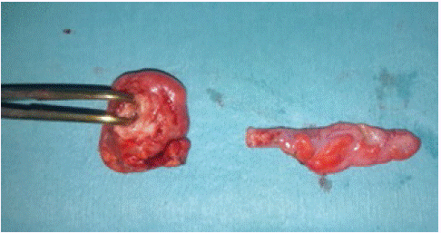
Figure 2: Image of the two appendices after resection.
Following surgery, the patient was stable and recovered well, and was discharged two days later. The histological examination of the appendix confirmed the diagnosis of acute purulent appendicitis for the first appendix, and a catarrhal form for the second one.
Keywords: Duplicate appendix; Acute Appendicites; Appendectomy