
Research Article
Austin J Surg. 2023; 10(2): 1299.
A Suture Tips Applied to Rotating Flaps in Reconstructing Facial Defects to Optimize Scarring: A Retrospective Study
Xiaojing Ge, MD#; Yute Sun, MD#; Youzhi Tang, MD; Fang Zhou, MD; Gang Yao, MD; Xin Su, MD*
Department of Plastic and Burn Surgery, The First Affiliated Hospital of Nanjing Medical University, China
*Corresponding author: Xin Su Department of Plastic and Burn Surgery, The First Affiliated Hospital of Nanjing Medical University, 300 Guangzhou Rd, Nanjing 210029, Jiangsu Province, China. Tel: +86 13914713233; Fax: +86 25 83718836 Email: suxin@jsph.org.cn
Received: March 08, 2023 Accepted: April 21, 2023 Published: April 28, 2023
Abstract
Background: Local flap reconstruction is the most common method to repair facial defects. In this study, authors have explored and introduced a modified suture technique with rotation flap to repair facial defects that makes scarring more discreet and easier to perform.
Methods: Patients with facial defects who underwent surgical treatment of the rotation flaps reconstructions and were admitted to our department between October 2014 and October 2019 were included in this retrospective study. The rotating flap is designed along the Relaxed Skin Tension Line (RSTLs) and contour lines, parts of patients received modified suture depending on the shape and size of the defect. Observe the flaps’ survival and complications. Assessments of the scars with Vancouver Scar Scale (VSS) regularly and follow-ups.
Results: This five-year retrospective study evaluated a total of 56 patients; of these, 45 patients underwent modified suture surgery, the average defects were 19.7±4.5×25.5±5.2mm, and the average duration of follow-up was 13.8±5.0 months. Scar appearance was evaluated using the VSS score, range from 0-5, with an average of 1.6±1.1, and mainly reflected in pigmentation and vascularity. Postsurgical scar concealment is made afterward, and all flaps survive. No hemodynamic disorder or necrosis was found.
Conclusion: The rotation flap was designed based on RSTLs/ contour lines to repair the facial defect and make the scarring well concealed. The modified suture method makes the flap line consistent with the RSTLs/ contour line of the facial skin to the maximum extent and leads to better facial aesthetics results.
Introduction
Facial defects are usually caused by trauma, cutaneous tumor excision, and tissue necrosis [1]. Different reconstruction methods based on age, defect size, and site include skin grafts, local flaps, and free flaps. Unlike other parts of the body, the facial has higher standards of cosmetic requirements and interferes as little as possible with the anatomical position of normal organs. Therefore, the choice of reconstruction method requires a case-by-case decision in order to ensure a natural appearance and full functionality [2]. The literature survey reveals [3], among the facial defect reconstructive modalities, the advanced flap is recognized as having overall functional as well as aesthetic results, even in the case of more significant defects [4,5]. Local flap can also achieve a better repair when the defect is too large, the surrounding skin is not too lax, or may involve the facial organs [6-8].
Rotation flap, with various refinements, is a classic method in plastic surgery, which also is an effective method commonly used to repair facial defects and can easily perform [9,10]. Sufficient arc length is an important factor to ensure proper reconstruction area and tension in the skin. A long incision arch allows tissue laxity immediately adjacent to the defect over a great distance. It has been demonstrated that when the circumference of the flap exceeds approximately five times the length of the defect, its tension-reducing effect becomes optimal [11] or changes the tension by designing a back cut [12]. However, the incision arch still brings unsightly scarring for facial restoration. Physicians have also thought of designing the incision line within the dermatoglyphic line, such as the Langer's line, Kraissl's line, RSTLs line, melolabial fold, alar sill, and philtral columns, which provide excellent cover and hides the surgical incision [9]. Based on simple and convenient classical rotation flap design, fulfilling the facial defects with minimal and most concealed scarring is still worth studying deeply in further.
Therefore, we designed the rotating flap with a more focus on following or parallel to the Relaxed Skin Tension Lines (RSTLs) and contour lines. However, such modifications alone still do not avoid the post-rotation incision line changes beyond the RSTLs or the additional scarring from the dorsal cut. We pre-designed a modified suture in the defect area to change the shape of the defect and by driving the tension moved so that the rotation flap rotates at a smaller angle, maintaining tension close to the RSTLs/contour lines, thus hiding the scar or parallel to that. In this study, we describe our experience and the follow-up with this suture technique.
Patients and Methods
This retrospective study is performed in a single center, patients with facial defects who underwent the rotational flap from October 2015 to October 2019. Photographs, surgical method, follow-up records, and the patient's opinion of the final functional or aesthetic outcome were examined. Smoking status and previous local treatment were not recorded and also not as exclusion criteria. Pre-/post-treatment and intraoperative photographs were taken, and all patients gave informed consent to used clinical photographs for research, educational, and publication purposes. This study was approved by the ethics committee (2021-SR-323) and conducted according to the ethical guidelines of the Declaration of Helsinki.
Data collection for analysis included patient demographic characteristics such as age, medical history, cause of the defect, type of cancer, and defect characteristics (e.g., size and sites). Complications, including infection, hematoma formation, flap necrosis, donor area complications, and cancer recurrence, were assessed in all cases. Two independent evaluators administered the VSS to all patients 6 months after surgery. The maximum follow-up of the study cases was approximately 4.5 years.
Surgical Technique
Local infiltration anesthesia was carried out among all cases. Using lidocaine (0.75%) and epinephrine (1:100000), nerve block and/or local infiltration anesthesia was accomplished in the area surrounding the defect left by primary tumor or trauma. Any injection into the part underneath the tumor base was avoided. Anesthetics were injected along the outline of the flap and did not reach the tissues underneath the flap. Intraoperative anesthesia achieved a good result.
For all tumors, either benign or malignant, the lesion tissues were thoroughly dissected. The cut edge extended by 1-2mm from the original lesion edge for benign tumors and 5-10mm for malignant tumors (Basal cell carcinoma or squamous cell carcinoma). The wound surface was shaped circular or triangular like in most conditions. The depth of excision was to the basilar part and the part underlying superficial fascia for benign tumors and to deep fascia or myolemma for malignant tumors; till the pathologist ensured that resection margins were free of tumor.
As shown by figure 1, Point A was set on the defect edge. Then, a straight line or an ARCc line parallel to RSTLs or contour lines was drawn to connect point B. The length of line AB was decided according to the defect size and the adjacent skin's elasticity, usually 2 to 5 times that of the defect diameter. A back cut of 1cm was made to connect point C, creating an angle of ≤45°. If the defect was circular or a long diameter, a chord EF of ≤1.0cm, perpendicular to AB was drawn as shown in figure 1.
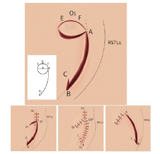
Figure 1: Our flap was designed in the conventional rotation flap; the length of line AB was typically 2 to 4 times the diameter of the defect and parallel to RSTLs or contour lines. If the defect was circular or had a significant diameter, E and F were designed with a spacing ≤1.0cm (Above). After EF two points were sutured, the shape of the defect area was changed so that point A did not have to reach the highest point of the defect, reducing flap tension and even avoiding point C back-cutting or minimizing it (Below Left). After suture, ideally, the majority of faults may be paralleled by RSTLs, which was more consistent with the principles of plastic surgery (Below Center). If a rotating flap was directly used to cover a significant defect, relatively large incision lines are applied to the RSTLs, increasing the tension on the wound edge (Below Right). In this circumstance, even if the original design made an incision parallel to the RSTLs. The incision scar can be more visible in long-term sequelae.
Following line AB, the skin was incised apart to expose its superficial or deep fascia. The flap was freed to the pedicle. Depending on the shape and size of the defect, during the procedure of raising the flap, three situations may be used:
1) The incision going along with line AB can be adjusted according to the defect size and adjacent tissue’s elasticity. It is not always necessary to continue the incision to point B. When being cut, the flap should be rotated and advanced simultaneously till it is large enough to cover the wound surface without any presence of tension.
2) The incision extends to point B, but the tension is still detected when the flap is rotated and advanced to the recipient site. In this case, a back cut incision of 1cm should be made to point C, with an intersection angle of ≤45°. Then V-Y advancement suture can be operated to repair the defect (Figure 2).
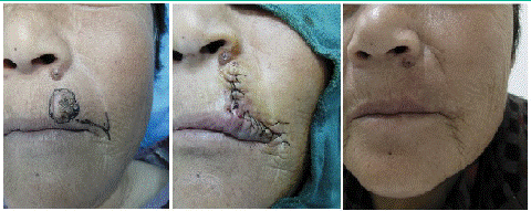
Figure 2: A 69-year-old woman with a 19mmx17mm defect on the left side of the upper lip (Left). An incision is made along the vermilion border of the lip, and a back cut incision in the nasolabial groove (Center). Ten-month follow-up (Right).
3) Modified suture before flap rotation. If the defect has a circular shape or a long diameter, subcutaneous undermining is implemented surrounding the defect area to free the defect’s adjacent tissues. Suture two points E- F according to the design of Figure 1. After the suture, one aspect is the wound surface change from circular to like-triangle. To match the shape of the flap, the area to be covered by the flap is reduced, thus relaxation of flap tension; on the other hand, with the E-F suture line, the tension of the flap incision arch changes as if it were stretched by a bowstring, making the intersection angle of the AB and AO lines with the RSTL or dermatoglyphic line after flap formation minimized and more in line with the principles of plastic surgery (Figure 1).
Combining methods A and C, or B and C, can parallel incision with the RSTLs or contour lines and minimize flap tension and scar size. Once prepared, the flap is rotated and advanced to the defect area. One tip of the flap is fixated firstly to distribute the tension evenly. The superficial fascia in the cut edge is interruptedly sutured with absorbable threads, achieving a tension-free flap-and-skin attachment. The use of drainage depends. If no obvious diffusion occurs, a pressure dressing is workable. The postoperative routine dressing is followed till stitches are taken out at 7 or 8 days.
Results
Over 5 years, a total of 56 patients with facial defects procedures were reviewed, of which 45 patients were treated in modified suture— ages from 16 to 80. The mean defect area was 19.7±4.5mm×25.5±5.2mm. 53(94.6%) fl aps survived with no hemodynamic disorder and necrosis was recorded, 3 cases (5.4%) were slightly necrotic due to infection at the tip and healed after dressing change. Both donor sites and recipient sites experienced I-phase healing. No secondary reconstruction was found. The mean length of follow-up was 13.8 months (range, 6 months to 4.5 years). The original defects showed flatness and normal shapes. Structures neighboring eyes, nose and mouth did not present secondary deformation. The flap pigmentation and texture went well with the surrounding skin. All patients were satisfied with the surgical outcome. Relevant patient characteristics and flap information are shown in Table 1.
![]()
Characteristic
N
%
Sex
Male
31
55.4
Female
25
44.6
Mean age ± SD, yr
53.9±14.4
Reasons for facial defect
Benign tumors
32
57.1
Malignant tumors
16
28.6
Trauma
8
14.3
Defect locations
Cheek
19
33.9
Anterior to ear
11
19.6
8
14.3
Surrounding nose
18
32.1
Defect area
Range, mm
15×15-30×40.
Mean defect area ± SD, mm
19.7±4.5×25.5±5.2
Table 1: Patients Data.
All patients completed VSS evaluation at 6 months after surgery, and VSS scores ranged from 0-5 with a mean of 1.6±1.1. Mainly in pigmentation and vascularity, height/thickness and pliability are mostly close to normal skin. All VSS scores in table 2.
![]()
    Mean    (SD)

    Range


    VSS

    1.6±1.1

    0-5


    Pigmentation

    0.7±0.5

    0-2


    Vascularity

    0.6±0.5

    0-2


    Height/thickness

    0.2±0.4

    0-1


    Pliability

    0.04±0.2

    0-1
Table 2: Vancouver Scar Scale.
Discussion
According to previous studies, when a surgical incision or wound parallels the tension line, the tension imposed on the wound edge can be relieved, and the scar size minimized [13]. So the incision is usually designed according to the directions of Langer’s lines [14]. After more than a century’s exploration, modern surgeons prefer to set incisions, especially on the face, by integrating RSTLs and Kraissl’s lines [14]. Kraissl’s lines refer to wrinkles produced by muscular contraction and RSTLs by the internal tension of relaxed skin. Normally, RSTLs parallel the expressional wrinkles. The scar can be minimized and hidden if a surgical incision is set through a thorough consideration of RSTLs, contour lines, hairline, wrinkles, or boundary of facial subunits.
Various flaps are commonly used in the clinic for facial defects, including Rhomboid flap [15], bilobe flap [16], hatchet skin flap [17], kite-shape flap [18,19], etc. To design these flaps, incision lines should go in the direction of RSTLs, but this only applies to additional cut lines. It is difficult to develop all incisions parallel to RSTLs. Therefore, after flap rotation and defect repairment, scars were exhibited in different directions, with at least one perpendicular to RSTL. The simple lines make the rotating flap easier to do in the same direction or parallel to the RSTL. The rotation arc can be adjusted according to skin elasticity and mobility [11]. A back cut incision can be made at the flap pedicle to facilitate rotation and advancement. But this incision can narrow the flap pedicle, making the whole flap easy to be twisted and impaired blood supply. The two incisions of the roFigure tation ARC are different in length, so skin wrinkles when rotated or “Dog-ears” may occur, usually at the site where the skin is thin, and the defect’s diameter is longer than 3cm, so Burow’s triangular incision should be added to lengthen the shorter edge. Some of that can be flattened through suture following the rule of halves. Also on this basis, homolateral double-rotation flap, bilateral “O-Z” flap in opposite directions, and axe-shape flap [20] can be created. This shows that the design of the rotation flap is flexible and varied, but the orientation of the improperly designed additional back cut line and incision arch closure line disrupts the harmony of the facial lines.
To solve this problem, we improved a suture skill, especially for round or larger defect areas, rotation will cause Burow’s triangular and the original design line to extend beyond the RSTLs or skin line. The trick and advantages of the modified suture included: (1) With the E-F sutured, the shape of the defect area fits the flap shape better and the flap rotation angle becomes smaller; avoiding the need to cut the flap to match the defect area causing greater tension. (2) Suturing the chord EF minimizes the intersection angle between AB and RSTLs. No line perpendicular to RSTLs appears, and the tension of the flap is reduced by large. (3) As the force of the string stretches the incision bow, the short back cut incision line also produces a slight angle with RSTLs as possible, which observes the principles of plastic surgery. Therefore, the modified suture can avoid the formation of Burow’s triangular and reduce the additional incision, even the dorsal incision; there is no reverse incision after the modification.
The flap can be flexibly and easily made. Figure 3 illustrates steps 2 and 3 of the surgery. Modified suture and back cut incision reduce the flap tension. The scar is hidden in the nasolabial fold. The blood of the flap is supplied by the vasculatures underneath the dermis and inside the superficial fascia. The pedicle is wide enough, and the back cut does not impair the regular blood supply. Modified suture changes the wound shape, decreases the tension inflicted on the flap in rotation, and safeguards the normal blood supply. Therefore, this technique can be used for any soft tissue defect in the body, either circular or triangular.
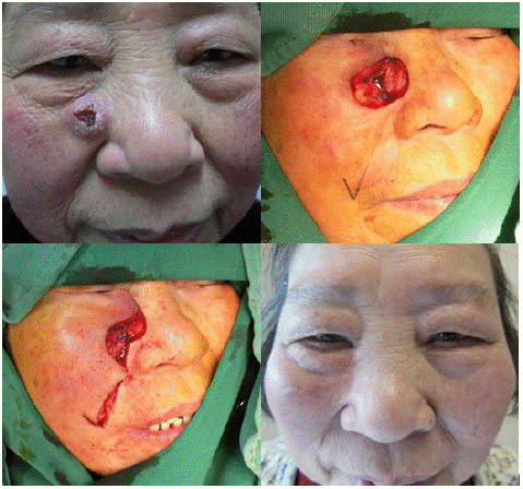
Figure 3: (A 66-year-old woman with a 25mmx23mm defect near the right nasal dorsum (Above, Left). An incision is designed along the nasolabial groove and a back cut incision in the tail end (Above, Right). Wound closure with the design of modified suture, thus reducing and modifying the shape of the defect (Below, Left). One-year follow-up (Below, Right).
Conclusion
In conclusion, the modified suture combined with rotation flap to repair the facial defect can minimize the flap’s tension and the angle between flap edge and RSTLs. Scars can be hidden in RSTLs or contour lines between facial subunits. This reconstruction strategy favors the facial defect with a circular shape or a long diameter of around 3 centimeters.
Author Statements
Funding
This research did not receive any specific grant from funding agencies in the public, commercial, or not-for-profit sectors.
Conflict of Interest
The authors declare that they have no competing interests.
Ethics Approval
All procedures performed were in accordance with the declaration of the ethical standards of the institutional research committee and with the 1964 Helsinki 387 Declaration and its later amendments. The ethics committee has approved this study of The First Affiliated Hospital of Nanjing Medical University between October 2015 to October 2019 in our department (No. 2021-SR-323).
Consent to Participate
All the pati ents provided informed consent upon this study.
Consent for Publication
Written informed was obtained from the patient for the publication of this retrospective study and any accompanying images. A copy of written consent is available for review by the Editor-in-Chief of this journal.
Authors’ Contributions
XJG, YZT, YTS collected the clinical data. XJG, XS drafted the manuscript. YTS helped to draft the manuscript and translation. JL manages data and follow-up patients. GY, FZ, and XS revised the manuscript for important intellectual content and translation. All authors read and approved the final manuscript.
References
- Heppt W. Skin tumors in facial plastic surgery. HNO. 2009; 57: 324-35.
- Riedel F, Hormann K. Plastic surgery of skin defects in the face. Principles and perspectives. HNO. 2005; 53: 1020-36.
- Schnabl SM, Breuninger H, Iordanou E, Scheu A, Kofler L, et al. Patient satisfaction in 1,827 patients following various methods of facial reconstruction based on age, defect size and site. J Dtsch Dermatol Ges. 2018; 16: 426-433.
- Koenen W, Schmieder A, Goerdt S, Breuninger H, Faulhaber J. Intracutaneous butterfly loop suture. J Dtsch Dermatol Ges. 2009; 7: 804-5.
- Breuninger H. Double butterfly suture for high tension: a broadly anchored, horizontal, buried interrupted suture. Dermatol Surg. 2000; 26: 215-8.
- Scholl L, Hessam S, Meier NM, Schmitz L, Bechara FG. The comet flap: an alternative technique for the reconstruction of facial defects. J Dtsch Dermatol Ges. 2016; 14: 442-4.
- Schultheis K, Kaufmann R, Meissner M. The double hatchet flap as a potential alternative closure technique for scalp defects. J Dtsch Dermatol Ges. 2015; 13: 73-5.
- Hafner J, Mohrle M, Loeser CR. Retroauricular pedicled flap for reconstruction of large helix and antihelix defects. J Dtsch Dermatol Ges. 2016; 14: 753-5.
- LoPiccolo MC. Rotation Flaps-Principles and Locations. Dermatol Surg. 2015; 41: S247-54.
- Mitkov M, Griffith J, Dyson M, Kimyai-Asadi A. Repair of Alar Defects Using the Alar Rotation Flap: Our Experience with 394 Patients. Plast Reconstr Surg. 2021; 147: 169-175.
- Lo CH, Kimble FW. The ideal rotation flap: an experimental study. J Plast Reconstr Aesthet Surg. 2008; 61: 754-9.
- Starkman SJ, Williams CT, Sherris DA. Flap Basics I: Rotation and Transposition Flaps. Facial Plast Surg Clin North Am. 2017; 25: 313-321.
- Borges AF, Alexander JE. Relaxed skin tension lines, Z-plasties on scars, and fusiform excision of lesions. Br J Plast Surg. 1962; 15: 242-54.
- Carmichael SW. The tangled web of Langer’s lines. Clin Anat. 2014; 27: 162-8.
- Becker FF. Rhomboid flap in facial reconstruction. New concept of tension lines. Arch Otolaryngol. 979; 105: 569-73.
- Monarca C, Rizzo MI, Palmieri A, Fino P, Parisi P, et al. Island pedicle and bilobed flaps in ala and back nose reconstruction: a prospective comparative analysis. Aesthetic Plast Surg. 2012; 36: 1168-74.
- Gurunluoglu R, Williams SA, Olsen A. Reconstructive outcomes analysis of lower eyelid and infraorbital skin defects using 2 hatchet flaps: a 6-year experience. Ann Plast Surg. 2014; 72: 657-62.
- Tomich JM, Wentzell JM, Grande DJ. Subcutaneous island pedicle flaps. Arch Dermatol. 1987; 123: 514-8.
- Yan M, Xiaobo Z, Zhaoqi Y, Xiuxia W, Rui J, et al. The kite flap for reconstructing tumour excision wounds in the middle and lower face: a retrospective study. J Wound Care. 2020; 29: 562-566.
- Hammond RE. Uses of the O-to-Z-plasty repair in dermatologic surgery. J Dermatol Surg Oncol. 1979; 5: 205-11.