
Research Article
Austin J Surg. 2023; 10(2); 1301.
Treatment of Complicated Pelvic Cystic Echinococcosis with Ureteral Stenosis
Chuang Yang*; Caihong Hao; Qiao Yuan; Pingping Yang
Department of Hepatobiliary Surgery, Fokind Hospital, Tibet University, PR China
*Corresponding author: Chuang Yang Hepatobiliary Surgery, Fokind Hospital, Tibet University, Tibet, No.14, Linkuo North Road, Lhasa City, Tibet Autonomous Region, China. Tel: +86 13458018352 Email: ycdocotr@sina.com
Received: April 12, 2023 Accepted: May 04, 2023 Published: May 11, 2023
Summary
Echinococcus cysts are found mostly in the liver and lung, but they can occur in any parts of the body. However, complicated pelvic echinococcosis is seen in China. We report cases of nine complicated echinococcal cysts with ureteral stenosis localized in the pelvis. These cases were diagnosed preoperatively by color Doppler ultrasound and computed tomography or magnetic resonance imaging. We pre-placed the double “J” tube 1-7 days before surgery, which improved the renal function of the patient, and at the same time used the double “J” tube for directional navigation during surgery to avoid ureteral injury. All patients were successfully treated and recovered, then discharged. The results showed that preimplantation of a ureteral double “J” tube before ureteral stenosis effectively prevented ureteral injury and reduced the occurrence of complications.
Keywords: Echinococcosis; Hydatid; Pelvis; Ureter; Surgery
Introduction
Cystic Echinococcosis (CE), also known as hydatid disease, is zoonotic and mainly prevalent in Tibet, Xinjiang, Qinghai, Inner Mongolia, and other provinces with developed animal husbandry in China [1]. In recent years, the survey of parasitic diseases has shown that the average prevalence of echinococcosis was 1.08% in the western region of China and could reach 6% in areas of endemic occurrence in the Qinghai-Tibet Plateau, with cystic echinococcosis as the main disease [1,2].
Although echinococcosis mainly occurs in the liver, it can also affect multiple organs in the body [3-5]. The incidence of pelvic echinococcosis is 0.20%~2.25% [6,7], however, complicated pelvic echinococcosis can invade pelvic organs, such as the ureter, bladder, rectum, uterine appendages, blood vessels, and even pelvis [8-10]. The pelvic position is relatively deep, so this results in great difficulty in surgical treatment, and easily results in organ or tissue damage.
The pelvic echinococcosis is usually caused by the spread of liver echinococcosis or celiac echinococcosis, and primary pelvic echinococcosis is rare [11]. The clinical manifestations of pelvic hydatid are often atypical, and there is no obvious symptom during the early stage. When the mass compresses or invades the surrounding tissues, there may be symptoms of urinary tractirritation such as lower abdomen bulging, pain, frequent micturition, painful micturition, and even palpable masses in the lower abdomen [12,13]. Surgery is the main treatment, supplemented with medical treatments to reduce complications, improve survival and the quality of life, and surgery to completely remove and kill hydatids to achieve a radical cure [5,14]. In the present study, we described our experiences in the treatment of complicated pelvic hydatid cysts with ureteral stenosis.
Materials and Methods
General Information
Nine patients with complicated pelvic hydatid cysts admitted to our hospital from January 2021 to July 2022 were collected. There were five males and four females. The age ranged from 24 to 65 years, with a mean age of 50.3 years. General patient demographics are shown in Table 1.
Diagnosis
History of epidemiology: Seven patients had a long-life history of 8-50 years in livestock areas. Two patients had worked with livestock and product processing, with 4 and 9 years of experience, respectively.
Clinical manifestations: All patients had abdominal pain, abdominal distension discomfort, and some patients had low back pain, urinary tract irritation symptoms, and pelvic distension discomfort. Abdominal masses were palpable in some patients. The clinical symptoms and signs are listed in Table 2.
Imaging: The diagnosis was confirmed by color Doppler ultrasound (US) and Computed Tomography (CT) or Magnetic Resonance Imaging (MRI), with the sensitivity of CT reaching 95-100% [15,16]. Pelvic hydatids involved multiple hydatid cysts that squeezed into each other to form typical cysts (Figure 1). US were simpler and faster, while CT or MRI had the advantages of multi-angle, multi-parameter, and high definition. Combined with Three-Dimensional (3D) computer-assisted imaging technology, echinococcosis was accurately located, and the relationships between mass and blood vessels and surrounding tissues were accurately determined (Figures 2,3).
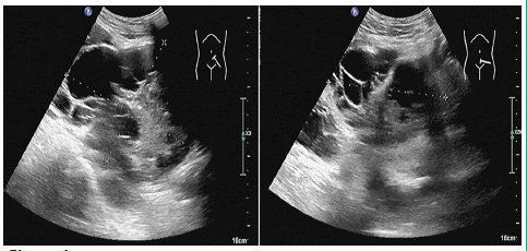
Figure 1:
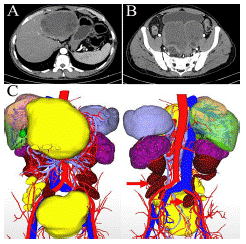
Figure 2:
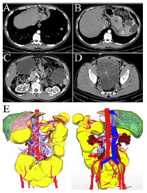
Figure 3:
Surgical Treatments
Preoperative general preparation: Albendazole tablets (15 mg/kg/d) were routinely orally administered 3-4 days before surgery. According to the patient’s renal functions, double “J” tubes were placed 1-7 days before surgery to relieve ureteral obstruction and prevent intraoperative ureteral injury. The night before surgery, oral electrolytes were used for bowel preparation, and a urinary catheter was placed 2h before surgery.
Surgical fundamentals: The purpose involved integrated removal of the hydatid cyst. When the cyst was located in the lower position of the pelvic cavity, the cyst volume was large. Under these circumstances, it was difficult to perform a complete resection of the cyst, so the surrounding tissues were carefully protected with hypertonic saline gauze when the intracapsular fluid was extracted, and hypertonic saline solution was injected into the capsule (≥15 min) to kill the daughter cyst. If part of the external capsule tightly adhered to the surrounding tissues, it was not necessary to forcibly remove it, to avoid organ damage.
Prevention of ureteral injury and urinary leakage of the bladder: Most of the complex pelvic hydatids adhered to the posterior wall of the bladder, so the bladder could be damaged during the separation process, leading to urinary leakage. Intravesical infusion with methylene blue was used to fill the bladder to determine whether there was urinary leakage caused by bladder wall injury. If this occurred, it was repaired in a timely manner. A double “J” tube was pre-placed before surgery, and this tube was palpated by hand during surgery to guide the course of the ureter. During the process of stripping the hydatid lesion, there was minimal ureteral injury.
Results
The number and size of hydatids, whether they invaded the ureter and were associated with hydatids in other organs, are shown in Table 3. Five cases (5/9) of double “J” tubes were placed in bilateral ureters and four cases (4/9) of double “J” tubes were placed in unilateral ureters (Figure 4), including three cases (3/4) on the right and one case (1/4) on the left.
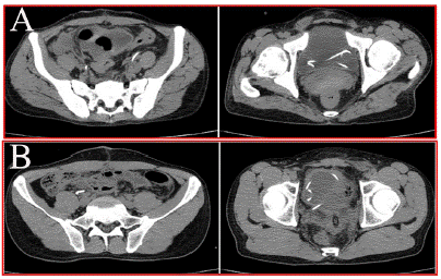
Figure 4:
After placing the double J tube, five patients with abnormal renal function returned to normal, meeting the surgical requirements. All cases were successfully treated, including complete removal of hydatid cysts in three cases, six cases of partial extracapsular extirpation combined with complete internal capsular extirpation, and six patients with combined hydatid excisions of other organs (Figure 5).
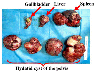
Figure 5:
There was no intraoperative iatrogenic injury of the ureter, and the postoperative pathology confirmed echinococcus granulosus disease. All cases were successfully treated and discharged, and the double “J” tube was removed 1-3 months after the postoperative follow-up. After surgery, albendazole tablets were orally administered for 6-12 months, and the liver and kidney functions were monitored during the followed-up.
Discussion
Echinococcosis mainly affects the liver, and other organs such as the lung and spleen may also be involved. With disease progression, the space occupying effect becomes increasingly obvious, which can cause organ dysfunction and eventually lead to death [17,18]. The best current treatment for this disease involves surgery, with traditional surgery as the main method of capsulectomy. With the development of surgical techniques, especially with the help of 3D imaging technology, the relationships between hydatidosis lesions and adjacent organs can be accurately identified [19,20]. The effect of radical surgical treatment of echinococcosis has become increasingly popular, the recurrence rate has been significantly reduced, residual cavity complications have been significantly decreased, and the quality of life of patients after surgery has been greatly improved [21,22].
The clinical symptoms of patients with pelvic hydatidosis are not obvious before complications occur, and the patient is seen because the lesion compresses the adjacent organs to develop corresponding clinical symptoms, or it sometimes contacts the abdominal mass. Primary pelvic hydatidosis is rare and is usually secondary [23]. Most of the secondary lesions are caused by rupture of the hydatid cyst, overflow of the source segment, or spontaneous rupture of the cyst under excessive pressure, resulting in the growth of a sac in the pelvic depression [24-26]. In this study, seven patients had a history of surgery for hydatidosis, four (4/9) for a single operation, two (2/9) for two operations, and one (1/9) for three operations. There was a history of liver surgery in five cases (5/9), and there was simultaneous removal of multiple organ hydatidosis in some cases. It is suggested that if surgical treatment of the liver or abdominal hydatid cysts is emphasized and reasonable measures are taken to prevent leakage of hydatid cyst fluid into the abdominal cavity, the occurrence of abdominal and pelvic hydatid cysts may be greatly reduced, especially those involving active hydatid cysts. The cases of two patients (2/9) involved no previous history of surgery: however, they had histories of hepatic hydatidosis or celiac hydatidosis, respectively. The occurrence of pelvic hydatidosis may have been related to spontaneous rupture of hydatid cysts and implantation metastasis, because the probability of hydatid cyst ruptures can reach 0.9-16% [16,27,28].
We believe that complex pelvic hydatidosis includes more than two hydatidosis foci, multiple hydatidal cysts squeezing and adhering to each other to form a cystic cyst, cysts connecting cysts, cysts more than 5cm in diameter, invasion of surrounding tissues, or combined with hydatidosis foci in other organs. In all cases, ureteral invasion was present, usually due to a space occupying effect, ureteral stenosis, or dilatation, combined with liver, spleen and abdominal lesions in seven cases, indicating that hydatidosis was characterized by multi-organ invasion, which involved wide distributions and large numbers [2,29].
It has previously been reported that complications of urinary leakage can reach up to 36.8% [30]. For the treatment of ureter invasion by hydatid lesions, ureter injury (e.g., tearing or cutting) may be caused by unclear identification of the ureter or dense adhesion of the ureter to the wall of the hydatid cyst. Preoperative US and CT can identify the location of ureteral dilatation and stenosis, and 3D imaging can also clearly show the location of ureteral stenosis (Figures 2C and 3E). Our experience indicated that double “J” tubes pre-placed 1-7 days before surgery eliminated the problem of ureteral obstruction, restored renal function, and guided the direction of the ureter during surgery, thus effectively avoiding ureteral injury. Immediate intraoperative ureteral injury, due to the double “J” tube support drainage, eliminated the risk of significant ureteral leakage leading to intra-abdominal infection. After surgery, the double “J” tube can be placed for a period of time. These measures fundamentally avoided ureteral injury and other complications such as urinary leakage and stenosis.
Due to the location of a pelvic hydatid cyst, it is easy to form dense adhesions with the posterior wall of the bladder, with the hydatid cyst being huge, the space narrow, and the boundary unclear. During surgical separation, it is also easy to injure the posterior wall of the bladder and cause urinary leakage. A relatively small bladder wall defect is not easy to find. Our experience suggested that intravesical instillation with methylene blue, usually 300-500mL, allowed pelvic filling to distinguish the border between the bladder and hydatid cyst, and to determine the presence of urinary bladder leakage. In case of urinary leakage, the lesion could be identified using methylene blue and repaired by sutures using an absorbable thread. In these cases, one case of bladder posterior wall injury was found during the operation, and was repaired in a timely manner. All patients successfully recovered and were discharged from the hospital, and the double “J” tubes were removed from 1-3 months after surgery, with no major discomfort to the patients.
Because complicated pelvic hydatid cysts involve compression, adhesion, and invasion to the surrounding organs, it did not require complete extirpation of the external capsule to ensure that the organs were not damaged. However, hydatid cyst cavities must be soaked in hypertonic saline, and attention should be directed to the possibility of hypernatremia.
Conclusion
Pelvic hydatid cysts are a serious hazard to patients’ health, so surgical treatment of complicated pelvic hydatidosis is the fundamental goal. Preimplantation of a ureteral double “J” tube before ureteral stenosis effectively prevented ureteral injury and reduced the occurrence of complications.
Ethical Standards
The FoKind Hospital Research Ethics Committee
Approved this retrospective analysis of anonymized data. The need for Informed consent was waived because of the retrospective nature of the study. We were confirmed that all methods have performed in accordance with the relevant guidelines.
References
- Hao W, Lucine V, Tuerhongjiang T, Li J, Vuitton DA, et al. Echinococcosis: Advances in the 21st Century. Clin Microbiol Rev. 2019; 32: e00075-18.
- Liying W, Gongsang Q, Min Q, Liu Z, Pang H, et al. Geographic distribution and prevalence of human echinococcosis at the township level in the Tibet Autonomous Region. Infectious Diseases of Poverty. 2022; 11: 10.
- Eckert J, Deplazes P. Biological, epidemiological, and clinical aspects of echinococcosis, a zoonosis of increasing concern. Clin Microbiol Rev. 2004; 17: 107-135.
- Atilgan TA, Abdullah U. Clinical characterization of unusual cystic echinococcosis in southern part of Turkey. Ann Saudi Med. 2014; 34: 508-516.
- Reza S, Amirhossein E, Mehrdad E, Rastegarian M, Sarkari B, et al. Uncommon Locations of Cystic Echinococcosis: A Report of 46 Cases from Southern Iran. Surg Res Pract. 2020; 2020: 2061045.
- Neeraj K, Raghav G, Ratnakar N. Primary pelvic hydatid cyst: A rare case presenting with obstructive uropathy. Int J Surg Case Rep. 2018; 53: 277-280.
- Kocaoglu C, Kocaoglu C, Yuvuz A, Akn A. Unusual localization and coexistence of primary hydatid cyst: A case report. Turk Pediatri Ars. 2019; 54: 192-195.
- Huma K, Adriano C, Majid FH, Afzal MS, Saqib MA, et al. A Retrospective Cohort Study on Human Cystic Echinococcosis in Khyber Pakhtunkhwa Province (Pakistan) Based on 16 Years of Hospital Discharge Records. Pathogens. 2022; 11: 194.
- Shiffali S, Shivani G, Swati A, Sharma S. Unusual Presentation of Hydatid Cyst. Euroasian J Hepatogastroenterol. 2022; 12: 31-34.
- Faisal I, Raza ER, Shoaib A, Ahmad R, Ahmad S. Pelvic Bone Hydatidosis: A Dangerous Crippling Disease. Cureus. 2019; 11: e4465.
- Sayanti P, Saumen M, Mansi U, Pramanik SR, Biswas SC, et al. Primary pelvic hydatid cyst in a postmenopausal fmale: a surgical challenge. Autops Case Rep. 2017; 7: 49-54.
- Tiaoying L, Akira I, Renqing P, Sako Y, Chen X, et al. Post-treatment follow-up study of abdominal cystic echinococcosis in Tibetan communities of Northwest Sichuan Province, China. PLoS Negl Trop Dis. 2011; 5: e1364.
- Saravanan A, Sathish M, Aditya T, Ramakrishnan E. Novel Multimodal Treatment Regimen for the Management of Primary Sacrococcygeal Cystic Echinococcosis. Journal of Orthopaedic Case Reports. 2021; 11: 22-26.
- Tarafdari A, Irandoost E, Jafari S, Vahed R, Hadizageh A, et al. Pelvic hydatid cyst: three cases with suspected adnexal masses. Int J Fertil Steril. 2022; 16: 61-63.
- Marija S, Kerstin R, Hans-Ullrich K, Junghanss T, Hosch W, et al. Diagnosing and Staging of Cystic Echinococcosis: How Do CT and MRI Perform in Comparison to Ultrasound? PLoS Negl Trop Dis. 2012; 6: e1880.
- Mouaqit O, Hibatallah A, Oussaden A, Maazaz K, Taleb KA, et al. Acute intraperitoneal rupture of hydatid cysts: a surgical experience with 14 cases. World J Emerg Surg. 2013; 8: 28.
- Nishit B, Hari K, Sumit S, Lingaiah P, Jaikumar K. Pelvic Hydatid Disease: A Case Report and Review of Literature. J Orthop Case Rep. 2017; 7: 25-28.
- Bita G. Unusual locations of the hydatid cyst: a review from Iran. Iran J Med Sci. 2013; 38: 2-14.
- Yang C, He JY, Yang XW, Wang W. Surgical approaches for definitive treatment of hepatic alveolar echinococcosis: results of a survey in 178 patients. Parasitology. 2019; 146: 1414-1420.
- Zheng JJ, Wang J, Zhao JQ, Meng X. Diffusion-weighted MRI for the initial viability evaluation of parasites in hepatic alveolar echinococcosis: comparison with positron emission tomography. Korean J Radiol. 2018; 19: 40-46.
- Nihat A, Metin K, Nuri O, Altuntas YE, Oncel M. Unusually located primary hydatid cysts. Ulus Cerrahi Derg. 2016; 32: 130-133.
- Nazim K, Mehmet SA, Tansel G, Mustafa G. Unusual Hydatid Cysts: Cardiac and Pelvic-Ilio femoral Hydatid Cyst Case Reports and Literature Review. Braz J Cardiovasc Surg. 2020; 35: 565-572.
- V Seenu, MC Misra, SC Tiwari, R Jain, C Chandrasekhar. Primary pelvic hydatid cyst presenting with obstructive uropathy and renal failure. Postgrad Med J. 1994; 70: 930-932.
- Bickers WM. Hydatid disease of female pelvis. Am J Obstec Gynecol. 1970; 107: 477-483.
- Abike F, Dunder I. Primary pelvic hydatic cyst mimicking ovarian carcinoma. Chin Med Assoc. 2011; 74: 237-239.
- Karakaya K. Spontaneous rupture of a hepatic hydatid cyst into the peritoneum causing only mild abdominal pain: a case report. World J Gastroenterol. 2007; 13: 806-808.
- Hamamci O, Besim H. Korkmaz A. Unusual locations of hydatid disease and surgical approach. ANZ J Surg. 2004; 74: 356-360.
- Sami A, Fatih O. Intraperitoneal rupture of the hydatid cyst: Four case reports and literature review. World J Hepatol. 2019; 11: 318-329.
- Bekdaulet A, Galina K, Temur Y, Daniyar T, Gulstan E. Practice of Surgical Treatment for Patients with Combined Echinococcosis of Chest and Abdominal Organs. Tanaffos. 2021; 20: 140-149.
- Huang M, Zheng H. Clinical and Demographic Characteristics of Patients with Urinary Tract Hydatid Disease. PLOS ONE. 2012; 7: e47667.