Case Report
Austin J Surg. 2023; 10(3): 1306.
Solid and Pseudopapillary Tumors of the Pancreas: A Case Report and Literature Review
Imade Elazzaoui1,3*; Hakim El Kaoui1,3; Marwa Sabur1,3; Mohamed Lamghari1,3; Mahmoud Dabbagh1,3; Imane Elmessaoudi1,3; Amine Maazouz1,3; Hind Hablaje1,3; Mohammed Najih1,3; Sidi Mohamed Bouchentouf1,3; Moutassir Moujahid1,3; Ahmed Bounaim1,3; Imane Tazi2,3; Amal Damiri2,3
¹Department of Visceral Surgery I, Mohamed V Military Instruction Hospital, Rabat, Morocco
²Department of Pathology, Mohamed V Military Instruction Hospital, Rabat, Morocco
³Mohammed V University, Faculty of Medicine and Pharmacy, Rabat, Morocco
*Corresponding author: Imade Elazzaoui Department of Visceral Surgery I, Mohamed V Military Instruction Hospital, Rabat, Morocco. Email: imad.elazzaoui@gmail.com
Received: May 08, 2023 Accepted: June 06, 2023 Published: June 13, 2023
Abstract
Solid and Pseudopapillary Tumors (SPT) of the pancreas are a rare entity, occurring essentially in young women, radiological examinations lead to the diagnosis while the positive diagnosis is based on immunohistological study. The primary treatment modality for SPTs condition is surgical intervention.
Our case aims to recall, when encountering a pancreatic mass in a young woman, that it is crucial for surgeons, radiologists, and pathologists to take into account the possibility of solid and pseudopapillary tumor. This is due to the favorable prognosis and unique treatment approaches associated with SPT, as opposed to other types of pancreatic tumors.
Keywords: Solid pseudopapillary tumor; Pancreas; Surgery; Left splenopancreatectomy
Introduction
Solid Pseudopapillary Tumors (SPTs) of the pancreas or Frantz tumor are rare neoplasms with low malignant potential. They represent less than 2% of pancreatic tumors [1] and occur mainly in young women, often in the second and third decade [2]. We report a new case of solid pseudopapillary tumor of the pancreas in a 39-year-old female patient.
Case Report
A female patient of 39 years old, with no medical history, is consulting for epigastric pain that radiates to the left hypochondrium, associated with vomiting, that has been progressing over several months. Abdominal examination reveals a palpable mass in the epigastric region.
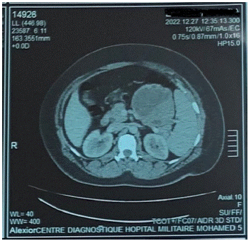
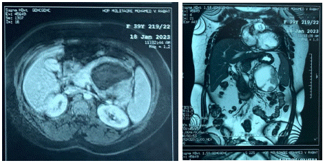
Abdominal ultrasound showed a solid cystic tumor process in the tail of the pancreas measuring 9x8.4cm. Abdominal CT scan (Figure 1) shows the presence of a well-encapsulated formation with dual cystic and tissular components enhanced after injection of contrast measuring 85x86x87mm, repressing the stomach, the splenic vein and the superior mesentericvein. MRI (Figure 2) shows large solid cystic mass in the same location, isosignal in T2, hypersignal in diffusion, enhanced after gadolinium injection measuring 90x84x89mm arriving in contact with the spleen and displacing the splenic vein and the splenic artery.

Figure 1: CT scan showing a mass of tail of pancreas with tissular and cystic components.

Figure 2: MRI showing a large solid cystic mass in the tail of the pancreas.
Surgical exploration discovered a tumor of 8cm of large axis at the expense of the posterior face of the tail of the pancreas. A left splenopancreatectomy was performed. The follow-up was uneventful, the patient received a post-splenectomy vaccination protocol before her discharge.

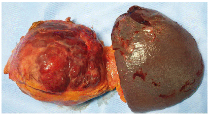
Figure 3: Tail of prancreas and spleen after resection.

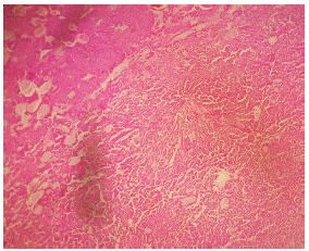
Figure 4: Histologic appearance of pseudopapillary cores.
The anatomopathological study showed an encapsulated tumor, half solid, half cystic, with large hemorrhagic foci. Immunohistochemistry was positive for vimentin, progesterone and β-catenin (Figure 5).

Figure 5: (Left) Diffuse nuclear positive staining for beta catenin (Right) the neoplastic cells membrane has mild positivity for vimentin.
Thus, showing a morphological and immunohistochemical aspect consistent with a solid pseudopapillary tumor of the pancreas.
Discussion
First described by Frantz in 1959, several names were attributed to this tumor until it was defined by the World Health Organization (WHO) in 1996 as a "solid pseudopapillary tumor” of the pancreas [3], This tumor is mainly located in the body or the tail of the pancreas.
The incidence of SPT is 0.13-2.7% of all pancreatic tumors [4]. This rare tumor seems to have a predilection for young Asian and African-American women, with a 10:1 predominance over men, at an average age of 24 years [5].
Vague abdominal pain and/or an enlarging abdominal mass may be the main clinical signs. In asymptomatic patients, SPTs of pancreas may be discovered as a palpable mass during a routine physical examination, a mass seen on casual abdominal examination, or an incidental finding on imaging for a non-tumor-related condition [6]. Occasionally the tumor, by increasing its size, leads to signs of compression of surrounding digestive, biliary or vascular structures [1].
On ultrasound, the tumor is seen as a large well-defined mass with heterogeneous appearances, due to its solid and cystic composition [7]. Abdominal CT scan shows a heterogeneous hypodense lesion with low peripheral enhancement and central calcification after contrast injection. MRI typically demonstrates a well-defined lesion and may show a pure solid consistency in ~80% of cases [8]. Reported signal characteristics include low to heterogeneous signal intensity on T1-weighted, and heterogeneous to high signal intensity in T2-weighted [8]. The value of echo-endoscopy is limited by the fact that the lesions are generally voluminous.
The positive diagnosis of this tumor remains pathological, Some defining histologic features of SPTs include the presence of solid cellular hypervascular regions without gland formation, and the presence of branching papillary fronds with sheets and degenerative pseudopapillae [9]. The immunophenotype stains positively for neuron-specific enolase, CD10, vimentin , and has atypical nuclear staining for β-catenin, which is generally a cytoplasmic protein [9]. SPTs often stain positive for progesterone receptors [10].
Given the unpredictable but real metastatic potential of these tumors, surgical resection is recommended in all cases. It involves a left pancreatectomy with conservation of the spleen whenever possible, a duodenopancreatectomy or even a partial pancreatectomy depending on the location [11], Enucleation is indicated in non-functioning tumors, TNM stages l-ll, <2cm, and with a safety margin between the tumor and the main pancreatic duct of 2-3mm [12]. Associated metastatic lesions and invaded organs should be resected, and tumor recurrences should be surgically removed [13,14]. On the other hand, extensive lymph node dissection, without visible macroscopic lesion, does not seem to be justified.
The prognosis of SPT is very good. Long-term survival after complete resection of a non-metastatic tumor is between 80 and 90%. Regardless of the type of resection, it is recommended to undergo prolonged observation through ultrasound and abdominal CT scan to detect locoregional or metastatic recurrence, as there is a potential for recurrence rates up to 15%.
Conclusion
SPT of pancreas is a rare tumor with low malignant potential, affecting mainly young women.
They may present as an abdominal mass or abdominal pain, prompting to perform an imaging (ultrasound, CT scan, MRI).
Surgery is the only curative treatment, and provides long-term survival.
Author Statements
Right to Privacy and Informed Consent
The authors declare that no patient data appear in this article.
Competing Interests
The authors declare that they have no ties of interest in relation to this article
References
- RCG Martin, DS Klimstra, MF Brennan, KC Conlon. Solid-pseudopapillary tumor of the pancreas: a surgical enigma?. Ann Surg Oncol. 2002; 9: 35–40.
- A Galvin, T Sutherland, AF Little. Part 1: CT characterisation of pancreatic neoplasms: a pictorial essay. Insights into Imaging. 2011; 2: 379–388.
- KM Coleman, MC Doherty, SA Bigler. Solid-Pseudopapillary Tumor of the Pancreas. 2008; 23: 1644–1648.
- BE Crawford. Solid and papillary epithelial neoplasm of the pancreas, diagnosis by cytology. South Med J. 1998; 91: 973–977.
- C Mao, M Guvendi, DR Domenico, K Kim, NR Thomford, et al. Papillary cystic and solid tumors of the pancreas: a pancreatic embryonic tumor? Studies of three cases and cumulative review of the world’s literature. Surgery. 1995; 118: 821–828, 1995.
- T Papavramidis, S Papavramidis. Solid pseudopapillary tumors of the pancreas: review of 718 patients reported in English literature. J Am Coll Surg. 2005; 200: 965–972.
- Y Weerakkody, A Zinaye. Solid pseudopapillary tumor of the pancreas | Radiology Reference Article | Radiopaedia.org. 2023. https://radiopaedia.org/articles/solid-pseudopapillary-tumour-of-the-pancreas-1?lang=us.
- MH Yu, JY Lee, MA Kim, SH Kim, JM Lee, et al. MR imaging features of small solid pseudopapillary tumors: retrospective differentiation from other small solid pancreatic tumors. AJR Am J Roentgenol. 2010; 195: 1324–1332.
- DS Klimstra, BM Wenig, CS Heffess. Solid-pseudopapillary tumor of the pancreas: A typically cystic carcinoma of low malignant potential. Seminars in Diagnostic Pathology. 2000; 17: 66–80.
- D. Santini, “Solid-papillary tumors of the pancreas: histopathology - PubMed”, Accessed: Mar. 20, 2023. Available: https://pubmed.ncbi.nlm.nih.gov/16407635/
- M Abid, KB Salah, MA Guirat, H Cheikhrouhou, M Khilif, et al. Tumeurs pseudopapillaires et solides du pancréas: deux observations et revue de la literature. Rev Médecine Interne. 2009; 30: 440–442.
- D Triguero, A Calero Amaro, I Caravaca Garcia, A Soler Silva, A Sanchis Lopez, et al. Parenchyma-Sparing Resection In A Non-Functioning Neuroendocrine Tumor of The Pancreatic Neck Ep170 Solid Pseudopapillary Neoplasm Of The Pancreas: Two Case Report. Int Hepato-Pancreato-Biliary Assoc. 2021; 23: S912.
- P Michel, P David, E Chatelain, T Anh Nguyen, D Oyeka Ibara, et al. Pseudopapillary tumor of the tail of the pancreas. J Chir (Paris). 2006; 143: 111–112.
- AL Mulkeen, PS Yoo, C Cha. Less common neoplasms of the pancreas. World J Gastroenterol. 2006; 12: 3180-5.