
Special Article: Laminectomy
Austin J Surg. 2023; 10(3): 1307.
Spondilolysthesis L5-S1: An Original Article
Saccomanni Bernardino, MD*
Department of Orthopaedic and Trauma Surgery, Asl Bari, Italy
*Corresponding author: Saccomanni Bernardino Department of Orthopaedic and Trauma Surgery, Asl Bari, Viale Regina Margherita, 70022, Altamura (Bari), Italy. Tel: 3208007854 Email: bernasacco@yahoo.it
Received: May 12, 2023 Accepted: June 23, 2023 Published: June 30, 2023
Abstract
Objectives: To assess the clinical, functional and radiological outcome of Posterior Lumbar Interbody Fusion (PLIF) by Banana cage with bone graft.
Material and Methods: This retrospective analytical study was carried out in unit of Orthopaedic surgery department from January 2010 to December 2020. We did PLIF by Banana cage with bone graft for High-grade Lumbar Spondylolisthesis at L5-S1. The follow up period ranges from 1 year to 2 years (average 18 months). Within these follow up period we have assessed the patients clinically, functionally and radiologically. All patients were assessed by Visual Analogue Score (VAS), Oswestry Disability Index (ODI), Waddell Disability Index (WDI), Spino-pelvic parameters, Modified Macnab’s Criteria to find out overall outcome and Hackenberge ctriteria for radiological fusion.
Results and Conclusions: Total 40 patients were included among them 16were male and 24 were female. The average age of the patients was 52.45±10.1 years. Maximum (60.0%) patients were housewife followed by 20.0%, 10.0%, 10% were day laborer, farmer and service holder respectively. Average pelvic tilt was 26.05±6.27° preoperatively and 24.10±6.26° at the final follow-up, average PI was 66.07±7.39° preoperatively and 61.19±7.08° at the final follow-up. Preoperative lumbar lordosis was 45.55±6.71° with postoperatively 37.29±6.19°at final follow-up. VAS score and ODI scales were improved significantly from preoperative 6.90±6.16 and 57.60±15.66, respectively, to postoperatively and final follow-up 2.0±0.8 and 7.60±2.40, respectively. Pre-operative Translation ratio, slip angle and disc height ratio were 21.96±10.25, -18.87±8.28, 11.03±4.36 respectively and postoperatively 13.17±6.57, -18.44±7.12, 19.60±3.36 respectively. Fusion was achieved in 36 cases (90%), 3 cases (7.5%) were fragmented and pseudoarthrosis showed only 1 case (2.5%). Most of the study population according to post operative clinical outcome showed excellent outcome (95%), 1 (2.5%) case had good and 1 (2.5%) case had fair outcome.
Conclusion: It can be concluded that, Posterior Lumbar Interbody Fusion (PLIF) by Banana cage with bone graft can be a very good option for the treatment of High-grade Lumbar Spondylolisthesis at L5-S1 levels.
Keywords: Spondylolisthesis; Banana cage; Patients
Introduction
Pain in the lower lumbar region is a socioeconomically serious medical illness worldwide. The main reason from physiological consideration is micro and macro instability of spine [1,2]. There are numerous causes for backache. Spondylolisthesis is one among them [3]. Spondylolisthesis is defined as a displacement of one vertebra over the next lower vertebra in the sagittal plane. High-Gradespondylolisthesis (HGS) is defined as greater than 50% slippage of a spinal vertebral body relative to an adjacent vertebral body AS per Meyerding classification, and most common location being down. Relationship of PT and SS is affected by lumbosacropelvic L5/S1 followed by L4/L5 [4]. Most commonly used classification flexion and extension. VRL, Vertical reference line. (From Jackson R, Kanemura T, Kawakami N, Hales C: Lumbopelvic lordosis and systems for spondylolisthesis were introduced by Wiltse et al and Marchetti and Bartolozzi which is most practical classification pelvic balance on repeated standing lateral radiographs of adult system in terms of prognosis and therapy [5,6].
In addition to the bony morphologic changes seen in high-dysplastic spondylolisthesis, spinopelvic balance plays an Oswestry Disability index important role in the development and progression of spondylolisthesis [7]. Altered biomechanical stresses found due to abnormal spino-pelvic balance at the lumbosacral junction and compensatory mechanisms used to maintain adequate posture and gait.
Degenerative Lumbar Spondylolisthesis (DLS) is always associated with facet joint degeneration and mostly observed in persons over the age of 50 years. Individuals may suffer from spinal stenosis with back and leg pain [8]. Decompression with fusion better than isolated decompression because it will further destabilize the spine, permitting further slip progression [9,10].
Vertebral Interbody Fusion (IBF), is relatively new set of technique, has become very popular in the treatment of symptomatic DLS. Interbody fusion provides a number of potential benefits forreliving symptoms. It improves the biomechanical stability of a construct mainly by stabilizing the anterior column. This can be proved important especially in patients with High-grade spondylolisthesis, Unstable slips, Degenerative type of scoliosis, and retained disc height) [11-14]. Insertion of interbody devices also improve sagittal alignment and restore disc and foraminal height as well, which ultimately provide indirect decompression of foraminal and canal stenosis and aiding in spondylolisthesis reduction [12,15].
Material and Methods
Posterior Lumbar Interbody Fusion (PLIF) surgery is performed by the standard posterior approach. Wide laminectomy is done first followed by partial bilateral facetectomy, and then the neural elements are retracted to either side, to make space for disc space preparation and finally insertion of a titanium interbody. This retrospective analytical study included 40 patients were device packed with autogenous bone graft within the inter-carried out in unit of Orthopaedic surgery department vertebral space [16,17] from January 2010 to December 2020 (Figure 1). PLIF by Banana cage with bone graft was done for High-grade Lumbar Spondylolisthesis only at L5-S1. The follow up period ranges from 1 year to 2 years (average 18 months). Within these follow up period we have assessed the patients clinically, functionally and radiologically. All patients were assessed pre and post operatively by Visual Analogue Score (VAS), Oswestry Disability Index (ODI), Waddell Disability Index (WDI), Spino-pelvic parameters, Modified Macnab’s Criteria to find out overall outcome and Hackenberge ctriteria for radiological fusion. IBM-SPSS V26 software was used for statistical analysis.
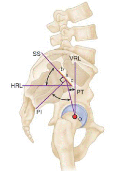
Figure 1: Sacral slope (SS) is angle subtended by Hori-zontal Reference Line (HRL) and sacral endplate line (bc). SS shares common reference line (bc) with Pelvic Incidence (PI) and Pelvic Tilt (PT). PI is measured from static anatomic structures. PT and SS depend on angular position of sacrum/pelvis in relation to femoral heads, which changes with standing, sitting, and lying down. Relationship of PT and SS is affected by lumbosacropelvic flexion and extension. VRL, Vertical reference line. (From Jackson R, Kanemura T, Kawakami N, Hales C: Lumbopelvic lordosis and pelvic balance on repeated standing lateral radiographs of adult volunteers and untreated patients with constant low back pain, Spine 25:575–586, 2000).
Selection of the Patients for Surgery
Patients with grade iii, iv and v spondylolisthesis.
Inclusion criteria:
Only at L5-S1 who had severe low pain, neurological deficit and restriction of movement with instability not responding to Sacral Slope (SS) is angle subtended by hori-zontal conservative treatment.
Exclusion criteria:
Patients with grade i and grade ii tilt (PT).
PI is measured from static anatomic structures of PT and spondylolisthesis. High grade spondylolisthesis patients with SS depend on angular position of sacrum/pelvis in relation to severe comorbidities and infection at local incision site. Femoral heads, which changes with standing, sitting, and lying Clinical, Functional and Radiological Outcome of Posterior Lumbar Interbody Fusion by Banana Cage with Bone Graft for the Treatment of High-Grade Lumbar Spondylolisthesis at L5-S1 Banana cage filled with bone graft, Per-operative picture of Figure 2 Figure 3, Figure 4, Figure 5.
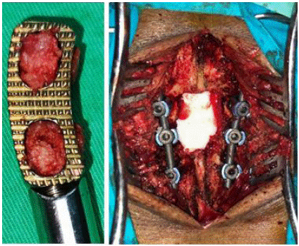
Figure 2: Banana cage filled with bone graft, Per-operative picture showing pedicle screw and rod.
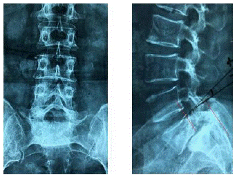
Figure 3: Pre-operative X-ray.

Figure 4: Per-operative C-ARM imaging.
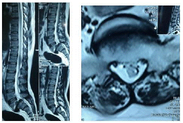
Figure 5: Pre-operative magnetic resonance imaging.
Operative technique
Patient is prone and two parallel pillows on a radiolucent table after General anesthesia ensure the abdomen hangs free. After proper cleaning, painting and draping of the operative area, a lower midline incision was made extending from spinus process Immediate Post operative -operative X-rayof L4 to S3.
Results
The skin, subcutaneous tissue was cut in a single line and the deep fascia and supra spinus ligaments cut in a same plane by diathermy. Then para spinal muscles made retracted the retrospective analytical study includes 40 patients who subperiosteally up-to tip of transverse processes bilaterally. Fulfilled the inclusion criteria, were operated by PLIF by Banana the space was identified under C-ARM guidance and partial or cage with bone graft for High-grade Lumbar Spondylolisthesis. Complete laminectomy of L5 was done. After identification of all patient were followed up from 1 year to 2 years (average 18 L5 nerve root, L5-S1 joint space clearance was done using end months) postoperatively. Plate curette and box curette. After that we put pedicle screw from L4 to S1 on either side. Then distraction between L4 and Pre operative and post operative comparison of clinical outcomes S1 done after putting pre bended rod on the pedicle screws after 24 months (n=40) According to VAS & WDI score slots, reduction of L5 achieved by either using a persuator with a traditional L5 pedicle screw or some times by a reduction screw.
Score Pre operative Final follow-up p value after reduction we again distract between L5-S1 to get enough space for introduction of proper sized Banana cage with bone graft. After putting cage with bone graft at space, we put gel foam over open cauda equina. Proper haemostasias was ensured pre operative and post operative comparison of clinical outcomes in every step of surgery. The wound was closed in anatomical after 24 months (n=40) According to ODI score layer by absorbable 1/0 cutting body vicryl and skin-by-skin stapler keeping a drain in situ (Table 1).
Score Pre-operative Final follow-up p value Mean pre- and post- operative (after 24 months) spinopelvic. In this study, age range of the patients were 41-72 years, mean parameters (n=40) age was 52.45±10.1 years and the male to female ratio was 8:12 which are comparable to the study of Audat et al, 2012. Who were housewives followed by 20.0%, 10.0%, 10.0% were day laborer, Pelvic Index (°) farmer and service holder respectively. In study of Sakeb et al., 2013, maximum 65.4% were house wife, 15.4% were manual worker and 19.2% were sedentary worker which is also comparable [18,19].
Radiological fusion status after 24 months of operation (n=40) All 40 (100%) patients had back pain, sciatica and neurogenic claudication (Audat et al., 2012). Sensory disturbance was present. According to Hackenberge criteria (Hackenberge et al.2005) in 38 (95%) patients, and motor weakness was observed in 31, 24 months after operation n (%) patients [18]. Only 1 (2.5%) patient had urinary retention. Which is also comparable to study of Sakeb et al., 2013 [19]. All Fused 36 were improved significantly (P<.005) in postoperative group. Probably fused 3 decrease in spinopelvic parameters were observed after Pseudo-arthrosis operation and remain almost unchanged throughout the follow up period (average pelvic tilt was 26.05±6.27° preoperatively.
Distribution of patients according to time taken for fusion (n=40) and 24.10±6.26° at the final follow-up, average PI was 66.07±7.39° preoperatively and 61.19±7.08° at the final follow-up. Pre-operative lumbar lordosis were 45.55±6.71° and 37.29±6.19° at final follow-up) [20].
Study carried out by Lengert et al, 2014, Significant improvement of VAS score for back pain and post operative score from 6.90±6.16 to 2.0±0.8 score with significant (p<0.001), and ODI was significant before operation 57.60±15.66 to 7.60±2.40 with (P<0.001) in at 12 months follow up. In a study of Sakeb et al., (2013) mean VAS reduced from 7.2 to 2.2 and mean ODI reduce from 60.7 to 11.2 at 12 months follow up.
WDI significantly reduced from 7.03±1.08 to 2.07±0.61 with a significant (P<0.001) value [19]. About 55% achieved radiological fusion at 6th month in our series which co-inside with 52% early fusion in Lee H et al. (2012) series [21]. Rate of spinal fusion with bone graft range from 46%-90% in Lowe GT et al. (2002) series [22]. In our series fusion evaluation by CT scan for 5 patient who showing doubt in x-ray fusion, 4 out of 5 (80.00%) fused and one was fragmented according Cristensen 18 month Post-operative X-ray showing good bony assessment scale after 18 months of follow-up (Figure 6 & 7). Fusion after 18 fusion months including X-ray & CT scan evaluation is 97.5%. Fusion was achieved 95.7% (45 of 47) cases in the study of AGAZZI et al. (1999) [23]. Radiographic fusion was present in 27 (88.9%) patients after one year in the study of Audat et al. (2012) [18].
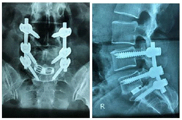
Figure 6: Immediate Post-operative X-ray.

Figure 7: 18th month Post-operative X-ray showing good bony Fusion.
According to Modified Macnab criteria (Macnab I et al.1971), Post operative wound infection was in 1 (2.5%) and pseudo-arthrosis was in 1 (2.5%) patient. Wound infection was managed comprehensive conservatively by antibiotics according to culture and sensitivity outcome report, improvement of nutritional status, removal of stitch, regular dressing secondary wound closure. Pseudo-arthrosis was managed conservatively as the symptoms reduced significantly and patient continues his daily life without any trouble. In our study 35 (87.5%) patients got satisfactory results and 4 (10.0%) patients got Good result in 1 (2.5%) case had fair outcome.
Discussion
Patients treated by PLIF with Banana cage and bone graft were followed up for the period of 24 months. Overall clinical outcome categorized as excellent, good, fair, poor according to modified Macnab criteria. For statistical analysis good and excellent were grouped as satisfactory and fair and poor as unsatisfactory. In this study, age range of the patients were 41–72 years, mean age was 52.45±10.1 years and the male to female ratio was 8:12 which are comparable to the study of Audat et al, 2012. Who found mean age 54.2±13.6; (range 36.0–66.0) [18]. Regarding occupation of the patient maximum (60.0%) patients were housewife followed by 20.0%, 10.0%, 10.0% were day laborer, farmer and service holder respectively. In study of Sakeb et al., 2013 maximum 65.4% were house wife, 15.4% were manual worker and 19.2% were sedentary worker which is also comparable [19]. All 40 (100%) patients had back pain, sciatica and neurogenic claudication (Audat et al., 2012). Sensory disturbance was present in 38(95%) patients, and motor weakness was observed in 31(77.5%) patients [18]. Only 1(2.5%) patient had urinary retention. Which is also comparable to study of Sakeb et al., 2013 [19]. All were improved significantly (P<.005) in postoperative group. Decrease in spinopelvic parameters were observed after operation and remain almost unchanged throughout the follow up period (average pelvic tilt was 26.05±6.27° preoperatively and 24.10±6.26° at the final follow-up, average PI was 66.07±7.39° preoperatively and 61.19±7.08° at the final follow-up. Pre-operative lumbar lordosis were 45.55±6.71° and 37.29±6.19°at final follow-up). Study carried out by Lengert et al., 2014 [20]. Showed similar improvement. Significant improvement of VAS score for back pain and post operative score from 6.90±6.16 to 2.0±0.8 score with significant (p<0.001), and ODI was significant before operation 57.60±15.66 to 7.60±2.40 with (P<0.001) in at 12 months follow up. In a study of Sakeb et., al (2013) mean VAS reduced from 7.2 to 2.2 and mean ODI reduce from 60.7 to 11.2 at 12 months follow up. WDI significantly reduced from 7.03±1.08 to 2.07±0.61 with a significant (P<0.001) value [19]. About 55% achieved radiological fusion at 6th month in our series which co-inside with 52% early fusion in Lee H et al. (2012) series [21]. Rate of spinal fusion with bone graft range from 46%-90% in Lowe GT et al. (2002) series [22]. In our series fusion evaluation by CT scan for 5 patient who showing doubt in x-ray fusion, 4 out of 5 (80.00%) fused and one was fragmented according Cristensen assessment scale after 18 months of follow-up. Fusion after 18 months including X-ray & CT scan evaluation is 97.5%. Fusion was achieved 95.7% (45 of 47) cases in the study of AGAZZI et al. (1999) [23]. Radiographic fusion was present in 27 (88.9%) patients after one year in the study of Audat et al. (2012) [18]. Post operative wound infection was in 1(2.5%) and pseudoarthrosis was in 1(2.5%) patient. Wound infection was managed conservatively by antibiotics according to culture and sensitivity report, improvement of nutritional status, removal of stitch, regular dressing secondary wound closure. Pseudo-arthrosis was managed conservatively as the symptoms reduced significantly and patient continues his daily life without any trouble. In our study 35(87.5%) patients got satisfactory results and 4(10.0%) patients got Good result in 1(2.5%) case had fair outcome.
Conclusion
Patients treated by PLIF with Banana cage and bone graft were followed up for the period of 24 months. Overall clinical outcome the results of the present study indicate that the outcomes were categorized as excellent, good, fair, poor according to modified significantly improved with surgery and the technique of Posterior Macnab criteria. For statistical analysis good and excellent were lumbar interbody fusion with Local bone graft and Cage produced grouped as satisfactory and fair and poor as unsatisfactory. satisfying clinical, functional and radiological improvement.
References
- Panjabi MM. Clinical spinal instability and low back pain. J Electromyogr Kinesiol. 2003; 13: 371-9.
- Lin Y, Chen W, Chen A, Li F. Comparison between minimally invasive and open transforaminal lumbar interbody fusion: A meta-analysis of clinical results and safety outcomes. J Neurol Surg A Cent Eur Neurosurg. 2016; 77: 2-10.
- Karla R, Kumar NP, Kalyan SS. A study on management of high grade spondylolisthesis. Int Arch Integr Med. 2017; 4: 41-8.
- Meyerding HW. Spondylolisthesis; surgical fusion of lumbosacral portion of spinal column and interarticular facets; use of autogenous bone grafts for relief of disabling backache. J Int Coll Surg. 1956; 26: 566-91.
- Wiltse LL, Newman PH, Macnab I. Classifi cation of spondylolysis and spondylolisthesis. Clin Orthop Relat Res. 1976; 23-9.
- Marchetti PG, Bartolozzi P. Classifi cation of spondylolisthesis as a guideline for treatment. In: Bridwell K, DeWald R, editors The textbook of spinal surgery. 2nd ed ed. Philadelphia: Lippincott-Raven; 1997; 1211-54.
- Vidal J, Marnay T. Morphology and anteroposterior body equilibrium in spondylolisthesis L5/S1. Rev Chir Orthop Reparatrice Appar Mot. 1983; 69: 17-28.
- Jacobsen S, Sonne-Holm S, Rovsing H, Monrad H, Gebuhr P. Degenerative lumbar spondylolisthesis: an epidemiological perspective: the Copenhagen osteoarthritis Study. Spine. 2007; 32: 120-5.
- Kepler CK, Vaccaro AR, Hilibrand AS, Anderson DG, Rihn JA, et al. National trends in the use of fusion techniques totreat degenerative spondylolisthesis. Spine. 2014; 39: 1584-9.
- Schroeder GD, Kepler CK, Kurd MF, Vaccaro AR, Hsu WK, Patel AA, et al. Rationale for the surgical treatment of LumbarDegenerative spondylolisthesis. Spine (Phila Pa 1976). 2015; 40: E1161-6.
- Oda I, Abumi K, Yu BS, Sudo H, Minami A. Types of spinal instability that require interbody support in posterior lumbar reconstruction:an in vitro biomechanical investigation. Spine. 2003; 28: 1573-80.
- McAfee PC, DeVine JG, Chaput CD, Prybis BG, Fedder IL, Cunningham BW, et al. The indications for interbody fusion cagesin the treatment of spondylolisthesis: analysis of 120 cases. Spine. 2005; 30: S60-5.
- Liao JC, Lu ML, Niu CC, Chen WJ, Chen LH. Surgical outcomes of degenerative lumbar spondylolisthesis with anterior vacuum disc: can the intervertebral cage overcome intradiscal vacuum phenomenon and enhance posterolateral fusion? J Orthop Sci. 2014; 19: 851-9.
- Ha K-Y, Na K-H, Shin J-H, Kim K-W. Comparison of posterolateralfusion with and without additional posterior lumbar interbody fusion for degenerative lumbar spondylolisthesis. J Spinal Disord Tech. 2008; 21: 229-34.
- Vamvanij V, Ferrara LA, Hai Y, Zhao J, Kolata R, Yuan HA. Quantitative changes in spinal canal dimensions usinginterbodydistraction for spondylolisthesis. Spine. 2001; 26: E13-8.
- Cole CD, McCall TD, Schmidt MH, Dailey AT. Comparison of lowback fusion techniques: transforaminal lumbar interbody fusion (TLIF) or posterior lumbar interbody fusion (PLIF) approaches. Curr Rev Musculoskelet Med. 2009; 2: 118-26.
- Lara-Almunia M, Gomez-Moreta JA, Hernandez-Vicente J. Posterior lumbar interbody fusion with instrumented posterolateralfusion in adult spondylolisthesis: description and association ofclinico-surgical variables with prognosis in a series of 36 cases. Int J Spine Surg. 2015; 9: 22.
- Audat Z, Moutasem O, Yousef K, Mohammad B. Comparison of clinical and radiological results of posterolateral fusion, posterior lumbar interbody fusion and transforaminal lumbar interbody fusion techniques in the treatment of degenerative lumbar spine. Singapore Med J. 2012; 53: 183-7.
- Sakeb N, Ahsan K. Comparison of the early results of transforaminal lumbar interbody fusion and posterior lumbar interbody fusion in symptomatic lumbar instability. Indian J Orthop. 2013; 47: 255-63.
- Lengert R, Charles YP, Walter A, Schuller S, Godet J, et al. Posterior surgery in high-grade spondylolisthesis. Orthop Traumatol Surg Res. 2014; 100: 481-4.
- Lee KH, Yue WM, Yeo W, Soeharno H, Tan SB. Clinical and radiological outcomes of open versus minimally invasive transforaminal lumbar interbody fusion. Eur Spine J. 2012; 21: 2265-70.
- Lowe TG, Tahernia AD, O’Brien MF, Smith DA. Unilateral transforaminal posterior lumbar interbody fusion (TLIF): indications, technique, and 2-year results. J Spinal Disord Tech. 2002; 15: 31-8.
- Agazzi S, Reverdin A, May D. Posterior lumbar interbody fusion with cages: an independent review of 71 cases. J Neurosurg. 1999; 91: 186-92.