Special Article: Sarcoma Surgery
Austin J Surg. 2023; 10(3): 1308.
Lung Sparing Bronchoplasty of Carcinoid Tumors with Single Lumen ETT in a Limited-Resource Hospital Setting
Maham Zehra Zaidi1*; Niaz Hussain2; Ayesha Siddiqua Hashmi3; Syed Suhail Qadri4#; Saima Imam5#; Shagufta Nasreen6#; Hina Khalid7#
1House Officer in Dow University of Health Sciences, Pakistan
2Associate Professor and Head of Department of Thoracic Surgery in Dow University of Health Sciences, Pakistan
3Post Graduate Resident in Dow University of Health Sciences, Pakistan
4Cardiothoracic Surgeon in Castle Hill Hospital, Pakisthan, Pakistan
5Consultant Anesthetist in Dow University of Health Sciences, Pakistan
6Post graduate Resident in Dow University of Health Sciences, Pakistan
7Post Graduate Surgical Resident in Dow University of Health Sciences, Pakistan
*Corresponding author: Maham Zehra Zaidi House Officer in Dow University Health Sciences, Pakistan. Email: mahamzehrazaidi@gmail.com
#These authors have contributed equally to this artcle.
Received: June 05, 2023 Accepted: July 05, 2023 Published: July 12, 2023
Abstract
Two patients, who presented with complaints of cough and hemoptysis, were diagnosed as having biopsy proven carcinoid tumor of the airway. Both patients underwent lung sparing surgery and bronchoplasty, with the aid of single lumen ETT in a limited resource, underprivileged hospital setting. This case report underscores the management of resectable tumors of major airways with single lumen ETT.
Keywords: Bronchoplasty; Lung sparing surgery; Bronchopulmonary carcinoid
Introduction
Carcinoid tumor is a slow growing [1] tumor originating in the cells of the neuroendocrine system. However, they can act aggressively and metastasize. It is estimated that approximately 5-6% of patients with a carcinoid tumor will develop carcinoid syndrome [2,3], which correlates with extensive pulmonary or hepatic metastases.
The majority of carcinoids are found to be within the gastrointestinal system (67.5%), and the bronchopulmonary system (25.3%). Nonlocalized lesions were mostly found to be cecal (81.5-83.2%) and pancreatic (71.9-81.3%) carcinoids, whereas localized disease involved the rectum (81.7%), stomach (67.5%), and bronchopulmonary system (65.4%). These neuroendocrine tumors may be functional (showing exaggerated hormone production) or non-functional [4], and may act benign or malignant.
In the past few decades, there has been a remarkable increment in the incidence of carcinoids. It has been reported that at the time of diagnosis, distant metastasis is already present in 12.9% of such cases; with only 67.2% survival among these patients at 5 years, bringing into question the presumptive benignity of this entity [5].
Thus, the need for prompt diagnosis and intent for curative surgery is being highlighted through these case reports, which can be achievable in a limited resources hospital setting.
Case Report
Case 01
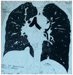
A young male, aged 30 years presented with complaints of cough, associated with hemoptysis and fever for the last 2.5 years. He did not have any history of flushing, diarrhea, shortness of breath etc. Chest examination revealed decreased breath sounds on the left side; rest of the examination was normal. Chest x-ray showed slight tracheal deviation towards the right. Further workup was done. CT scan showed a well demarcated, ovoid, hypodense lesion in the left main bronchus measuring 1.5x0.9cm, with hyper-expansion of the left lung and concomitant infection, without any evidence of metastatic lesions elsewhere in the body.
Bronchoscopy was performed, and the mass was identified in the distal part of left main bronchus, which was biopsied. Biopsy results concluded that it was a typical (low grade) carcinoid tumor and plan was made for curative surgery.

Figure 1: Preoperative CT scan (coronal view) of Case 01, demonstrating an ovoid mass in the left main bronchus.

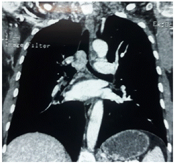
Figure 2: Preoperative CT scan (coronal view) of Case 02, demonstrating mass in trachea and right main bronchus.
Left posterolateral thoracotomy was done followed by Bronchotomy of left main bronchus. ETT which was initially placed in the trachea was directed into the right main bronchus for single lung ventilation by the surgeon, with the assistance of the anesthetist.
The tumor was then resected, followed by bronchoplasty to spare lung resection. Post-operatively, the patient had an uneventful recovery, and was discharged after 2 weeks. Further plan was to have regular follow ups of the patient on outpatient basis.
Case 02
35 years’ old female presented with complaints of cough and hemoptysis for the past 5 years, associated with infrequent spells of shortness of breath.
On chest examination, there were decreased breath sounds on the right side. CT scan revealed a well-defined, homogenous soft tissue density mass with lobulated margins measuring 3.1x2.8x1.3cm, seen involving the trachea in the region of carina and right main bronchus, causing widening and obliteration of the lumen of right bronchus.
Bronchoscopy showed a mass arising from the right main bronchus extending up to carina; vascular, bled on touch. Left main bronchus was spared completely. Biopsy was taken which showed it to be a well differentiated carcinoid tumor.
Thoracotomy was done, followed by tracheotomy and bronchotomy of right main bronchus, and the ETT formerly placed in the trachea- just below the vocal cords- was directed into the right main bronchus for single lung ventilation by the surgeon, with the assistance of the anesthetist.
The mass was then resected completely from the trachea and right main bronchus, followed by tracheobronchoplasty to spare lung resection.
Post-operatively, the patient had an uneventful recovery, remained vitally stable and was mobilized early. Patient was called for regular follow ups on outpatient basis.
Discussion
Bronchopulmonary carcinoids constitute up to 25.3% of all carcinoid tumors [2]. Among the spectrum of bronchopulmonary Neuroendocrine Tumors (NETs), which encompasses small cell lung cancer as its most malignant form, as well as several other forms such as carcinoid lung tumors representing the more benign counterpart [6].
Histologically, typical carcinoid tumors contain “uniform cells with small round to oval nuclei, few nucleoli, and granular eosinophilic cytoplasm [6].” The cells are most commonly in islands interweaving with one another in a mosaic pattern, as interconnecting ribbons of cells in a trabecular pattern, or in an adenopapillary pattern; mixed forms are also seen. In a typical carcinoid, nuclear pleomorphism and mitoses are rare [6].
Typical carcinoid tumors have a 5-year Overall Survival (OS) of 90% to 95% after surgical resection [7,8].Recurrence is seldom seen in patients with typical carcinoids without any nodal involvement [8].
The most favorable 5-year survival rates were found with rectal (88.3%), bronchopulmonary (73.5%), and appendiceal (71.0%) carcinoids; among which the bronchopulmonary carcinoids showed invasive growth or metastatic spread in up to 27.5% of patients [5].
Most patients present with complaints of cough and hemoptysis, with other signs of carcinoid syndrome included in patients with metastasis at the time of presentation.
Detterbeck FC concluded that typical carcinoids are characterized by young age, central tumor, and no nodal enlargement [7].
Surgery is the modality of choice for resectable carcinoid tumors without any metastatic spread. Surgical resection of thesetumors is associated with a survival advantage as compared to nonoperative management [9].
In the past two decades, a lung parenchyma-preserving approach was assumed. Bronchoplastic surgery is being adapted for carcinoids of central origin and minimal resection of one segment or less reserved for peripheral carcinoids. The prognosis of patients in whom a more extensive resection has been done has shown no excessive benefit as compared to a more conservative, lung preserving approach. Locoregional lymph node involvement, or positive resection margins does not have influence on prognosis [10].
In patients with typical bronchial carcinoid, even limited surgery, such as bronchoplastic surgery, has become an accepted treatment modality [10-12].
Okike N. et al reported successful treatment of “typical” carcinoids with simple wedge tracheobronchotomy without lung resection [12].
The optimal method of ventilation should provide sufficient alveolar ventilation and oxygenation, while ensuring unobstructed access to the circumference of the trachea and bronchi for anatomical alignment of an airtight repair [12].
The single-lumen endotracheal tube can be utilized for endobronchial intubation in two ways, which is essential in procedures involving resection of the airway, either by intubating the contralateral main bronchus, or by making use of another anesthesia circuit and intubating through a separate endobronchial tube through the operative site [13].
A double lumen endotracheal tube provides satisfactory ventilation but poses the disadvantage of constrained access to the operative field in carinal surgery. Furthermore, double-lumen tubes have a limited application during major airway surgery [14].
In both reported cases, single lumen ETT was used with confidence, without any adverse events.
References
- Maroun J, Kocha W, Kvols L, Bjarnason G, Chen E, et al. Guidelines for the diagnosis and management of carcinoid tumours. Part 1: the gastrointestinal tract. A statement from a Canadian National Carcinoid Expert Group. Curr Oncol. 2006; 13: 67-76.
- Fox DJ, Khattar RS. Carcinoid heart disease: presentation, diagnosis, and management [presentation]. Heart. 2004; 90: 1224-8.
- Warrell, et al. Oxford Textbook of Medicine. 8th ed. Oxford University Press. ISBN 0-19-262922-0; 2010.
- Wood DE. National Comprehensive Cancer Network. National Comprehensive Cancer Network (NCCN) Clinical Practice Guidelines for Lung Cancer Screening. Thorac Surg Clin. 2015; 25: 185-97.
- Modlin IM, Lye KD, Kidd M. A 5-decade analysis of 13,715 carcinoid tumors. Cancer. 2003; 97: 934-59.
- Sheppard MN. Neuroendocrine differentiation in lung tumours. Thorax. 1991; 46: 843-50.
- Detterbeck FC. Management of carcinoid tumors. Ann Thorac Surg. 2010; 89: 998-1005.
- Lou F, Sarkaria I, Pietanza C, Travis W, Roh MS, et al. Recurrence of pulmonary carcinoid tumors after resection: implications for postoperative surveillance. Ann Thorac Surg. 2013; 96: 1156-62.
- Raz DJ, Nelson RA, Grannis FW, Kim JY. Natural history of typical pulmonary carcinoid tumors: a comparison of nonsurgical and surgical treatment. Chest. 2015; 147: 1111-7.
- Schreurs AJ, Westermann CJ, Van den Bosch JM, Vanderschueren RG, Brutel de la Rivière A, et al. A twenty-five-year follow-up of ninety-three resected typical carcinoid tumors of the lung. J Thorac Cardiovasc Surg. 1992; 104: 1470-5.
- Arrigoni MG, Woolner LB, Bernatz PE. Atypical carcinoid tumors of the lung. J Thorac Cardiovasc Surg. 1972; 64: 413-21.
- Okike N, Bernatz PE, Payne WS, Woolner LB, Leonard PF. Bronchoplastic procedures in the treatment of carcinoid tumors of the tracheobronchial tree. J Thorac Cardiovasc Surg. 1978; 76: 281-91.
- El-Baz N, Jensik R, Faber LP, Faro RS. One-lung high-frequency ventilation for tracheoplasty and bronchoplasty: a new technique. Ann Thorac Surg. 1982; 34: 564-71.
- Bjork VO, Carlens E, Crafoord C. The open closure of the bronchus and the resection of the carina and of the tracheal wall. J Thorac Surg. 1952; 23: 419-28.