Review Article
Austin J Surg . 2024; 11(2): 1323.
Percutaneous Endoscopic Gastrostomy (PEG) With Gastropexy New Placement Tech New Placement Techniques at a Glance
Christian Bojarski¹*; Arno Dormann²*
¹Main Practice Gastroenterology, Josef-Haubrich-Hof 5, 50676 Cologne, Germany
²Director of the Center for Interdisciplinary Visceral Medicine, Kliniken der Stadt Köln gGmbH, Campus Merheim and Holweide, Neufelder Str. 34, 51067 Cologne, Germany
*Corresponding author: Christian Bojarski Main Practice Gastroenterology, Josef-Haubrich-Hof 5, 50676 Cologne, Germany;
Arno Dormann, Director of the Center for Interdisciplinary Visceral Medicine, Kliniken der Stadt Köln gGmbH, Campus Merheim and Holweide, Neufelder Str. 34, 51067 Cologne, Germany. Email: c.bojarski@gastroenterologie-koeln.de; dormanna@kliniken-koeln.de
Received: May 28, 2024 Accepted: June 19, 2024 Published: June 26, 2024
Abstract
Percutaneous Endoscopic Gastrostromy (PEG) was introduced in 1980 as an interventional endoscopically procedure and was applied for many years as a Pull-Through Technique (PTT). Although the technical success rate of PEG placement is near 100% and therefore very high, this procedure had the highest peri-interventional complication rates among all interventional endoscopy techniques. In 1999, the first gastropexy device for the introducer PEG was approved to the European market and allowed direct puncture of the ventral gastric wall under endoscopic control. In patients with oral colonization with multi-resistant germs, pharyngeal/esophageal stenosis and those with ascites, gastropexy allowed direct fixation of the gastric and the abdominal wall to avoid leakage and prevent infectious complications. A gastrotube with a blockable inner balloon served as the final PEG. After several years of use, gastric tubes with balloons instead of classical pull-through PEGs showed some complications, mostly induced by misuse. A combination of a PEG applied as a gastropexy which is followed by PTT is called a hybrid PEG and is superior over the placement of each single technique. This short review present background data and give a current review on different PEG techniques and their related complications.
Approach to PEG Placement
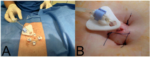
Over the years and depending on the expertise of each individual center or interventional endoscopy unit, the approach to patients with an indication for PEG placement may vary widely. The first description of endoscopic placement of a PEG was in 1980 [1]. The initial procedure was slightly improved and modified and has become a standard procedure worldwide. A modification, the so-called introducer PEG, was not successful due to missing fixation of the anterior wall. The majority of procedures today are performed endoscopically, however, in rare cases a pure surgical or radiologicial (sonography/CT) [2] procedure may be an alternative [3]. In most countries, the PEG is placed under conscious sedation [4] with either administration of intravenous applied propofol or midazolam with or without additional analgesics, and a personal team of at least three persons (two physicians and one nurse, one physician and two nurses, ideally one of them exclusively skilled in PEG placement techniques), pre- or periinterventional antibiotics are mandatory [5]. The Pull-Through Technique (PTT) is the most widespread procedure used for placement. After inflating the gastric cavity with CO2, the puncture of the ventral abdominal and gastric wall ensures a stable access to the stomach. A long thread is pushed through a trocar and removed orally by grabbing with a biopsy forceps through the endoscope. After mounting the inner plastic plate to the thread outside the patient, the thread is pulled gently through the upper mouth, hypopharynx and esophagus into the stomach. The fixation of the outer plastic plate and the continuous and stable adaption of the ventral abdominal and gastric wall is essential within the first 24 hours after PEG insertion to guarantee adhesion of these different layers and prevent complications. In 2000, Dormann et al. first described modified introducer PEG with gastropexy in 457 patients, mostly with esophageal stenosis, and presented first experience with direct punction system Cliny PEG 13 and gastropexy [6]. The encouraging results of this report and the following studies thereafter opened the door for the development of general available systems for direct punction and safe placement of gastric nutritional tubes. The first Europe-wide approval of gastropexy device was introduced in 2003 as Freka® Pexact I with gastropexy Device I by Fresenius Kabi. New products were additionally presented in the following years (Table 1). However, the administration of gastric tubes with blockable balloons can cause bleeding, malfunction, leakage or rupture, mostly due to misuse of these systems. Balloon-associated problems may occur up to 20% [7]. The combination of PTT and a gastropexy is called a hybrid PEG. Although many clinical centers use this combination routinely or for special indiations, the term hybrid PEG was first described as a secondary use in leakage after PTT-PEG by Wejda et al. in 2005 [8] and then established by Grund et al [9]. This technique has the potential to significantly reduce complications by the total summation of the advantages of each technique. Three crucial steps are defined for generating a safe procedure with a minimum of complications: a) correct handling of the pexy device with application of at least 3 sutures, b) adequate knotting technique for safe apposition of the wall layers and c) possible use of mini ball-swaps to underlay the knots which may protect the skin and reduce pain [9]. A characteristic view of four sutures with mini ball-swaps is shown in Figure 1A.

Figure 1: Hybrid-PEG with initial applying three or four sutures (gastropexy) followed by a classical pull-through PEG (15 CH). In (A) mini ball swaps have been used, which can relieve abdominal pain by avoiding excessive tightening of the sutures. (B) shows Hybrid-PEG without ball swaps.
Complications
The complication rates of PEG procedures are varying widely depending on the technique and the expertise of the individual center [3]. It is important to clearly define minor and major complication rates (Table 2) and to determine a follow-up period of at least 60 days after PEG placement. In the literature we find major complication rates between 2% [10] and 9.8% [11] and overall rates up to 26% [12]. If any AE is carefully documented and evaluated, overall complication rate can also reach 92% in a highly selected group of patients with Parkinson’s Disease [13]. These different rates mainly depend on the underlying patients collective, their co-morbidities and the technique used for PEG placement. A large review with presentation of numerous complications and their management was recently published [14]. In a large retrospective cohort of 1197 patients receiving a PEG we found major and minor complications of 9.8% and 23.3%, respectively, and showed that direct puncture techniques were associated with a 50% reduction in minor and 85.7% reduction in major complication rates [11]. In the following study, which actually is submitted but not published yet, we demonstrated the same results for the newer hybrid technique. These encouraging data should be confirmed by other groups in a prospective trial to exclude single center effects and to further help improving the outcome values for PEG procedures in the future.
Year of Introduction
Product
2003
Freka® Pexact I (with Gastropexy Device I)
2014
Pexact II
2014
Gastropexy Device I + II as single products
2018
Freka® Pexact with new connectors
(ENFit™ conversion New Placement Tech tube changed)2024
2 new Sets Freka® Hybrid PEG with Gastropexy Device II
(15/16 CH)
Table 1: Time-line for the Europe-wide introduction of gastropexy devices by Fresenius Kabi.
    Minor    Compliations
    Major    Complications


    Follow-up    exams
    Large infection


    Pain
    Acute abdomen


    Small    bleeding
    Subcutaneous abscess


    Small wound    infection/Secretion
    ICU hospitalization


    Peristomal    leakage
    Ventilation / Aspiration


    GI-symptoms
    Large bleeding


    Nausea /    emesis
    Surgery
Table 2: Definition of minor and major complications.
Education, Hands-On-Training
The PEG is one of these endoscopic procedures with the highest rates of possible complications due to the conditions of the patients. For Physicians a specific training is recommended or necessary before starting with their first procedures. In Germany, nurses and further assistant endoscopic staff, are regularly trained in certified training courses [15] as a collaboration of the German Society of Gastroenterology (DGVS) with the German Society of Endoscopic Nurses (DEGEA). In these 3-day-training courses a distinct curriculum with a large amount of hands-on-training allows acquisition of PTT, gastropexy and hybrid PEG as well as learning perfect suturing on the abdominal wall. Unpublished data of our own group demonstrated that nurses safely and effectively perform PEG punctures in 500 patients and that complication rates differ not significantly compared to PEG puncture performed by physicians (Reich et al., submitted).
Conclusion
PEG is a relatively safe and worldwide established procedure for over 40 years. However, centers with a remarkable rate of post-interventional complications after PEG placement should think about changing current practice and may gain expertise in newer techniques for more safety, standardized follow-up and reduced complication rates. If our data can be confirmed by other research groups the hybrid PEG has the potential to become a new standard procedure for PEG puncture. The industry meanwhile has shown reaction to the recent development and introduced a hybrid PEG system in 2024 (Fresenius Kabi Deutschland GmbH, 61352 Bad Homburg, Deutschland, Freka® PEG Hybrid Set, CH 15 and CH16, Table 1).
References
- Gauderer MW, Ponsky JL, Izant RJ. Gastrostomy without laparotomy: a percutaneous endoscopic technique. J Pediatr Surg. 1980; 15: 872-5.
- Hermush V, Berner Y, Katz Y, Kunin Y, Krasniansky I, Schwartz Y, et al. Gastrostomy Tube Placement by Radiological Methods for Older Patients Requiring Enteral Nutrition: Not to be Forgotten. Front Med (Lausanne). 2018; 5: 274.
- Dormann AJ, Deppe H. [Tube feeding - who, how and when]. Z Gastroenterol. 2002; 40: S8-S14.
- Gkolfakis P, Arvanitakis M, Despott EJ, Ballarin A, Beyna T, Boeykens K, et al. Endoscopic management of enteral tubes in adult patients - Part 2: Peri- and post-procedural management. European Society of Gastrointestinal Endoscopy (ESGE) Guideline. Endoscopy. 2021; 53: 178-95.
- Denzer UW. Quality Assurance in Endoscopy: Which Parameters? Visc Med. 2016; 32: 42-51.
- Dormann AJ, Glosemeyer R, Leistner U, Deppe H, Roggel R, Wigginghaus B, et al. Modified percutaneous endoscopic gastrostomy (PEG) with gastropexy--early experience with a new introducer technique. Z Gastroenterol. 2000; 38: 933-8.
- Chan SC, Chu CW, Liao CT, Lui KW, Ko SF, Ng SH. Complications of fluoroscopically guided percutaneous gastrostomy with large-bore balloon-retained catheter in patients with head and neck tumors. J Formos Med Assoc. 2010; 109: 603-8.
- Wejda BU, Deppe H, Huchzermeyer H, Dormann AJ. PEG placement in patients with ascites: a new approach. Gastrointest Endosc. 2005; 61: 178-80.
- Grund KE, Zipfel A, Duckworth-Mothes B, Jost WH. Optimised endoscopic access for intrajejunal levodopa application in idiopathic Parkinson’s syndrome. J Neural Transm (Vienna). 2023; 130: 1383-94.
- Vujasinovic M, Ingre C, Baldaque Silva F, Frederiksen F, Yu J, Elbe P. Complications and outcome of percutaneous endoscopic gastrostomy in a high-volume centre. Scand J Gastroenterol. 2019; 54: 513-8.
- Schuhmacher L, Bojarski C, Reich V, Adler A, Veltzke-Schlieker W, Jurgensen C, et al. Correction: Complication rates of direct puncture and pull-through techniques for percutaneous endoscopic gastrostomy: Results from a large multicenter cohort. Endosc Int Open. 2022; 10: E1454-E1461.
- Pih GY, Na HK, Ahn JY, Jung KW, Kim DH, Lee JH, et al. Risk factors for complications and mortality of percutaneous endoscopic gastrostomy insertion. BMC Gastroenterol. 2018; 18: 101.
- Fernandez HH, Standaert DG, Hauser RA, Lang AE, Fung VS, Klostermann F, et al. Levodopa-carbidopa intestinal gel in advanced Parkinson’s disease: final 12-month, open-label results. Mov Disord. 2015; 30: 500-9.
- Boeykens K, Duysburgh I. Prevention and management of major complications in percutaneous endoscopic gastrostomy. BMJ Open Gastroenterol. 2021; 8: e000628.
- Engelke M, Grund KE, Schilling D, Beilenhoff U, Kern-Waechter E, Engelke O, et al. [Interprofessional knowledge and skills training of the insertion technique for the PEG placement on simulators - development and testing of a national curriculum for physicians and nurses]. Z Gastroenterol. 2021; 59: 1163-72.