Case Report
A Case Report. 2024; 11(4): 1334.
Gallbladder Agenesis Discovered During Surgery: A Case Report
Zouhry Ibrahim1,2*; Rebbani Mohammed1; Halim Elmustapha1; Bahi Achraf1; Rahali Anwar1; Ghannou Nizar1; Njoumi Nourddine1; Ait Ali Abdelmounaim1
¹Department of Visceral Surgery, Morocco
²HMIMV, Rabat, Morocco
*Corresponding author: Zouhry Ibrahim Department of Visceral Surgery, HMIMV, Rabat, Morocco. Email: ibrahim.zouhry@um5r.ac.ma
Received: September 16, 2024 Accepted: October 03, 2024 Published: October 10, 2024
Summary
Agenesis of the gallbladder is a rare congenital anomaly. The aim of this study is to explore, through the case we present, the epidemiological aspects of this anomaly as well as the specificities of diagnosis and therapeutic management. The patient is a 35-year-old individual with no significant medical history, who presented with hepatic colic and an ultrasound showing a sclerotic and atrophic gallbladder. A laparoscopic cholecystectomy was indicated. Intraoperatively, the gallbladder was not visualized, even after examining various ectopic sites for the gallbladder.
Keywords: Agenesis, Gallbladder, gallbladder imaging
Introduction
Gallbladder agenesis is a very rare congenital condition defined by the congenital absence of the gallbladder, which may or may not be associated with the absence of the cystic duct [1]. This condition is attributed to an embryonic developmental anomaly, likely of genetic origin. It can occur in isolation or in conjunction with other congenital anomalies. Most patients are asymptomatic, and radiological assessments rarely lead to a definitive diagnosis, often interpreting the observed images as a sclerotic-atrophic gallbladder [2]. We report, through this case, the epidemiological aspects of gallbladder agenesis as well as the specificities of diagnosis and therapeutic management.

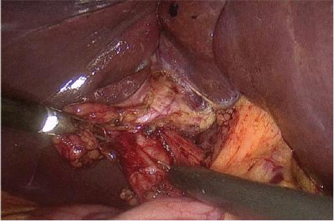
Figure 1: Coelioscopic picture showing a principal biliary duct without any presence of the gallbladder.
Clinical Case
The patient is a 35-year-old man with a history of hypertension, presenting with hepatic colic for four months, accompanied by vomiting, while maintaining a stable general condition. Biological tests were normal, showing no cholestasis, cytolysis, or leukocytosis. The CRP was also normal. An abdominal ultrasound revealed a sclerotic-atrophic gallbladder with a common bile duct measuring 10 mm in diameter. The diagnosis of symptomatic gallbladder lithiasis was made, and laparoscopic cholecystectomy was indicated. During surgery, the hepatic pedicle was well-defined, but the gallbladder could not be visualized in its anatomical position, even after examining various ectopic sites.Postoperative recovery was uncomplicated, and the patient was discharged after 24 hours. A magnetic resonance cholangiopancreatography was requested.
Discussion
Gallbladder agenesis is a rare embryological anomaly with an incidence of approximately 10 per 100,000 individuals. Its prevalence is estimated to be between 1 and 10 per 10,000 people in the general population, occurring more frequently in women [3]. This anomaly is due to a developmental defect of the caudal bud of the hepatic diverticulum, occurring around the mid-third week of gestation in the distal part of the foregut. It is noteworthy that gallbladder agenesis is often associated with cystic duct agenesis [4]. Most patients with this agenesis are asymptomatic. Occasionally, symptoms may include right upper quadrant pain, nausea, vomiting, intolerance to fatty foods, dyspepsia, choledocholithiasis, or dilated common bile duct compensating for the absence of the gallbladder by taking over the bile storage function [3]. The diagnosis of gallbladder agenesis is rarely established preoperatively. Ultrasounds performed to investigate hepatic colic often find a sclerotic-atrophic gallbladder, similar to our case. Currently, magnetic resonance cholangiopancreatography is considered the best examination for diagnosing gallbladder agenesis and can also locate a gallbladder in an ectopic site [5]. Discovery of gallbladder agenesis can occur either preoperatively or intraoperatively. In preoperative cases, patients present with biliary pain without the gallbladder being identified on ultrasound, or it is reported as sclerotic-atrophic. In such cases, additional exploration via magnetic resonance cholangiopancreatography is necessary to confirm the diagnosis with certainty, and surgical intervention is not indicated. For agenesis discovered intraoperatively in patients mistakenly operated on for symptomatic gallbladder lithiasis, the gallbladder should be sought in various possible ectopic sites. Conversion of the procedure is not beneficial, and the utility of cholangiography should be reconsidered due to the high risk of bile duct injury [5]. In all cases, a follow-up with magnetic resonance cholangiopancreatography is essential to confirm the diagnosis.
Conclusion
Gallbladder agenesis is a very rare condition resulting from an aberration in embryological development. It can be isolated or associated with other congenital anomalies. It should be considered when the gallbladder is not visualized on ultrasound, or when a sclerotic-atrophic appearance is noted, and confirmed with magnetic resonance cholangiopancreatography to avoid unnecessary and risky surgical interventions.
References
- Cavazos-García R. Agenesia de la vesícula biliar. Reporte de caso. Cirugia y Cirujanos. 2015; 83: 424-428.
- Chouchaine A, Fodha M, Abdelkefi MT, Helali K, Fodha M. Gallbladder agenesis: about three cases. Pan Afr Med J. 2017; 28: 114.
- Chouchaine et al. - Gallbladder agenesis about three cases.
- Hepatobiliary and Pancreatic Agenesis of the gall.pdf.
- Malde - 2010 - Gallbladder agenesis diagnosed intra-operatively .pdf.