Case Series
Austin J Surg. 2024; 11(5): 1340.
Malignant Retro Rectal Tumor: Two Compelling Case Reports
Dabbagh M*; El Mouhib S; Maazouz A; El Azzaoui I; Lamghari M; El Mouhib R; Hablaj H; Bouzroud M; Najih M; EL Kaoui H; Bouchentouf SM; Bounaim A; Moujahid M
Department of General Surgery, Mohammed V Military Hospital, Rabat, Morocco
*Corresponding author: Dabbagh M, Department of General Surgery, Mohammed V Military Hospital, Rabat, Morocco. Tel: 212668699621 Email: mahmoud.dabbagh@um5r.ac.ma
Received: November 21, 2024; Accepted: November 27, 2024; Published: December 04, 2024
Abstract
Background: Retrorectal tumors are a challenging pathology in terms of diagnosis and surgical management. They are usually asymptomatic and present a variety of histopathological presentations.
Case Presentation: We report two cases of malignant presacral tumors, a leiomyosarcoma, and a hemangiopericytoma. An asymptomatic rectal mass was discovered during a routine rectal digital examination in a 45-year-old male with a history of anal fistula. After an imaging confirmation, a complete surgical resection was conducted and pathological analysis revealed a leiomyosarcoma. The second patient was a 42-year-old female admitted for a 2-year persistent right buttock and low back pain, imaging detected a presacral tumor extending to S1 and S2 that was completely resected and followed with radiotherapy.
Conclusion: Our cases highlight Malignant retrorectal tumors’ rarity, diagnostic challenge, and surgical management.
Keywords: Retrorectal; Presacral; Tumor; Leiomyosarcoma; Hemangiopericytoma; MRI
Introduction
Retrorectal Tumors (RRT) are an extremely rare entity in adults. Most of the lesions are benign and asymptomatic but malignant cases account for 21%-50% of patients and therefore all require aggressive surgical management [1]. Most tumors can be detected during a digital rectal examination, and once identified, pelvic Magnetic Resonance Imaging (MRI) plays a vital role in surgical planning [1].
Case Presentation
Case 1
A 45-year-old male whose medical and surgical history included an anal fistula treated in 1997, and an inguinal hernia operated on in inguinal hernia in 2003. The patient reported an intermittent purulent anal discharge accompanied by tenesmus and false needs, accompanied by urinary symptoms combining pollakiuria and dysuria.
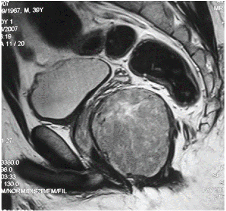
A routine digital rectal examination identified a painless, soft, and mobile mass laterally-located anal to the left of the gluteal fold, with a diameter of 4 to 5 cm, showing no local inflammatory signs. Rectoscopy showed a left-sided retro rectal mass pushing the rectum without invading the mucosa. Pelvic MRI revealed a tumoral mass with a size of 70mm/70mm, retro-prostatic lateral-rectal extending anteriorly into the rectum and anal canal and extending to the fourth sacral vertebra S4 (Figure 1).

Figure 1: MRI image revealed a presacral tumoral mass (blue arrow).
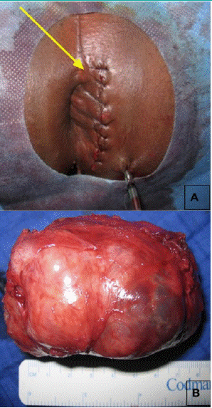
A perineal approach was conducted (Figure 2A) and revealed a deeply embedded mass in the left ischial fossa, extending towards the coccyx and the posterior rectum. Complete surgical resection was performed without effraction (Figure 2B), and pathological analysis confirmed a low-grade leiomyosarcoma. No adjuvant chemotherapy was proposed.

    Figure 2: Operative photos of the surgery approach (A, yellow arrow) and
the surgical specimen (B) of the first case.
The patient’s postoperative recovery was uneventful with no signs of recurrence after a final clinical check-up at 4 years.
Case 2
A 42-year-old female patient with a 2-year history of persistent right buttock and low back pain since initial admission to the department. for which she had received symptomatic treatment. The evolution was marked by progressive worsening of the pain, which took the form of S1-type lumbosciatica. The patient also reported paresthesias affecting the right leg.
Abdominal examination found no palpable mass however, a rectal examination revealed a painless mass located in the right lateral pelvic region, with its lower portion situated approximately 5 cm from the anal margin. The neurological assessment demonstrated an antalgic gait and discomfort during ambulation and a muscle atrophy throughout the right lower extremity, as well as a sensory deficit corresponding to the S1-S2 dermatome.
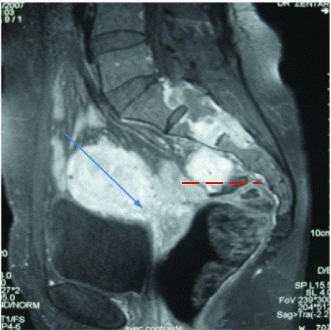
Pelvic MRI imaging demonstrated a presacral tumor extending into the right S1 and S2 vertebral areas (Figure 3). The lesion exhibited a poorly defined margin and heterogeneous appearance, with intermediate signal intensity on both T1 and T2 sequences, and demonstrated intense and rapid enhancement following the administration of gadolinium.

    Figure 3: Pelvic MRI in sagittal section showing a sacral tumoral mass (red
arrow) with an intra-pelvic retrorectal extension (blue arrow).
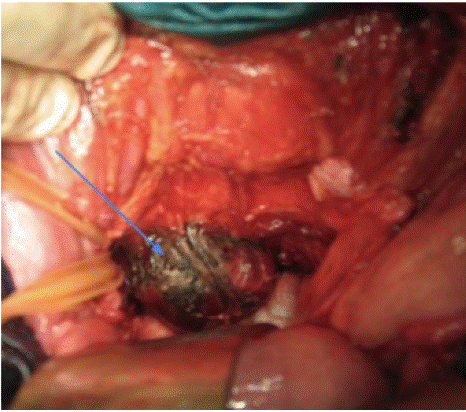
Following a multidisciplinary case review, a transabdominal surgical exploration confirmed the presence of a presacral tumor invading the middle sacral artery and presacral fascia (Figure 4). Surgical resection was performed, removing the tumor mass and surrounding the affected bone. Pathological analysis revealed a hemangiopericytoma. As an adjuvant therapy, the patient received 45 Gy of radiotherapy.

    Figure 4: Operative photo, of the second case, after dissection of the retrorectal
space, showing an encapsulated tumor mass (blue arrow).
Postoperative monitoring revealed no abnormalities with complete relief of the antalgic gait. At the final 4-year follow-up, the patient exhibited no evidence of recurrence.
Discussion
Retrorectal tumors occupy the presacral region, which is the anatomical space between the posterior rectal wall and the sacrum. It extends from the peritoneal reflection above to Waldeyer's fascia below which lies 3 to 5 cm proximal to the anorectal junction [2]. Their annual incidence in the general population is estimated at between 0.0025% and 0.014%, with a female predominance and an average age of 30 years [3-6].
Although they are uncommon, most general surgeons can expect to encounter at least one patient with a retrorectal tumor during their careers. The most common symptom of retrorectal masses is vague pain in the perineal area, which can also present with chronic constipation, incontinence, or sexual dysfunction. The presence of a midline sinus or fistula may indicate an underlying retro rectal mass, though these tumors are often silent and discovered incidentally during a routine digital rectal examination [1]. The histopathological varieties of retrorectal result from the multiple embryological remnants [7].
Imaging via transrectal or suprapubic ultrasonography verifies the cystic character and retrorectal position of the lesion. Ct-scan and MRI corroborate the location of the lesion, and its locoregional extension [8]. Rectoscopy and fistulography may supplement the investigative process [8].
Retrorectal masses are typically classified into five categories: congenital, inflammatory, neurogenic, osseous, and miscellaneous. Almost 50% of presacral tumors are malignant, and the most common malignancy is sacrococcygeal chordoma. Leiomyosarcoma and hemangiopericytoma are malignant types of miscellaneous tumors and account for 10 to 25% of all retrorectal tumors [9].
Excision of a retrorectal tumor is essential, even in asymptomatic patients. A wide en bloc resection must be performed to improve prognosis and survival and decrease the risk of recurrence. A multidisciplinary case review involving colorectal surgeons, neurosurgeons, orthopedics, and plastic surgeons is essential to decide on the operative approach, which depends on the extension of the tumor and the sacral involvement [1].
The prognosis of the presacral tumors depends on the nature of the tumor and the successful surgical resection with clear margins achieved in the first operation. Malignant tumors can metastasize to the liver, lung, and brain, which are all associated with significantly worse prognosis [1].
Conclusion
Malignant retrorectal tumors are a rare entity. Diagnosis begins with a digital rectal examination followed by a pelvic MRI. The surgical approach requires a multidisciplinary case review and depends on the sacral limits and the size of the lesion, but the posterior route is the preferred method. The prognosis depends on the histopathological of the tumor and the « en bloc » resection.
Author Statements
Conflicts of Interest Statement
The author has no conflicts of interest to declare. The author alone is responsible for the content of this manuscript.
Consent
Written informed consent was obtained from the patient for publication of this case report and any accompanying images.
Ethical Approval
Ethical approval is not required at our institution for an anonymous case report.
Conflicts of Interest Statement
The author has no conflict of interest to report. The author is entirely liable for the manuscript’s content.
Informed Consent
The patients provided written informed consent for the publishing of these case reports and any related pictures.
References
- D Lev-Chelouche, Gutman M, Goldman G, Even-Sapir E, Meller I, Issakov J, et al. Presacral tumors: a practical classification and treatment of a unique and heterogeneous group of diseases Surgery. 2003; 133: 473-8.
- Mirilas P, Skandalakis JE. Surgical anatomy of the retroperitoneal spaces part II: the architecture of the retroperitoneal space. Am Surg. 2010; 76: 33–42.
- S Belkacem, et al. Le syndrome de Currarino. Feuill Radiol. 2010.
- G Chêne, et al. Tératome bénin mature pré-sacré et formations kystiques vestigiales rétro-rectales chez l’adulte J Chir (Paris). 2006.
- J Ghosh, et al. Presacral tumors in adults Surg J R Coll Surg Edinb Irel. 2007.
- Hobson KG, Ghaemmaghami V, Roe JP, Goodnight JE, Khatri VP. Tumors of the retrorectal space. Dis Colon Rectum. 2005; 48: 1964–1974.
- Hassan I, Wietfeldt ED. Presacral tumors: diagnosis and management. Clin Colon Rectal Surg. 2009; 22: 84–93.
- Liessi G, Cesari S, Pavanello M, Butini R. Tailgut cysts: CT and MR findings. Abdom Imaging. 1995; 20: 256–8.
- Balci B, Yildiz A, Leventoglu S, Mentes B. Retrorectal tumors: A challenge for the surgeons. 2021; 13: 1327–1337.