
Special Article: Plastic Surgery
Austin J Surg. 2024; 11(6): 1342.
Patient with Deeply Burned Head Due to Electrical Injury: Case Report
Lengyel P¹*; Frišman E¹; Eliáš E¹; Hyseniová S¹; Demčák T¹; Michalíková M²
¹Burns and Reconstructive Plast. Surg. Clinic AGEL Hospital Košice šaca, Slovak Republic
²Department of Biomedical Engineering and Measurement, Mechanical Engineering Faculty Technical University of Košice, Slovak Republic
*Corresponding author: Peter Lengyel, PhD, Burns and Reconstructive Plastic Surgery Clinic AGEL Hospital Košice šaca, Lučna 57, 040 15 Košice, Slovak Republic. Tel: +421911500551 Email: peter.lengyel@nke.agel.sk
Received: November 18, 2024; Accepted: December 09, 2024; Published: December 16, 2024
Abstract
61-year-old man suffered an electric shock at a low voltage alternant current while repairing electrical wiring at work. The patient was admitted to regional hospital and after stabilizing his healthy status and regaining consciousness, he was transferred to our workplace.
On admission, the patient was conscious, on the head there were an entrance and exit wounds in left frontotemporal and frontoparietal regions of IIIIVth degree electric current burns, a sutured linear wound. Necrectomy of deep burns by electric current on the head in the frontoparietal area on the left side as well as the frontotemporal area on the left, including part of the left upper eyelid and eyebrow, was performed under general anesthesia. Tangential removal of pathologically changed parts of the frontal bone, the lateral edge of the orbit, M.Orbicularis OC.l.Sin, m. temporalis was performed. The defect of soft tissue in the left temporoparietal area was solved by reconstructive surgery. rotational: 1 fasciocutaneous and 1 musclecutaneous flap. Defect in the left frontoparietal area was solved by a rotational fasciocutaneous flap from the biparietal area.
In postoperative course necrotic tip of the flap near left lateral eyedge was solved by local plasty respectivelly skin grafting. After this the healing of the wounds was continued and finished and the patient was discharged from our workplace after 20 days of hospitalisation.
Keywords: Electric shock; Burns
Introduction
Electrical burns represent in our workplace in average 3-4 % of all burn injuries. They are rare, but if occur they destroy large amount of tissues and organs. The surgical treatment of these burn wounds must be tailored according the extent, localisation and depth with goals to achieve maximum functionality and aesthetic result.
Case Presentation
61-year-old man suffered an electric shock from direct contact at a low voltage of 380 V alternant current while repairing electrical wiring at work. He was admitted to the Department of Intensive Medicine in Banská Bystrica unconscious. After stabilizing his condition of general status and regaining consciousness, he was treated at the trauma center of that hospital, from where he was transferred after telephonical consultation to our workplace.
On admission, the patient was conscious, breathing was spontaneous, neurologically normal, heart rate regular, normotensive. On the head there were an entrance and exit wounds in left frontotemporal and frontoparietal regions of a III-IVth degree electric current burn, a sutured linear wound, the area around the necrotic burns was inflamed (Figure 1).
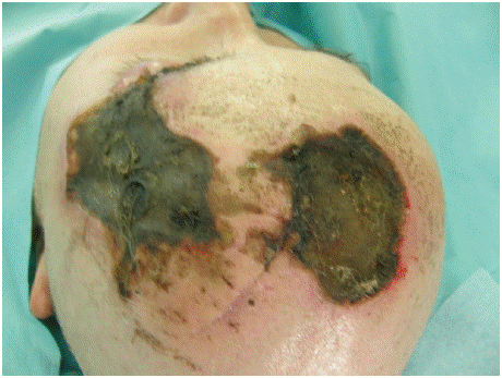
Figure 1:
We performed neurological examination, ophthalmological examination, x-ray examination of the skull and internal preoperative examination. The results from these examinations were negative, without of need some neurological of ophthalmological therapy. On the x- ray examination with findings of missing soft tissue in the deeply burned regions of the head and calcificates in epiphysis. Due to deep burn wound we indicated surgery.
Necrectomy of deep burns by electric current on the head in the frontoparietal area on the left side as well as the frontotemporal area on the left, including part of the left upper eyelid and eyebrow, was performed under general anesthesia. Tangential removal of pathologically changed parts of the frontal bone, the lateral edge of the orbit, M.Orbicularis oculi l.Sin., m. temporalis was performed (Figure 2 & 3).
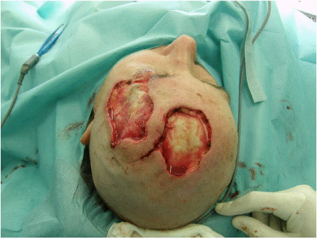
Figure 2:
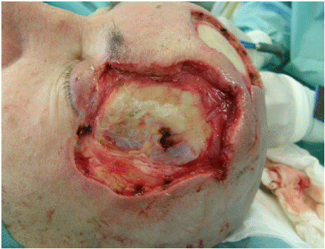
Figure 3:
After thorough hemostasis, the defect of soft tissue in the left temporoparietal area was solved by reconstructive surgery. rotational: 1 fasciocutaneous and 1 musclecutaneous flaps. Defect in the left frontoparietal area solved by a rotational large fasciocutaneous flap from the biparietal area (Figure 4-6). Donor sites with exposed galea aponeurotica were covered by split thickness skin autografts from the left thigh (Figure 7 & 8).
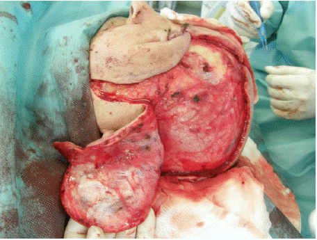
Figure 4:
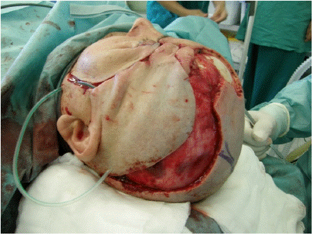
Figure 5:
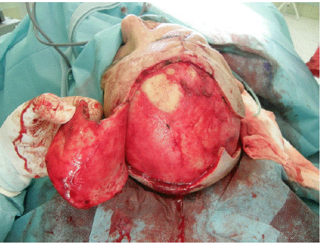
Figure 6:
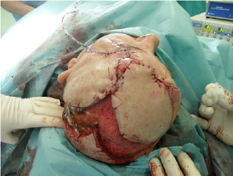
Figure 7:
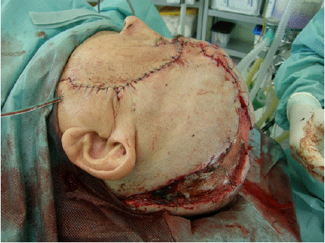
Figure 8:
In postoperative course necrotic tip of the flap near left lateral edge of eye as the small /1 cm/ complication was solved by local plasty respectivelly skin grafting. After this the healing of the wounds was continued and the patient was discharged from our workplace after 20 days of hospitalisation. After 2 months of treatment the patient was complete healed (Figure 9 & 10).
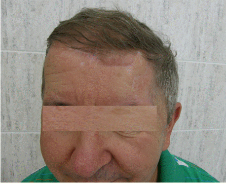
Figure 9:
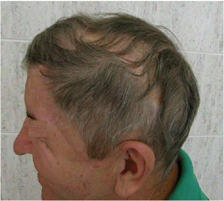
Figure 10:
Discussion
In our workplace electrical injury was present in 3,14 % of all treated hospitalised burn patients in last 20 years. This is in accordance with the data of most other studies from burn centers in different parts of the world [1,2], but in some the incidence was higher (9.1 and 16.4 % ) [3,4]. However, the true incidence in the general population can be considerably higher because minor accident are not referred to medical institutions and/or are treated in local outpatient clinics. The outcome of high voltage injuries even in profesionals is very dubious and often lead to invalidisation of worker as reported [5]. The danger of electrical injuries as compared to burns of other causes lies in the possibility of hidden damage of internal tissues and organs, especially the heart. We did not encountered with such a type of contact electrical injury to the head with very close lying to each other of the entrance and the exit wounds yet.
Conclusion
The treatment to electrical injury is based on multidisciplinary cooperation to save the patient, allow him recovery with minimum mutilation and maintain the functions [6]. The long time must be given for special rehabilitation and splinting as a prevention of scar and joint contractures. From social point of view is very important the psychological support to patient from the family backgrorund and friends. These factors can help the patient find the way back to life.
Author Statements
Acknowledgments
This work was supported by the Slovak Research and Development Agency under the contract No. APVV-22-0340.
References
- Luz DP, Millan LS, Alessi MS, Uguetto WF, Paggiaro A, Gomez DS, et al. Electrical burns: a retrospective analysis across a 5-year period. Burns. 2009; 35: 1015-9.
- Kym D, Seo DK, Hur GY, Lee JW. Epidemiology of electrical injury: Differences between low- and high-voltage electrical injuries during a 7-year study period in South Korea. Scand J Surg. 2015; 104: 108-14.
- Haberal M, Kaynaroglu V, Oner, Golay K, Bayraktar U, Bilgin N. Epidemiology of electrical burns in our centre. Ann Mediteran Burns Club. 1989; 2: 14-16.
- Vierhapper MF, Lumenta DB, Beck H, Keck M, Kamolz LP, Frey M. Electrical injury: a long-term analysis with review of regional differences. Ann Plast Surg. 2011; 66: 43-6.
- Piotrowski A, Fillet AM, Perez P, Walkowiak P, Simon D, Corniere MJ, et al. Outcome of occupational electrical injuries among French electric company workers: a retrospective report of 311 cases, 1996-2005. Burns. 2014; 40: 480-8.
- Lipový B, Rihová H, Kaloudová Y, Suchánek I, Gregorová N, Hokynková A, et al. The importance of a multidisciplinary approach in the treatment of mutilating electrical injury: a case study. Acta Chir Plast 2010 Vol.52; 61-4.