Clinical Image
Austin J Surg. 2025; 12(1): 1348.
Surprising Image of a Meckel’s Diverticulum and an Abscessed Appendix
Moujahid B*, Bahi A, Zouhry I, Abdelilah H, Rahali A, Ali AA
Mohammed V Military Teaching Hospital, Department of General Surgery, Rabat, Morocco
*Corresponding author: Badr Moujahid, Department of General Surgery, Mohammed V Military Teaching Hospital, Rabat, Morocco Email: badrmouj96@gmail.com
Received: February 17, 2025; Accepted: March 11, 2025; Published: March 12, 2025;
Abstract
Meckel’s diverticulum (MD), a congenital abnormality resulting from incomplete obliteration of the proximal omphalomesenteric duct during the 7th week of gestation (making it the most prevalent congenital malformation of the gastrointestinal tract) [1].
shares overlapping clinical features with acute appendicitis, a common surgical emergency, making simultaneous presentation diagnostically challenging. Given its diverse clinical manifestations, MD can be challenging to diagnose.
Symptomatic MD commonly presents with bleeding, followed by intestinal obstruction, diverticulitis, intussusception, and, rarely, neoplasm or perforation [2].
In this context, we present an image of MD and an abcessed appendix diagnosed during laparotomy and confirmed postoperatively.
Keywords: Diverticulum; Appendix; Appendectomy; Diverticulectomy
Intraoperative Image
Meckel's diverticulum (MD) has a prevalence of 1 to 2% in the general population. It is most often discovered incidentally during abdominal surgery [3].
In adults, the necessity for preventive resection of an asymptomatic MD to prevent acute complications or degeneration remains debated [4,5].
A 53-year-old female patient with a history of hypothyroidism was admitted for suspected appendicitis. Her symptoms began 20 days earlier with pain in the right iliac fossa, along with nausea and vomiting in a febrile context.
The abdominal exam revealed tenderness in the right iliac fossa with psoitis and a positive Rovsing sign. The infectious workup showed elevated markers, and ultrasound revealed a swollen, laterocaecal appendix complicated by an appendiceal abscess.
Initial management included fluid resuscitation, analgesia, and antibiotic prophylaxis. Subsequently, a laparotomy was performed, during which peritoneal effusion was aspirated. Intraoperative exploration revealed two distinct structures:
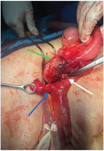
1. The MD originating from the last ileal loop (Figure 1, blue arrow), attached to the cecal base (Figure 1, white arrow).

    Figure 1: Intraoperative image showing a Meckel’s diverticulum (blue arrow)
and an abscessed appendix (green arrow).
2. The appendix, which was latero-caecal, swollen, and abscessed (Figure 1, green arrow).
We proceeded with an appendectomy and diverticulectomy while preserving the carrying ileal loop. Histological analysis of the two surgical specimens confirmed the diagnosis of Meckel's diverticulum and an abscessed appendix.
Postoperative recovery was marked by early rehabilitation and the resumption of digestive transit on the 3rd postoperative day.
References
- Ihedioha U, Panteleimonitis S, Patel M, Duncan A, Finch G. An unusual presentation of Meckel’s diverticulum. J Surg Case Rep. 2012; 2012: 4.
- Ding Y, Zhou Y, Ji Z, Zhang J, Wang Q. Laparoscopic Management of Perforated Meckel’s Diverticulum in Adults. Int J Med Sci. 2012; 9: 243–247.
- Zani A, Eaton S, Rees CM, Pierro A. Incidentally Detected Meckel Diverticulum: To Resect or Not to Resect? Ann Surg. 2008; 247: 276.
- Park JJ, Wolff BG, Tollefson MK, Walsh EE, Larson DR. Meckel Diverticulum: The Mayo Clinic Experience With 1476 Patients (1950–2002). Ann Surg. 2005; 241: 529.
- Ruscher KA, Fisher JN, Hughes CD, Neff S, Lerer TJ, Hight DW, et al. National trends in the surgical management of Meckel’s diverticulum. J Pediatr Surg. 2011; 46: 893–896.