Case Report
Austin J Surg. 2025; 12(2): 1350.
Hypercondylism: A Case Report
Boukhlouf O#*, Benwadih S#, Harmali K#, Dani B# and Boulaadas M#
Maxillofacial Surgery and Stomatology Departement, Rabat, Morocco
#Specialties Hospital, Rabat, Morocco
*Corresponding author: Oumaima Boukhlouf, Maxillofacial Surgery and Stomatology Departement, Rabat, Morocco Email: boukhloufoumaima@gmail.com
Received: June 19, 2025 Accepted: July 18, 2025 Published: July 21, 2025
Introduction
Hypercondylism is a rare and progressive pathology characterized by hypertrophy of the mandibular condyle, often associated with deformities of the mandible, occlusion and temporomandibular joint.
It results from dysregulation of prechondroblastic cells in the condylar cartilage. This condition can lead to facial asymmetries, joint pain and dental malocclusion, affecting the patient's function and aesthetics [1-5].
The mainstay of treatment is condylectomy, often accompanied by orthodontic management, aimed at correcting these anomalies and restoring optimal occlusal and aesthetic function [3,4].
Early identification of the pathology and surgical planning are essential to avoid long-term complications.
Case Presentation
Mrs. Y.S., a 16-year-old female patient, presented with temporomandibular joint pain, aggravated during mastication and mouth opening, associated with progressive mandibular laterodeviation.
Clinical examination revealed facial asymmetry, with laterodeviation of the mandible and chin to the left, jugal elongation on the right side compared with the left, and prominence of the mandible on the right.
Palpation of the TMJs revealed a swelling on the right, with a cracking sound when the jaw was moved.
Endobuccal examination revealed a tilted occlusal plane on the left, with deviation of the lower midline to the left, and a partial crossbite on the right, with limited mouth opening.
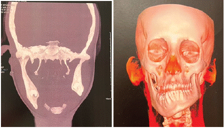
Preoperative imaging, including dental panoramic and computed tomography (CT), showed vertical and horizontal hypertrophy of the right mandibular condyle, with deviation of the lower midline and asymmetry of the ratio between the two arches, confirming a diagnosis of hypercondylism (Figure 1).

    Figure 1: Frontal section and 3D reconstruction of a CT scan of the facial
mass, showing on the right a hypertrophied mandibular condyle with vertical
and horizontal elongation of the ramus and horizontal branch, pinching of
the joint space with left mandibular latero-deviation.
Mrs. Y.S. underwent a right condylectomy under general anesthesia, using a pre-tracheal approach to expose the TMJ and condyle, with resection of the condyle using a surgical burr, as well as excess bone and remodeling of the residual condyle surface to ensure proper TMJ function.
The post-operative course was uncomplicated, the patient regained a satisfactory mouth opening, and was referred to the orthodontist for further treatment.
Discussion
Mandibular hypercondylism is a rare but significant pathology characterized by progressive hypertrophy of the mandibular condyle. Although relatively uncommon, this condition has a major impact on facial morphology, occlusion and temporomandibular joint (TMJ) function. Historically, the condition was first described in 1836 by Robert Adams in a clinical case of progressive facial deformity, laying the foundations for the early understanding of hypercondylism [1]. It occurs mainly after puberty, a period when condyle growth is more marked. Symptoms often appear between the ages of 20 and 40, although some cases can be diagnosed as early as adolescence, due to excessive growth of the condyle [3].
The condition appears to be more prevalent in women, with a sex ratio favouring female patients, suggesting a possible role for hormonal factors in the genesis of hypercondylism [6].
The exact pathophysiology remains poorly understood, but is generally attributed to dysregulation of prechondroblastic cells in the condylar cartilage, leading to abnormal cell proliferation and progressive hypertrophy of the condyle, thus altering the morphology of the mandible [4].
This condition can affect the morphology of the mandible and, indirectly, that of the maxilla, alter dental occlusion, or lead to dysfunctional joint disorders such as pain and crackling in the TMJ [5].
There are several types of hypercondylism, classified as vertical, horizontal and mixed, depending on the orientation and extent of the condylar deformity.
Vertical forms result in an increase in condylar height, altering facial height, while horizontal forms cause lateral expansion of the condyle, resulting in deviation of the mandible. Mixed forms combine these two phenomena, creating more complex deformities [6].
Clinical symptoms include facial asymmetries, malocclusion, joint pain and crackling in the TMJ, as well as masticatory dysfunction due to mandibular deviation [5]. Unilateral condylar involvement is often responsible for jaw deviation, creating visible asymmetries in the patient's profile [7].
Diagnosis is based on clinical examination and complementary tests such as bone scan, computed tomography (CT) and MRI.
Orthopantomogram, CT and MRI are crucial for visualizing bony and cartilaginous changes in the condyle, facilitating diagnosis and surgical treatment planning [1].
Bone scintigraphy can be used to measure osteoblastic activity in the condyle and monitor the progression of the pathology over time, although it is not diagnostic in itself [2].
Differential diagnosis should include similar conditions such as osteochondroma, TMJ osteoarthritis, or other mandibular condyle malformations [8]. Osteochondromas present an exostotic growth of the condyle and must be distinguished from hypercondylism, as therapeutic approaches differ according to diagnosis.
The treatment of hypercondylism is mainly surgical, with condylelectomy, which involves removal of the enlarged condyle, being the treatment of choice in the majority of cases [4]. The aim of this procedure is to halt the progression of the deformity and alleviate functional and aesthetic symptoms.
After condylectomy, orthodontic treatment is often required to correct the malocclusion and restore normal occlusal function.
In some cases, orthognathic procedures may be required to realign the mandible and correct facial asymmetries [8].
Post-operative complications are rare, but may include infection, recurrence of deformity, and functional disorders of the TMJ.
Long-term post-operative follow-up is essential to assess the stability of results and prevent future alterations in TMJ function [5].
Post-operative follow-up is also important to ensure stable functional and aesthetic results. Regular check-ups, including X-rays and clinical consultations, are necessary to detect any recurrences or complications. Orthodontic follow-up is crucial to ensure stable, functional occlusion [6].
Conclusion
Hypercondylitis is a rare but serious pathology that requires early diagnosis to prevent functional and aesthetic deformities. Complementary examinations are essential to establish a precise diagnosis and guide the treatment plan. Surgical management is necessary to halt the progression of the pathology, while rigorous post-intervention follow-up ensures long-term stability of functional and aesthetic results.
References
- Abaouz A, Benider H, Amellouk S, Bennani H, El Guensi A. Place de la TEMP/TDM dans le diagnostic des hypercondylies mandibulaires: à propos de 5 cas. Médecine Nucléaire. 2022; 46: 91-92.
- Lecocq G, Ferri J, Doual-Bisser A. L’hypercondylie: apport de la scintigraphie du diagnostic au traitement. L’Orthodontie Française. 2005; 76: 165-173.
- Nicot R, Raoul G, Ferri J. Hypercondylies. Hypercondylies. 2019.
- Ferri J, Raoul G, Potier J, Nicot R. Articulation temporomandibulaire (ATM): hypercondylie et condylectomie. Revue de Stomatologie, de Chirurgie Maxillo-faciale et de Chirurgie Orale. 2016; 117: 259-265.
- Rodrigues DB & Castro V. Condylar hyperplasia of the temporomandibular joint: types, treatment, and surgical implications. Oral and Maxillofacial Surgery Clinics. 2015; 27: 155-167.
- Wolford LM, Movahed R, Perez DE. A classification system for conditions causing condylar hyperplasia. J Oral Maxillofac Surg. 2014; 72: 567–595.
- Jones RH, Tier GA. Correction of facial asymmetry as a result of unilateral condylar hyperplasia. J Oral Maxillofac Surg. 2012; 70: 1413–1425.
- Vezeau PJ, Fridrich KL, Vincent SD. Osteochondroma of the mandibular condyle: literature review and report of two atypical cases. J Oral Maxillofac Surg. 1995; 53: 954–963.