
Case Report
Austin J Surg. 2025; 12(2): 1353.
Uncommon Bony Fusion: A Rare Case of Pseudoankylosis Between the Coronoid Process and Zygomatic Bone
André M Eckardt¹*, K Hakki Karagozoglu² and Gary Parker³
¹Department of Oral and Maxillofacial Surgery, Hannover Medical School, 30625 Hannover, Germany
²Department of Oral and Maxillofacial Surgery/ Oral Pathology, Amsterdam UMC, Vrije Universiteit Amsterdam, The Netherlands
³Volunteer Maxillofacial, Head and Neck Surgeon, M/V The Global Mercy, The Mercy Ships, West Africa
*Corresponding author: André M. Eckardt, Department of Oral and Maxillofacial Surgery, Hannover Medical School, Carl-Neuberg-Strasse 1, 30625 Hannover, Germany Email: prof.eckardt@gmx.net
Received: July 30, 2025 Accepted: August 15, 2025 Published: August 19, 2025
Abstract
Extraarticular pathologies of various causes associated with restriction of mandibular movement and mouth opening are summarized under the term pseudoankylosis. Pseudoankylosis may be of myogenic, osteogenic, neurogenic, or psychogenic origin. Treatment is directed to relieve the restricted mouth opening and restore adequate masticatory function. Our clinical case report of a bony fusion between the zygomatic bone and the coronoid process documents the need for precise clinical and radiological diagnosis. Plain radiographs are usually of limited diagnostic advantage. Therefore, a computed tomography was employed for necessary diagnostic information. Once the diagnosis has been made, surgical resection is the treatment of choice. Our patient was surgically managed with intra- / extraoral coronoidectomy. Microscopic examination of the specimen revealed dense lamellar bone with bone marrow space and trabeculae, allowing the diagnosis of osteoma of the coronoid process. Intensive postoperative physiotherapy is advised to gain adequate mouth opening and jaw function.
Keywords: Coronoid process; Pseudoankylosis; Osteoma; Coronoidectomy
Introduction
By definition, pseudoankylosis of the temporomandibular joint (TMJ) is a persistent restriction of mandibular mobility caused by various extraarticular pathologies [1,2]. This extraarticular TMJ affliction can be of myogenic, osteogenic, neurogenic, or psychogenic origin. Compared to the true form of ankylosis, pseudoankylosis is much less frequent. Pain symptoms are usually not reported. However, some degree of facial deformity can occur.
The extent of restricted mandibular mobility in pseudoankylosis can range from partial to complete restriction in relation to the amount of involved fibrous tissues. Lateral and protrusive excursions of the mandible are restricted in most cases.
Complete restriction of mandibular mobility indicates a bony union between the coronoid process and the zygomatic arch caused by any bony pathology [3]. Complete bony union in the coronoidzygomatic arch region, long duration of restricted mobility with no pain symptoms can be caused by a slowly growing osteoma of the coronoid process [4,5]. In such a case of pseudoankylosis, the use of CT scans is strongly advised [6]. Plain radiography such as panoramic radiography can also be used to detect morphological changes of the coronoid process, but the diagnostic advantage is limited if any surgical intervention is planned [7].
Osteoma of the coronoid process is a rare, slowly growing benign tumor that can cause pseudoankylosis. The first case of compact osteoma of the coronoid process was reported by Lewars in 1959 [8]. To our knowledge, only 12 cases of this entity, including ours, have been reported in the literature [9-14]. As benign bone-forming tumors, osteomas occur predominantly in the craniofacial region, with peripheral osteoma as the most frequently described sub-type.
Other bone-forming pathologies such as osteochondroma or condylar hyperplasia have to be included in the differential diagnosis [15-17]. Therefore, histopathological evaluation is a key tool to establish a correct diagnosis.
Surgical management of osteoma of the coronoid process depends on several patient-related factors, such as the amount of functional restriction of the mandible, occlusal discrepancy, and possible impact on psychosocial status. Surgical planning is also affected by the duration of restricted mobility of the mandible [7,18].
This paper describes the surgical management of a rare case of osteoma of the coronoid process and will highlight some aspects regarding the differential diagnosis, and surgical management of pseudoankylosis of the temporomandibular joint.
Case Presentation
A 43-year-old female patient presented in Freetown/Sierra Leone on board the hospital ship “Global Mercy” of the American NGO “Mercy Ships” with a suspected diagnosis of left temporomandibular joint ankylosis. A clinical examination revealed a restricted mouth opening of no more than 5 mm and some minor facial asymmetry of the left zygoma (Figure 1a). On inquiry, it was reported that the mouth-opening restriction had been present for about ten years. The enlargement of the left coronoid process but was inconclusive. Further CT diagnostics revealed a well-defined radiopaque lesion between the left zygomatic bone and the coronoid process caused by a large bony tumor instead of a true left temporomandibular joint ankylosis (Figure 1b). Based on clinical presentation and preoperative radiologic features, a provisional diagnosis of coronoid osteoma was established.
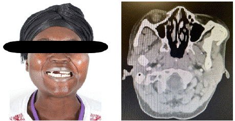
    Figure 1a, b: Preoperative frontal view shows minor facial asymmetry of
the left zygoma and restricted mouth opening (1a). Axial CT scan showing
bony mass extending from the left coronoid process towards the left
zygoma suggesting bony fusion (1b).
The surgical procedure planned was left coronoidectomy under general anesthesia. Therapeutically, bony resection of the osteoma was performed through a combined intra- and extraoral approach (Figure 2). Because of the size of the osteoma and bony fusion with the inner aspect of the left zygoma, a single intraoral approach was not sufficient. Immediately after resection of the osteoma and left coronoid, a spontaneous mouth opening of 25 mm could be achieved intraoperatively (Figure 3). The intraoperative bony defect of the outer cortical bone plate of the zygoma was reconstructed using a titanium mesh (Figure 4a,b). The suspected diagnosis of “osteoma” was finally confirmed by histological diagnosis. After an uneventful postoperative course with no facial nerve deficits, the patient was discharged on the 8th postoperative day with stable wound conditions, no occlusal discrepancy, and a constant mouth opening of 30 mm. The patient was advised to continue with physiotherapy using regular wooden tongue blades and regular follow-up.
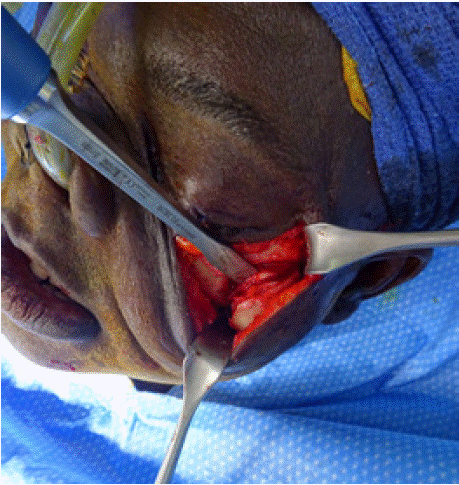
    Figure 2: Surgical access to the left zygoma using subciliar incision with
lateral extension.
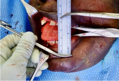
    Figure 3: Immediate intraoperative mouth opening after left coronoidectomy
using combined intra-/extraoral approach.
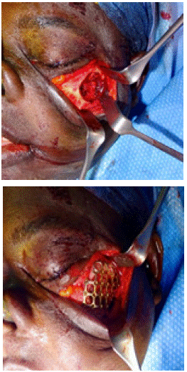
    Figure 4a, b: Bony mass results in a limited bony defect of the left outer
cortical plate of the zygoma (4a). A titanium mesh was used to cover the
bony defect of the left zygoma (4b).
Discussion
Pseudoankylosis is a clinical diagnosis characterized by restricted mandibular mobility with no pain. Because of minimal clinical symptoms, a correct clinical diagnosis can be challenging. Compared to the true form of TMJ ankylosis, pseudoankylosis is caused by multiple extraarticular pathologies and other conditions, of which maxillofacial trauma, infections, and various iatrogenic causes have been most commonly reported [19].
If pseudoankylosis is caused by any bone pathology, we need to establish a differential diagnosis between idiopathic hypertrophic coronoid, Jacob’s disease, or benign bone tumors such as osteoma or osteochondroma [1,16,17,20].
Panoramic radiography can be useful to detect elongated coronoids, a condylar mass, or the true form of TMJ ankylosis. However, if there is suspicion of a coronoid mass and bony fusion with the zygoma, a CT or CBCT scan is preferable [6,7]. A CT scan allows differentiating pseudoankylosis from true TMJ ankylosis and it is useful for any surgical planning [7]. In our case, the CT scan showed a bony fusion of a left coronoid bony mass with the zygoma.
If bony fusion is present, any conservative intervention will be ineffective in relieving symptoms and there is general agreement that a surgical approach is the treatment of choice. With restricted mouth opening and interincisal distance of 5mm in our patient, we decided to approach pseudoankylosis surgically. Based on CT findings with a bony mass of 3x3cm and bony fusion of the left coronoid with the inner surface of the zygoma, a combined intra- and extraoral surgical approach was chosen. The surgical approach to address pseudoankylosis should always be case-specific and depends on the size of the bony mass and any accompanying facial deformity [7]. Although most authors recommend an intraoral approach because of less morbidity [17], our combined extra- and intraoral approach was very effective in performing left coronoidectomy and complete removal of the osteoma. A subciliar incision with lateral extension gave adequate access to the zygomatic area, and resulted in an excellent cosmetic outcome with no facial injury or ectropium of the lower eyelid. Immediate release of the pseudoankylosis allows for an intraoperative mouth opening of 25mm.
The immediate postoperative period is the most critical time for the successful relief of TMJ pseudoankylosis. There is general agreement that immediate postoperative intensive physiotherapy is required to maintain the mobility obtained during surgery.
The authors emphasize the need for early postoperative physiotherapy to achieve a satisfactory and stable mandibular function and to avoid a relapse.
References
- Kumar S, Charllu AP. Extraarticular joint ankylosis: a rare presentation. BMJ Case Rep. 2021; 14: e244616.
- Baraldi CE, Martins GL, Puricelli E. Pseudoankylosis of the temporomandibular joint caused by zygomatic malformation. Int J Oral Maxillofac Surg. 2010; 39: 729-732.
- Lee SM, Baek JA, Kim Y. Ankylosis of the coronoid process to the zygomatic bone: A case report and review of the literature. J Oral Maxillofac Surg. 2019; 77: 1230.
- Collini M, Bozzetti A, Ravasini G. Limitation of mouth opening due to osteoma of the coronoid process. Riv Odontostomatol Implantoprotesi. 1983; 5: 23-24.
- Iwai T, Izumi T, Baba J, Maegawa J, Mitsudo K, Tohnai I. Peripheral osteoma of the mandibular notch: report of a case. Iran J Radiol. 2013; 10: 74-76.
- de Bont LG, van der Kuijl B, Stegenga B, et al. Computed tomography in the differential diagnosis of temporomandibular joint disorders. Int J Oral Maxillofac Surg. 1993; 22: 200-209.
- Spijkervet FKL, de Bont LGM, Boering G. Management of pseudoankylosis of the temporomandibular joint: Report of a case. J Oral Maxillofac Surg. 1994; 52: 1211-1217.
- Lewars PH. Osteoma of the mandible. Br J Plast Surg. 1959; 12: 277-283.
- Chen YK, Lin LM, Lin CC. Osteoma of the mandibular coronoid process. Report of a case. Int J Oral Maxillofac Surg. 1998; 27: 222-223.
- Plezia RA. Osteoma of the coronoid process. Oral Surg Oral Med Oral Pathol. 1984; 57: 111.
- Wesley RK, Cullen CL, Bloom WS. Gardner’s syndrome with bilateral osteomas of the coronoid process resulting in limited opening. Pediatr Dent. 1987; 49: 753-756.
- Ord RA, Rennie JS, MacDonald DG, Moos KE. Cancellous osteoma of the coronoid process: Report of a case. Br J Oral Surg. 1983; 21: 49-55.
- Kurita K, Kawai T, Ikeda N, Kameyama Y. Cancellous osteoma of the coronoid process: report of a case. J Oral Maxillofac Surg. 1991; 49: 753-756.
- da Costa Araujo FA, Melo Barbalho JC, de Farias ON, de Vasconcellos RJH, do Egito Vasconcelos BC. Pseudo-ankylosis caused by osteoma of the coronoid process. Ann Maxillofac Surg. 2014; 4: 208-210.
- Amary F, Flanagan AM, O’Donnell P. Benign Bone-forming Tumors. Surg Path Clin. 2021; 14: 549-565.
- D’Ambrosio N, Kellman RM, Karimi S. Osteochondroma of the coronoid process (Jacob’s disease): an unusual cause of restricted jaw motion. Am J Otolaryngol Head Neck Med Surg. 2011; 32: 52-54.
- Mohan Choontharu M, Buch SA, Babu SA, Castellino RL, Rao S, Rao K. A rare clinical presentation of an osteochondroma of coronoid process of mandible. J Dent Shiraz Univ Med Sci. 2018; 19: 325-330.
- Saikrishna D, Das A, Jha C. Management of a case of osteoma of coronoid: A rare case report. Natl J Maxillofac Surg. 2021; 12: 276-279.
- Ostrovsky MK, Lownie JF. Zygomatico-coronoid ankylosis. J Oral Surg. 1977; 35: 752-754.
- Larrea-Oyarbide N, Valmaseda-Castellón E, Berini-Aytés L, Gay-Escoda C. Osteomas of the craniofacial region. Review of 106 cases. J Oral Pathol Med. 2008; 37: 38-42.