
Case Report
Austin J Surg. 2025; 12(3): 1356.
Maxillary Ameloblastoma: A Case Report
Zeine El Abidine Baba El Hassene*, Anagam Manal, Oussalem Amine, Dani Bouchra and Boulaadas Malik
Maxillo-Facial Surgery and Stomotology Departement, University Hospital of Ibn Sina, Rabat, Morocco
*Corresponding author: Zeine El Abidine Baba El Hassene, Maxillo-Facial Surgery and Stomotology Departement, University Hospital of Ibn Sina, Rabat, Morocco Email : zeynelabidine2018@yahoo.com
Received: August 11, 2025 Accepted: September 09, 2025 Published: September 11, 2025
Abstract
Maxillary ameloblastoma is a rare, benign but locally aggressive odontogenic tumor arising from remnants of the dental lamina. While ameloblastomas predominantly affect the mandible, maxillary involvement constitutes a small fraction of cases and presents unique clinical challenges due to the anatomical complexity of the maxilla and its proximity to vital structures such as the orbit, nasal cavity, and cranial base. Clinically, maxillary ameloblastomas often manifest as painless swelling, facial asymmetry, or nasal obstruction, but they may remain asymptomatic until reaching a considerable size. Radiographically, they typically appear as multilocular radiolucencies with ill-defined borders, often mimicking other maxillary pathologies. Histologically, they exhibit diverse patterns, with the follicular and plexiform subtypes being the most common. Due to their aggressive behavior and tendency for local recurrence, especially when not completely excised, management requires wide surgical resection with clear margins. Reconstruction and long-term follow-up are crucial to monitor for recurrence and to restore function and aesthetics. Given their rarity and complex behavior, multidisciplinary management is often essential for optimal outcomes. We report a case of a patient diagnosed with maxillary ameloblastoma treated successfully.
Keywords: Ameloblastoma; Maxillary; Tumor; Oral cavity; Radical surgery
Introduction
Ameloblastoma is a benign, slow-growing odontogenic tumor that predominantly arises in the mandible or, less commonly, the maxilla. It typically presents in individuals between 30 and 60 years of age, with an equal incidence in males and females.
Clinically, ameloblastomas are characterized by a painless swelling that gradually enlarges over time. Although pain is not a hallmark feature, it may occur in cases involving hemorrhage within or around the lesion. Other associated symptoms can include malocclusion, facial asymmetry, soft tissue invasion, and tooth mobility [1].
Approximately 80% of cases occur in the mandible, particularly in the posterior region. In contrast, maxillary ameloblastomas are most often located in the posterior molar area and are frequently associated with unerupted third molars.
Maxillary ameloblastomas tend to exhibit more aggressive behavior compared to their mandibular counterparts. This increased severity is partly due to the lack of early clinical symptoms, often leading to delayed diagnosis at a more advanced stage, sometimes with local invasion. Anatomical differences also contribute to this behavior; the mandible consists of dense, compact bone, which limits tumor spread, whereas the maxilla is composed of cancellous bone, allowing easier infiltration into adjacent structures such as the nasal cavity, paranasal sinuses, orbit, parapharyngeal space, and the skull base.
When symptoms eventually manifest, they often include unilateral facial deformity, intraoral ulceration, dental pain, headache, nasal obstruction, epistaxis, and visual disturbances [2].
Case Presentation
A 55-year-old male patient presented with a complaint of a swelling on his upper left cheek. He reported that the mass had first appeared approximately five years ago as a small lesion, which gradually increased in size over time.
There was no associated loss of appetite or weight.
Diagnostic imaging was performed, including a dental panoramic radiograph, and a cranial CT scan.
Extraoral examination revealed facial asymmetry due to a mass in the left maxillary region. The mass had diffuse borders, matched the color of the surrounding tissues, and showed no visible edema, ulceration, or fistulas (Figure 1).
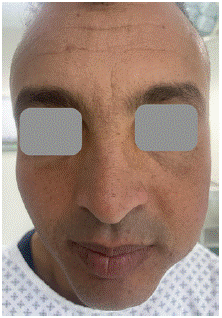
Figure 1: Left facial swelling.
On palpation, the mass exhibited well-defined margins, a firm consistency, and was associated with both palpable paresthesia and tenderness.
Intraoral examination identified a swelling extending from tooth 24 to tooth 28, measuring approximately 3 cm in diameter. The lesion had a rubbery consistency posteriorly, well-defined borders, and was similar in color to the adjacent oral mucosa. Buccopalatal expansion was observed. An ulcer was present over the lesion, but no fistulas were detected. The teeth in the region were stable, and no tenderness was noted during intraoral palpation (Figure 2).
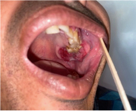
Figure 2: Intraoral photograph of the maxillary lesion.
A CT scan of the facial skeleton was performed, revealing a lytic lesion of the left maxillary bone with a multilobulated appearance and a soap bubble image. After evaluation of all the examinations conducted, the diagnosis of a benign but locally invasive ameloblastoma was made (Figure 3).
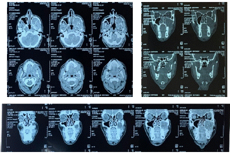
Figure 3: Facial CT scan showing the left maxillary tumor.
The patient underwent a complete resection of the tumor in the left maxilla with resection margins of 1cm (Figure 4,5). Postoperative, he had nasogastric tube placed. Post-operative prescription included antibiotics, appropriate analgesia with frequent and regular antiseptic mouthwash.
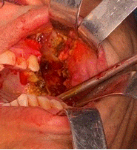
Figure 4: Per operating picture of the maxillary lesion after resction.
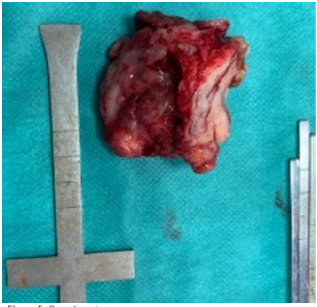
Figure 5: Operating piece.
The follow-up on day 7 and after 2 months was sartisfactory. The nasogastric tube was removed on the 7th day. The patient showed no signs of infection or recurrence and was in good general condition, with no significant facial asymetry (Figure 6).
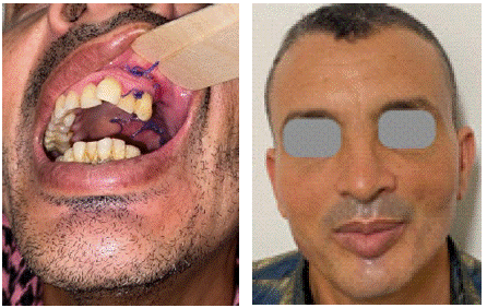
Figure 6: 1 week and 2 months post-operative images.
Discussion
Ameloblastoma, while relatively common among odontogenic tumors, is considered rare when accounting for all tumors and cysts of the maxillofacial bones. It represents approximately 1% of all bone tumors and between 25.2% to 58.6% of benign odontogenic tumors, depending on the study [3].
Its geographic distribution varies with country and ethnicity. Unlike environmental factors such as climate or region of residence, ethnicity appears to play a stronger role in prevalence. A significantly higher incidence is reported in sub-Saharan African populations, where it can reach up to 80.1% in some series [4]. Ameloblastoma typically occurs between the third and fourth decades of life, with a slight predilection for individuals aged 20 to 35 years [5,6]. Both genders are equally affected [5,7,8].
Ameloblastomas are often characterized by a long asymptomatic phase and are frequently discovered incidentally during radiographic examinations for unrelated dental issues. Clinically, the swelling is usually hard, firm, with a bony consistency, and lacks signs of local inflammation. The most frequently reported dental signs in the literature include tooth mobility, displacement, disturbances in the eruption of premolars and molars, and delayed alveolar healing following premature extractions. Two important negative findings should be noted: the absence of regional lymphadenopathy and no alteration of cutaneous or mucosal sensation.
Medical imaging plays a critical role in the diagnosis of ameloblastoma. Conventional imaging, as well as advanced modalities such as CT and MRI, are used to better characterize the lesion. However, radiographic appearance is variable and nonspecific, serving primarily to guide differential diagnosis. A definitive diagnosis is based on a combination of clinical, epidemiological, and especially histopathological findings. In the maxilla, three typical radiographic patterns may be observed: unilocular or multilocular radiolucencies, and in some cases, maxillary sinus opacification — the latter being the most suggestive feature.
Ameloblastoma is a benign tumor of the jaws that, like all odontogenic tumors, requires surgical management. Treatment may be conservative or radical, depending on several factors, including the patient's age, anatomical location of the lesion, its extent, radiographic characteristics, growth potential, and likelihood of long-term followup. Despite being histologically benign, ameloblastomas exhibit locally aggressive behavior similar to that of malignant tumors. Because of this aggressiveness and high recurrence rate, they are seldom treated conservatively. Wide surgical excision is often preferred to reduce recurrence, even at the cost of postoperative sequelae [9].
There are two main surgical approaches: conservative methods such as marsupialization, enucleation, and curettage, and radical surgery involving bone resection with margins extending into healthy tissue. The choice between these approaches is challenging, as recurrence tendencies heavily influence therapeutic decisions. Recurrence rates after conservative treatment have been reported as high as 45–90%. However, these recurrent lesions are often smaller and more easily treated than the original tumor, meaning recurrence is not necessarily synonymous with therapeutic failure.
Conversely, several authors advocate for immediate wide resection with adequate safety margins [10]. A study by Ghandi and al. reported an 80% recurrence rate after enucleation and curettage, compared to less than 50% following radical treatment among 50 cases studied [11].
Lau and Samman conducted a study to determine the lowest recurrence rates among different treatment modalities for unicystic ameloblastoma. They reported a 3.9% recurrence rate after resection, 30.5% after enucleation alone, 16% after enucleation with Carnoy’s solution, and 18% after marsupialization with or without adjunctive therapy [12].
In summary, recurrence is significantly higher following conservative treatment compared to radical approaches. For this reason, most authors favor radical surgery, which remains the most reliable method for preventing recurrence.
Conclusion
The standard treatment for ameloblastoma typically involves surgical resection of the affected bone. In this case, a maxillectomy was performed. However, maxillectomy can result in significant facial and oral cavity defects, leading to both aesthetic deformities and functional impairments. Consequently, maxillary reconstruction becomes essential to restore the form and function of the maxilla following partial or total surgical removal.
References
- Ghai S. Ameloblastoma: An Updated Narrative Review of an Enigmatic Tumor. Cureus. 2022.
- Evangelou Z, Zarachi A, Dumollard Jm, Peoc’h M, Komnos I, Kastanioudakis I, et al. Maxillary ameloblastoma: A review with clinical, histological and prognostic data of a rare tumor. In Vivo. International Institute of Anticancer Research. 2020; 34: p. 2249–2258.
- Chomette G, Auriol M. Histopathologie buccale cer- vico-faciale Masson, Paris, 1986; 51-57.
- Simon EN, Merkx MA, Vuhahula E, Ngassapa D, Stoelinga PJ. A 4 - year prospective study on epide- miology and clinicopathological presentation of odontogenic tumours in Tanzania. Oral surg. Oral Pathol Oral Radiol Endod. 2005; 99: 598-602.
- Ladeinde AL, Ogunlewe MO, Bamgbose BO, Adeyemo WL, Ajayi OF, Arotiba GT, Akinwande JA. Ameloblastoma analysis of 207 cases in Nigerian teaching hospital. Quintessence Int. 2006; 37: 69-74.
- Arotiba GT, Ladeinde AL, Arotiba JT, Ajike SO, Ugboko VI, Ajayi OF. Ameloblastoma in Nigerian children and adolescents; a review of 79 cases. J Oral Maxillofac Surg. 2005; 63: 747-751.
- Ruhin B, Guilbert F, Fouret P, Ghoul S, Berdal A, Bertrand JC. Tumeurs des maxillaires; ameloblastome: donnees actuelles et perspectives. Rev Stomatol Chir Maxillofac. 2005; 106: 1s64.
- Regezi A, Sciubba J. Oral pathology: clinical patholo- gic correlations. Saunders Company, St- Louis (USA), 1989; 337-348.
- Olazoji H, Enwere O. Treatment of ameloblastoma - a review. Niger J Med. 2003; 12: 7-11.
- Ruhin B, Guilbert F, Bertrand J. Traitement des kystes, tumeurs et pseudotumeurs benignes des maxillaires. Encycl Med Chir. Paris, 22-062- K-10. 2005.
- Ghandhi D, Ayoub AF, Pogrel MA, MacDonald G, Brocklebank LM, Moos KF. Ameloblastoma: surgeon’s dilemma. J Oral Maxillofac Surg. 2006; 64: 1010- 1014.
- Lau SL, Samman N. Reccurence related to treatment modalities of unicystic ameloblastoma: a systematic review. Int J Oral Maxillofac Surg. 2006; 35: 681-690.