
Special Article - Minimally Invasive Surgery: Current & Future Developments
Austin J Surg. 2015;2(2): 1051.
Endovascular Sclerotherapy for Male Varicoceles: A Retrospective Analysis of 1619 Patients
Grosso M1, Balderi A1*, Antonietti A1, Pedrazzini F1, Sortino D1, Vinay C1, Giovinazzo G1, Caridi J2 and Manenti M3
1Department of Radiology, Hospital S. Croce and Carle, Italy
2Department of Radiology, Shands at the University of Florida, USA
3Department of Gynecology Endocrinology Hospital, University of Turin, Italy
*Corresponding author: Balderi A, Dipartimento Radiologico - S.C. Radiodiagnostica Azienda Ospedaliera “S.Croce e Carle” via M.Coppino 26 - 12100 Cuneo, Italy
Received: November 25, 2014; Accepted: January 05, 2015; Published: January 07, 2015
Abstract
Purpose: To retrospectively evaluate safety and efficacy (resolution of pain, recurrence rate, improvement in sperm counts and motility) after transcatheter sclerotherapy using Lauromacrogrol 400 in 1619 consecutive patients.
Material and Methods: The institutional review board approved the study and informed consent was obtained.
A retrospective analysis was conducted on 1646 procedures using sclerotherapy performed on 1619 male patients (mean age 29.4 years; range 12.3−65.5 years) with varicoceles during twelve years (from 1998 to 2010). Suspicious testicular veins were catheterized via a right femoral vein approach. Sclerosis of affected veins was induced by a slow injection of a liquid sclerosing agent (Lauromacrogol 400; Kreussler Pharma, Wiesbaden, Germany). Patients were discharged after 6 hours of observation. Follow-up was performed with ultrasound (US)-Doppler 30 days after the procedure. Post-procedure semen analysis was available for 56.4 % of patients.
Results: Catheterisation and sclerotherapy were possible in 1613 of 1646 procedures (98%). No major periprocedural complications occurred. Recurrence was found on US-Doppler follow-up in 30 patients (1.9%). All were retreated: 27 (90%) by using the same percutaneous technique and three by surgery. Post-sclerotherapy semen analysis (available for 56.4% of patients) showed significant improvements in sperm counts (mean 7.9 +/- 3.8 million sperm/mL; p<0.005) and motility (7.2 +/- 2.1%; p<0,005).
Conclusion: Percutaneous sclerotherapy is a safe and minimally invasive technique treating for varicoceles in men.
Keywords: Varicocele; Endovascular sclerotherapy
Introduction
Varicoceles are abnormal dilatations of the spermatic veins. The primary variety develops before puberty (around 12−15 years old), rarely during childhood and adulthood [1]. Primary varicoceles, resulting from reflux into testicular veins, are common (up to 18%) in paediatric subjects [2]. Secondary varicoceles, which are rarer, are caused by external compression of the renal and/or spermatic veins (pelvic or abdominal malignancy, “nutcracker syndrome”), resulting in an increase in hydrostatic pressure.
In 95% of cases varicoceles involve the left spermatic venous plexus (pampiniform) and are attributable to vascular anatomic factors, congenital weakness of the venous walls or incontinent valves. In 4% of cases varicoceles are bilateral, and in 1% are isolated to the right side [3]. Varicoceles are almost always asymptomatic: only a few patients report mild testicular discomfort that is worse on standing or during prolonged effort.
Up until adolescence, normal development of the left testis can be altered by compression by the dilated veins [4]. Furthermore, blood stagnation around the epididymis increases the local temperature from 35° to 37°C, interfering with normal maturation of sperm.
In addition, it has been suggested that reflux of adrenal metabolites, with consequent vasoconstriction, hypo-oxygenation and testicular damage can occur [5]. As a result of these factors, varicoceles are the commonest identifiable cause of infertility in men, being present in 8−22% of the general population and 21−39% of men attending infertility clinics [6]. Commonly associated seminal alterations include oligospermia, asthenospermia, teratospermia and increased numbers of spermatogenetic cells [7].
In Italy, compulsory military service used to permit evaluation of young men: clinical examinations revealed that approximately 15% of recruits had varicoceles. Because this useful screening no longer occurs, military conscription having been abolished, evaluation of teenagers is recommended. In fact, several studies have demonstrated the importance of early management of varicoceles during adolescence to prevent development of adult infertility [8,9].
Traditional therapy for varicoceles includes surgical ligation and exclusion of the spermatic vein in the inguinal region [10]. This can be performed with or without local injection of sclerosing agents. Interventional radiology offers a minimally invasive alternative, potential benefits including, according to comparative studies, lower complication rates [11,12] and quicker recovery of full activity [13], with no statistically significant differences in seminal values or pregnancies achieved. The aim of this study was to assess and report on our twelve year experience of male varicocele sclerotherapy in 1619 patients.
Materials and Methods
During the twelve years from January 1999 to June 2011, 1646 varicocele percutaneous sclerotherapy procedures were performed in our centre on 1619 patients whose ages ranged from 12.3 to 65.5 years (mean ± sd: 29.4 ± 7.7 years). The procedure was the first therapy for 1565 patients (173 of them had symptoms such as scrotal pain) and the second treatment for 54 patients in whom surgical treatment had failed.
In 1587 patients (98%) we performed unilateral left-side treatment instead of in 32 patients we performed a bilateral treatment.
The main indications for treatment were infertility, sub fertility or symptomatic varicoceles. In paediatric patients, the indications were enormous symptomatic varicoceles with the potential to cause testicular atrophy [13].
All patients admitted to our department with varicoceles underwent clinical examination (according to the Dubin and Amelar classification [13]) and scrotal ultrasound (US)-Doppler evaluation during which the veins were studied both at rest and during Valsalva manoeuvres.
Reflux was classified according to the Sarteschi Doppler classification [14-18]:
Grade 1: Prolonged reflux in the vessels of the inguinal canal only during the Valsalva manoeuvre, while scrotal varicosity is not evident in the grey-scale study.
Grade 2: Small posterior varicosities reach the upper pole of the testicle and increase in diameter during the Valsava manoeuvre. Doppler demonstrates evident venous reflux in the supratesticular region only during the Valsalva manoeuvre
Grade 3: Vessels appear enlarged to the inferior pole of the testis when the patient is standing, whereas no dilatation is evident with the patient in the supine position. Colour Doppler demonstrates evident reflux only during the valsalva manoeuvre.
Grade 4: Venous dilatation identifiable with the patient both standing and supine. The dilatation increases with the patient standing and during the Valsalva manoeuvre
Grade 5: Presence of patent venous dilatation in both the prone and supine position. Colour Doppler shows significant baseline venous reflux which does not increase after the Valsalva manoeuvre.
In our study, US-Doppler showed grade I−II varicoceles in 49 patients (3%) grade III in 701 (43.3%) and grade IV−V in 869 (53.7%).
Semen analysis was performed in all patients except for prepubescent children (57 patients were aged < 14 years).
The institutional review board approved the study. Before starting the procedure, an informed consent was obtained from all subjects or both of their parents in the case of minors.
The percutaneous interventions were performed in an angiography suite as day patients (routinely 8 hour same day hospitalization). Atropine (0.5 mg intramuscularly) was administered to minimise the risk of bradycardia during the Valsalva manoeuvre. Steroids/ antihistamines were administered to all reportedly allergic patients.
Following induction of local anaesthesia, the right femoral vein was accessed percutaneously and a 5 Fr sheath inserted through which a catheter with a curve specifically shaped for spermatic vein catheterisation was introduced (VSC catheter; Cook Medical, Bjaeverskov, Denmark). Next, the left renal vein was cannulated, using a spermatic (VSC 1, 2 or 3, Cook) or Cobra catheter (Terumo Europe, Leuven, Belgium) while right spermatic vein was catheterized using Simmons right catheter (Terumo Europe, Leuven, Belgium) under fluoroscopic guidance (MultiDiagnost 3; Philips, Best, The Netherlands). With the patient performing the Valsalva manoeuvre, a renal vein venogram was then obtained (Figure 1 a−d).
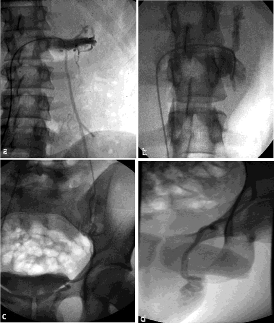
Figure 1 a-d: Renal vein venogram obtained during Valsalva manouvre using a spermatic catheter.
Subsequently, the spermatic vein was catheterized using a hydrophilic wire (Radifocus guide wire angled, 0.035, Terumo Europe); in cases of difficult catheterisation (135 patients, 8.4%; tortuous anatomy, continent valve), micro catheters, usually a Pro great (Terumo Europe) were used (Figure 2 a−c).
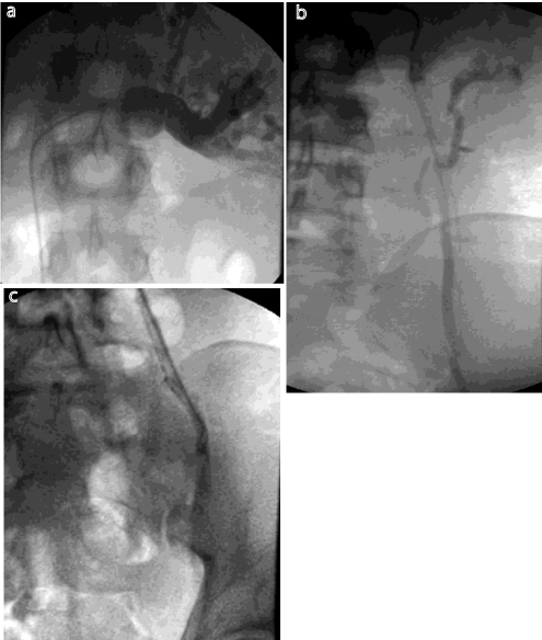
Figure 2 a-c: Case of difficult catheterisation: procedure performed with micro catheter.
Selective phlebography was performed, during the Valsalva manoeuvre, the aim being to demonstrate venous incontinence by detecting evidence of contrast reflux along the internal spermatic vein.
A selective catheter was then advanced into the internal spermatic vein and placed at the level of the hip joint [19,20]. The table was tilted into the inverted Trendelenburg position and the patient instructed to apply manual compression to the spermatic cord at the level of the inguinal ligament for 40 seconds for each 1 mL of sclerosing agent to prevent caudal diffusion of the sclerosant solution. The sclerosing agent (Atossisclerol 2% [Lauromacrogol 400 40 mg] 2 mL vials at 2%; Kreussler Pharma) mixed 50:50 with contrast medium (Ioversol 300 mg/mL, Optiray, Covidien Italia Spa) was slowly injected in small boluses, care being taken to avoid reflux downstream of the compression, until our primary goal was achieved: a steady cessation of flow. The total dose of Atossisclerol was 120 mg (three vials at 2%)
in patients with grade I−III varicoceles and 160 mg (four vials at 2%) for those with grade IV-V varicoceles.
The puncture site was dressed with a sterile compressive bandage after cleansing. No post procedural drugs were required. The patients were discharged after 6 hours of observation. Post procedure instructions included abstinence from sports and sex for 2 weeks.
Follow-up consisted of testicular US-Doppler 1−2 month and semen analysis 3−6 months after the procedure. Testicular USDoppler was re-performed 1 year post-procedure in patients with persistent infertility. If the US-Doppler demonstrated varicocele resolution, no further follow-up was performed.
Results
Varicocele sclerotherapy was performed in 1619/1646 procedures (98%). Failures were due to inability to achieve spermatic vein catheterisation (23/33, 75%), anatomic variations (7/33, 14%, Figure 3), and vessel rupture (3/33, 11%). In cases of failure, antegrade sclerotherapy was not performed. The mean injection time of sclerosant was 30 minutes, the total treatment time being approximately 1 hour. There were no major complications that required additional hospitalization or surgical intervention. Identified minor complications included left groin pain (26 patients, 1.6%) and puncture site haematoma (2 patients, 0.1%).
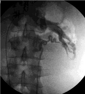
    Figure 3:  Failure of the procedure due to inability to achieve spermatic vein
catheterisation because of anatomic variations.
All patients were discharged on the day of the procedure. USDoppler follow-up evaluation showed varicocele persistence in 30 patients (1.9%), 27 of whom (90%) were retreated using the same percutaneous transfemoral technique. The remaining three patients were treated surgically: in two of these the second percutaneous treatment was aborted because of complete lack of testicular vein reflux during phlebography; in the third patient, we were unable to catheterise the left internal spermatic vein.
We treated two different cohorts of patients; namely, prepubescent patients with clinical varicoceles and medium to high reflux (to prevent future infertility) and patients with clinical/subclinical varicoceles associated with fertility problems and seminal abnormalities. Most of the 57 prepubescent patients (aged < 14 years) did not undergo semen analysis. Patients with fertility problems underwent semen analysis before and 3−6 months after sclerotherapy (913 patients available at follow-up, 56.4%). Significant improvements in sperm counts (7.9 +/- 3.8 million sperm/mL; p<0.005) and motility (7.2 +/- 2.1%; p<0.005) were observed. There was no statistically significant improvement in normal sperm morphology. For statistical analysis we used T - test.
Discussion
Although usually clinically silent, varicoceles can play a significant, preventable role in male infertility. Spermiograms are the most common test for assessing male fertility. In patients with varicoceles, Fobbe F. et al. identify the effects on reproductive potential [19]. Nevertheless, for ethical and practical reasons (normal spermatogenesis only develops years after completion of pubertal development), semen analysis is not indicated in peripubertal subjects. Eliminating varicose vessels prevents their negative effects on testes. These negative effects such as atrophy, increasing of temperature caused by venous stagnation, are especially significant during pubertal development [20,21]. In a review regarding varicocele repair and male sub fertility [22], the authors assert: “Varicocele repair does not seem to be an effective treatment for male or unexplained sub fertility”. On the contrary, other researchers have concluded that treatment of varicoceles is an effective means of treating male infertility [23-26]. We consider that the most important difference between our cases and those reviewed by Evers and Collins is patient selection. Most of the patients treated in this study had clinical (rather than subclinical) varicoceles and seminal abnormalities (rather than normal semen).
Traditional surgical varicocele correction consists of the ligature and section of all varicose veins of the funicular plexus, one by one, preserving only a few normal vessels for venous drainage. Microsurgical interventions are performed through small infrainguinal incisions under local anaesthesia as same-day procedures. Anterograde injection of sclerosing agents can complete the ligature [27,28]. Unsuccessful occlusion is often attributable to overlooked smaller vessels and anatomic variants.
Percutaneous sclerotherapy is a minimally invasive, safe and effective alternative treatment for varicoceles. Unlike other centres [29,30], we usually use a transfemoral vein approach, rather than a jugular or brachial approach. Although jugular access allows treatment of bilateral varicoceles with the same catheter, it is not our first choice because it involves traversing the right atrium. Catheterisation of the left spermatic vein by femoral access is usually easy, requiring microcatheterisation only when anatomic variations [31] or continent valves are present. If spermatic vein laceration occurs, we recommend waiting for several minutes before attempting a new catheterisation. When, even in the inverted Trendelenburg position, there is severe renal vein reflux, an occlusion balloon (5−6 mm in diameter) can be utilized (Figure 4 a-b) for spermatic vein obliteration.
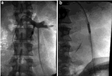
Figure 4 a-b: Case of severe renal vein reflux: procedure performed using an occlusion balloon (5−6 mm in diameter).
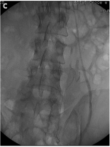
Figure 4 c: Completion venogram as final control shows contrast stasis.
We prefer to employ Atossisclerol 2% (Lauromacrogol 400; Kreussler Pharma) because other sclerosant materials, such as sodium tetradecyl sulphate [32] or alcohol, tend to induce greater pain during injection. In particular, alcohol causes transitory peritoneal irritation because of its transudation through the vessel wall. In fact in our study no patients felt pain during procedure. Bradycardia is the only complication associated with the use of Atossisclerol , and to avoid this fact we injected Atropin intramuscular [33].
Coil embolization is a good alternative to sclerotherapy, but this procedure is significantly more expensive and more complex and has complications, including coil migration and nontarget embolization [34]. In our opinion, coils are useful and cost-effective as second line treatments for large spermatic veins recurrences. The use of plugs has also been reported (Amplatzer Vascular Plug 4, St. Jude Medical St. Paul, Minnesota, USA), mainly for recurrent varicoceles [35].
Lauromacrogol 400 (Kreussler Pharma ) acts chemically: it has tensioactive effects on the lipoid structures of initial surfaces, causing thrombotic apposition and platelet adhesion. Because of its high molecular weight, Lauromacrogol 400 (Kreussler Pharma) does not pass through the endothelium. Our patients experienced no drug intolerance or drug-related complications. Diffusion of the sclerosing agent to the epididymus was successfully prevented by the patients compressing the spermatic cord. A definite advantage of sclerosing agents is that they are able to reach even the smallest plexus′ collateral branches, which are cannot be visualised by phlebography [36]. These vessels and variant anatomy can lead to unsuccessful occlusion during surgery. This may partially explain why, in some comparative studies [37-39], fewer recurrences reportedly occurred after percutaneous sclerotherapy than surgery. Moreover percutaneous sclerotherapy is performed as an outpatient procedure, requires only local anaesthesia and does not preclude future additional percutaneous or surgical treatments. Additionally, the percutaneous approach has the advantage of treating, albeit rarely, bilateral varicocele from a single access point.
After the procedure only few patients needed therapy for low grade of pain well controlled with FANS.
Our study has a number of limitations. First it was a single centre retrospective study, in which all patients were subjected to the same technique without any control group (surgical approach or different embolization technique). In addition, because about half of our patients came from different cities or regions, pre and post-procedural examinations (Doppler, spermiograms) were not performed by the same practitioners and evaluation criteria differed. Finally, the study subjects were heterogeneous, with significant differences in age distribution between patients treated for uncomfortable symptoms (peak age 21 years) and patients treated for subfertility (peak age 34 years).
Conclusion
In our cohort of patients, percutaneous sclerotherapy of varicocele proved to be a minimally invasive technique with a high technical success rate. It did not require hospitalisation, resulted in no major complications and daily activities could be resumed quickly. The few minor complications did not necessitate any medical treatment. With the additional advantage of its cost-effectiveness, percutaneous sclerotherapy is a safe and efficacious treatment for varicocele and should be considered as first line therapy.
References
- Braedel HU, Steffens J, Ziegler M, Polsky MS, Platt ML. A possible ontogenic etiology for idiopathic left varicocele. J Urol. 1994; 151: 62-66.
- Meacham RB, Townsend RR, Rademacher D, Drose JA. The incidence of varicoceles in the general population when evaluated by physical examination, gray scale sonography and color Doppler sonography. J Urol. 1994; 151: 1535-1538.
- Kubal A, Nagler HM, Zahalsky M, Budak M. The adolescent varicocele: diagnostic and treatment patterns of pediatricians. A public health concern? J Urol. 2004; 171: 411-413.
- Kim WS, Cheon JE, Kim IO, Kim SH, Yeon KM, Kim KM, et al. Hemodynamic investigation of the left renal vein in pediatric varicocele: Doppler US, venography, and pressure measurements. Radiology. 2006; 241: 228-234.
- Camoglio FS, Zampieri N, Corroppolo M, Chironi C, Dipaola G, Giacomello L, et al. Varicocele and retrograde adrenal metabolites flow. An experimental study on rats. Urol Int. 2004; 73: 337-342.
- Belker AM. The varicocele and male infertility. Urol Clin North Am. 1981; 8: 41-51.
- Chehval MJ, Purcell MH. Deterioration of semen parameters over time in men with untreated varicocele: evidence of progressive testicular damage. Fertil Steril. 1992; 57: 174-177.
- Kurgan A, Nunnelee JD, Zilberman M. The importance of the early detection of varicocele in adolescent males. Nurse Pract. 1994; 19: 36-37.
- Wyllie GG. Varicocele and puberty--the critical factor? Br J Urol. 1985; 57: 194-196.
- Bassi R. Trattamento chirurgico Del varicocele. Minerva Chir. 1999; 54: 367-371.
- Shlansky-Goldberg RD, VanArsdalen KN, Rutter CM, Soulen MC, Haskal ZJ, Baum RA, et al. Percutaneous varicocele embolization versus surgical ligation for the treatment of infertility: changes in seminal parameters and pregnancy outcomes. J Vasc Interv Radiol. 1997; 8: 759-767.
- Dewire DM, Thomas AJ Jr, Falk RM, Geisinger MA, Lammert GK. Clinical outcome and cost comparison of percutaneous embolization and surgical ligation of varicocele. J Androl. 1994; 15 Suppl: 38S-42S.
- Pinto KJ, Kroovand RL, Jarow JP. Varicocele related testicular atrophy and its predictive effect upon fertility. J Urol. 1994; 152: 788-790.
- Sarteschi LM, Menchini Fabris GM. Ecografia andrologica. Modena: Athena audiovisuals. 2003.
- Bechara CF, Weakley SM, Kougias P, Athamneh H, Duffy P, Khera M, et al. Percutaneous treatment of varicocele with microcoil embolization: comparison of treatment outcome with laparoscopic varicocelectomy. Vascular. 2009; 17: 129-136.
- Dubin L, Amelar RD. Varicocele size and results of varicocelectomy in selected subfertile men with varicocele. Fertil Steril. 1970; 21: 606-609.
- Sarteschi LM, Paoli R, Bianchini M. Lo studio Del varicocele con eco-color- Doppler. G Ital Ultrasonologia. 1993; 4: 43-49.
- Chiou RK, Anderson JC, Wobig RK, Rosinsky DE, Matamoros A Jr, Chen WS, et al. Color Doppler ultrasound criteria to diagnose varicoceles: correlation of a new scoring system with physical examination. Urology. 1997; 50: 953-956.
- Liguori G, Trombetta C, Garaffa G, Bucci S, Gattuccio I, Salame L, et al. Color Doppler ultrasound investigation of varicocele. World J Urol. 2004; 22: 378-381.
- Fobbe F, Hamm B, Sorensen R, Felsenberg D. Percutaneous transluminal treatment of varicoceles: where to occlude the internal spermatic vein. AJR Am J Roentgenol. 1987; 149: 983-987.
- Jarow JP, Ogle SR, Eskew LA. Seminal improvement following repair of ultrasound detected subclinical varicoceles. J Urol. 1996; 155: 1287-1290.
- Zini A, Buckspan M, Berardinucci D, Jarvi K. The influence of clinical and subclinical varicocele on testicular volume. Fertil Steril. 1997; 68: 671-674.
- Evers JL, Collins JA. Assessment of efficacy of varicocele repair for male subfertility: a systematic review. Lancet. 2003; 361: 1849-1852.
- Flacke S, Schuster M, Kovacs A, von Falkenhausen M, Strunk HM, Haidl G, et al. Embolization of varicocles: pretreatment sperm motility predicts later pregnancy in partners of infertile men. Radiology. 2008; 248: 540-549.
- Galfano A, Novara G, Iafrate M, De Marco V, Cosentino M, D'Elia C, et al. Improvement of seminal parameters and pregnancy rates after antegrade sclerotherapy of internal spermatic veins. Fertil Steril. 2009; 91: 1085-1089.
- Cozzolino DJ, Lipshultz LI. Varicocele as a progressive lesion: positive effect of varicocele repair. Hum Reprod Update. 2001; 7: 55-58.
- Hawkins CM, Racadio JM, McKinney DN, Racadio JM, Vu DN. Varicocele retrograde embolization with boiling contrast medium and gelatin sponges in adolescent subjects: a clinically effective therapeutic alternative. J Vasc Interv Radiol. 2012; 23: 206-210.
- Tauber R, Johnsen N. Antegrade scrotal sclerotherapy for the treatment of varicocele: technique and late results. J Urol. 1994; 151: 386-390.
- May M, Johannsen M, Beutner S, Helke C, Braun KP, Lein M, et al. Laparoscopic Surgery versus Antegrade Scrotal Sclerotherapy: Retrospective Comparison of Two Different Approaches for Varicocele Treatment. Eur Urol. 2006; 49: 384−387.
- Pieri S, Agresti P, Morucci M, De'Medici L, Fiocca G, Calisti A. [A transbranchial approach for the percutaneous therapy of pediatric varicocele]. Radiol Med. 2003; 106: 221-231.
- Pieri S, Minucci S, Morucci M, Giuliani MS, De' Medici L. [Percutaneous treatment of varicocele. 13-year experience with the transbrachial approach]. Radiol Med. 2001; 101: 165-171.
- Gandini R, Konda D, Reale CA, Pampana E, Maresca L, Spinelli A, et al. Male varicocele: transcatheter foam sclerotherapy with sodium tetradecyl sulfate--outcome in 244 patients. Radiology. 2008; 246: 612-618.
- Wunsch R, Efinger K. The interventional therapy of varicoceles amongst children, adolescents and young men. Eur J Radiol. 2005; 53: 46-56.
- Pieri S, Agresti P, Fiocca G, Regine G. Phlebographic classification of anatomic variants in the right internal spermatic vein confluence. Radiol Med. 2006; 111: 551-561.
- Chomyn JJ, Craven WM, Groves BM, Durham JD. Percutaneous removal of a Gianturco coil from the pulmonary artery with use of flexible intravascular forceps. J Vasc Interv Radiol. 1991; 2: 105-106.
- Cil B, Peynircioglu B, Canyigit M, Geyik S, Ciftci T. Peripheral vascular applications of the Amplatzer vascular plug. Diagn Interv Radiol. 2008; 14: 35-39.
- Iaccarino V, Venetucci P. Interventional radiology of male varicocele: current status. Cardiovasc Intervent Radiol. 2012; 35: 1263-1280.
- Abdulmaaboud MR, Shokeir AA, Farage Y, Abd El-Rahman A, El-Rakhawy MM, Mutabagani H. Treatment of varicocele: a comparative study of conventional open surgery, percutaneous retrograde sclerotherapy, and laparoscopy. Urology. 1998; 52: 294-300.
- Falappa P, Cotroneo AR, De Cinque M, De Santis M. Long term results of percutaneous sclerotherapy of varicocele. Ann Radiol (Paris). 1984; 27: 316-318.