Special Article - Surgical Case Reports
Austin J Surg. 2015;2(2): 1052.
Foreign Body Penetration: New Solutions for Classic Problems
Costa D*, Romero M, Caravaca I, Domenech E and Saeta R
Department of Surgery, Alicante University General Hospital, Spain
*Corresponding author: Costa D, Department of Surgery, 9th floor, Alicante University General Hospital, 12, Pintor Baeza Avenue, 03010 Alicante, Spain
Received: November 05, 2014; Accepted: January 27, 2015; Published: January 29, 2015
Abstract
The aim of this paper is to present two extremely rare cases of bowel perforation due to ingested foreign bodies: the first case entailed acute abdominal wall cellulitis; in the second case, the foreign body penetrated the duodenum and extended into the hepatic artery. These have not been previously reported in the literature. In both cases, we propose the possibility of laparoscopic management for such emergencies.
Keywords: Intestinal perforation; Meckel diverticulum; Duodenum; Laparoscopy; Foreign body in alimentary tract
Introduction
Foreign body perforation of the bowel occurs occasionally, in less than 10% of the cases consulting for foreign body ingestion. Surgical treatment by means of open surgery is commonly reported [1-3].
Clinical Cases
We present two extremely rare cases of foreign body penetration of the bowel with extension to neighboring organs treated by laparoscopic surgery.
Case 1
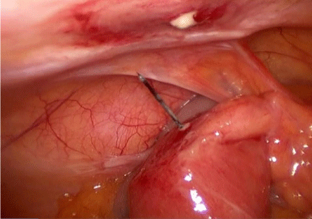
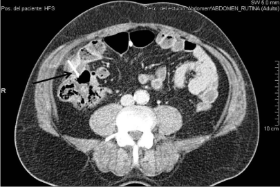
A 54-year-old male reported pain in the right lower quadrant which he had experienced for three days along with fever in the previous 24 hours. Blood tests determined leukocytosis (20,000/3) and left shift. Furthermore, abdominal examination revealed signs of cellulitis in the abdominal wall affecting the right lower quadrant. Computer Axial Tomography scan (CAT-scan) showed the presence of a foreign body that had penetrated a bowel loop and had extended to the abdominal wall. Laparoscopy was carried out and revealed the presence of a Meckel’s diverticulum perforated by a nail that had also pierced the abdominal wall (Image 1). The foreign body was removed and the portion of bowel, including the Meckel’s diverticulum, underwent resection by means of an endoscopic linear stapler (EndoGIA, Covidien®, and Mansfield, Massachusetts). Subsequently, semi-mechanical anastomosis was performed by using an endoscopic linear stapler (EndoGIA, Covidien®) and 3/0 synthetic monofilament absorbable suture. The cellulitis of the abdominal wall was treated by administering the patient broad-spectrum antibiotics over a seven day period. The postoperative course was unremarkable and the patient was discharged on the fourth postoperative day. Following surgery it was ascertained that the patient had swallowed the nail accidentally; nevertheless, he mentioned that he frequently puts nails in his mouth due to his job as a picture framer. At a subsequent review in the outpatients´ clinic two weeks later, the cellulitis had completely disappeared. Two years later, the patient remains non-symptomatic.

    Image:  Operative view of the Meckel’s diverticulum perforated by a nail
after detachment from the abdominal wall.
Case 2
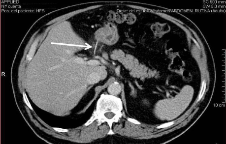
A 62-year-old male attended the emergency department reporting epigastric abdominal pain persisting for four hours, the onset occurring one hour after having lunch. Physical examination revealed pain and abdominal tenderness in the epigastrium and right upper quadrant. Laboratory tests determined a white blood cell count of 16,000/mm3, left shift (86%) and reactive C protein (CRP) levels of 5.8 mg/L. CAT-scan revealed the presence of a high density foreign body passing through the wall of the first duodenal angle and piercing the hepatic artery. CAT-scan showed neither pneumoperitoneum nor free liquid (Figure 1). Emergency laparoscopy was carried out: proximal and distal control of the hepatic artery was achieved and the foreign body was then removed (it appeared to be a very fine fish bone). No hemorrhage was observed, but a single stitch of 5/0 synthetic monofilament absorbable suture was performed the artery and covered by Tachosil® (Takeda Pharmaceuticals International GmbH, Zurich, Switzerland). Also, suture of the duodenal perforation by means of a 3/0 synthetic monofilament absorbable suture was performed and gentle irrigation with warm normal saline solution was carried out. The patient was given 3 postoperative extradoses of broad-spectrum antibiotics. The postoperative outcome was unremarkable and the patient was discharged on the fourth postoperative day. A follow up CAT-scan one month later showed no complications.

    Figure 1:  Foreign body (arrow) penetrating the duodenum and extended to
the common hepatic artery.
Discussion
The ingestion of foreign bodies is a frequent clinical entity, but penetration occurs in less than 10% of the cases. The ileocecal region is one of the most frequently affected areas [4-6]. Penetration can occur anywhere in the digestive tract, but the most commonly affected areas are the ileocecal, rectosigmoidal and oesophageal regions or any narrow areas of the gastrointestinal tract [1-7]. Materials are varied, but sharp metallic objects (45%), small bones (40%), toothpicks and splinters (5%) are the most frequent foreign bodies that cause either perforation or penetration [4]. Extension of the foreign body to neighboring organs may sometimes occur; penetration of the duodenum with extension to the colon, pancreas and liver [1-3] has been published, but extension to the hepatic artery has never been reported (Figure 2). Abdominal pain is the most common primary symptom and CAT-scan is the most sensitive test for the diagnosis [1,8,9]. Cellulitis represents anecdotal evidence. Delay in diagnosis can occur in up to 50% of the cases and as much as 2 weeks; in such cases complications may occur (particularly septic complications) [4- 6]. In the first case that we are presenting, an immediate diagnosis was made. A delay in this case could have led to an abscess formation or, even worse, a life-threatening hemorrhage. The treatment of choice for these cases is surgery, but few papers report the possibility of a laparoscopic approach and none of them as the first possibility [10- 12]. The continuing advances in laparoscopy allow many emergency procedures to be performed this way, thereby gaining impetus for a combined approach using diagnostic and therapeutic laparoscopy for the management of the acute abdomen, provided that this approach brings major benefits for the patient such as, less pain and a faster recovery among others [13,14]. The vascular control of the hepatic pedicle performed in case 1 and the intestinal resection and anastomosis performed in case 2 require surgical skills in advanced laparoscopy. In both cases, laparoscopy led to a very quick patient recovery with low morbidity.

    Figure 2:  Foreign body (arrow) penetrating a Meckel´s diverticulum with
extension to the abdominal wall.
Conclusion
Ingestion of foreign bodies is common but penetration, perforation and extension to other organs are much less frequent. Fish bone duodenal penetration with extension to the hepatic artery has not been previously reported. Laparoscopic approach is feasible and safe in the majority of cases, albeit with the intervention of experienced surgeons in advanced laparoscopy.
References
- Yasuda T, Kawamura S, Shimada E, Okumura S. Fish bone penetration of the duodenum extending into the pancreas: report of a case. Surg Today. 2010; 40: 676-678.
- Okuma T, Nagamoto N, Tanaka E, Yoshida Y, Inoue K, Baba H. Repeated colon penetration by an ingested fish bone: report of a case. Surg Today. 2008; 38: 363-365.
- Clarencon F, Scatton O, Bruguiere E, Silvera S, Afanou G, Soubrane O, et al. Recurrent liver abscess secondary to ingested fish bone migration: report of a case. Surg Today. 2008; 38: 572-575.
- Rodriiguez Hermosa JI, Farres Coll R, Codina Cazador A, Olivet Pujol F, Girones Vila J, Roig Garcia J, et al. Perforaciones intestinales causadas por cuerpos extranos. Cir Esp. 2001; 69: 504-506.
- Martinez Turnes A, Gonzalez Sanz-Agero P, Segura Cabral JA, Conde Gacho P, Olveira Martín A, Alvarez de la Marina JR, et al. [Intestinal perforation caused by a foreign body]. Rev Esp Enferm Dig. 1998; 90: 731-732.
- Ochoa LM, Gonzalez MD, Caparros R. Absceso subfrenico tras ingestion de cuerpo extrano. Tratamiento quirúrgico. Cir Esp. 1999; 66: 361-362.
- Moya Forcen PJ, Martinez Blasco A, Cansado P, Calpena R. [Foreign body ingestion: a common problem, an original treatment]. Cir Esp. 2012; 90: e13.
- Masunaga S, Abe M, Imura T, Asano M, Minami S, Fujisawa I. Hepatic abscess secondary to a fishbone penetrating the gastric wall: CT demonstration. Comput Med Imaging Graph. 1991; 15: 113-116.
- Gonzalez JG, Gonzalez RR, Patino JV, Garcia AT, Alvarez CP, Pedrosa CS. CT findings in gastrointestinal perforation by ingested fish bones. J Comput Assist Tomogr. 1988; 12: 88-90.
- Aarabi S, Stephenson J, Christie DL, Javid PJ. Noningested intraperitoneal foreign body causing chronic abdominal pain: a role for laparoscopy in the diagnosis. J Pediatr Surg. 2012; 47: e15-17.
- Thatigotla B, Vattipally V, Farkas D. Minimally invasive management of bowel perforation due to a foreign body in a super obese individual: a less morbid and safe approach. Am Surg. 2012; 78: E17-18.
- Barto W, Yazbek C, Bell S. Finding a lost needle in laparoscopic surgery. Surg Laparosc Endosc Percutan Tech. 2011; 21: e163-165.
- Di Saverio, S. Emergency laparoscopy: a new emerging discipline for treating abdominal emergencies attempting to minimize costs and invasiveness and maximize outcomes and patients' comfort. J Trauma Acute Care Surg. 2014; 77: 338-350.
- Paterson-Brown S. Emergency laparoscopic surgery. Br J Surg. 1993; 80: 279-283.