Special Article - Surgical Case Reports
Austin J Surg. 2015;2(2): 1054.
Mucocele of Appendix Causing Intussusception Managed by Drainage and Appendisectomy
Tayade SH, Pandya JS*, Vutha RS, Thakre M and Waghmare S
Department of General Surgery, TNMC & BYL Nair Hospital, India
*Corresponding author: Pandya JS, Department of General Surgery, Department of General Surgery, Topiwala National Medical College and BYL Nair Hospital, Mumbai Central, Mumbai-400 008, India
Received: December 08, 2014; Accepted: March 09, 2015; Published: March 19, 2015
Abstract
Appendiceal mucocoele presenting as an intussusceptions is a rare phenomenon, and can lead to various complications such as bleeding per rectum, gangrenous changes perforation and pseudomyxoma peritonei. The pre operative diagnosis of a mucocolele of appendix or its presentation as an intussusception may aid in deciding the surgical approach and the extent of surgery to be performed ranging from simple appendisectomy to a right hemicolectomy. We present such a case presenting as an acute abdomen managed by a simple appendisectomy after reduction of the intussusceptions and the use of a `frozen section’ to rule out a malignant pathology. The absence of warning signs of complicated nature of the appendiceal mucocoele ie rupture, intraperitoneal seeding, involved margins of appendisectomy (on frozen section)served in aiding the decision of a simple appendisectomy as against a more radical approach.
Keywords: Mucocele of appendix; Appendiceal intussusceptions; Appendisectomy
Case Presentation
Appendiceal intussusceptions are rare presentation found only in 0.01% who had undergone appendisectomy [1]. The patient presents with pain, vomiting, and palpable lump in right iliac fossa and infrequently with symptoms of intestinal obstruction. We report a case of acute abdomen where computed tomography was suggestive of appendiceal intussusceptions. Based on the intraoperative findings such as the existence of gangrenous changes of the caecum or the appendiceal base, suspected malignancy of the appendix leading to a mucocoele, various treatment modalities are proposed ranging from simple appendectomy to a more radical right hemicolectomy. The case is discussed here due to its rarity in presentation and management decision.
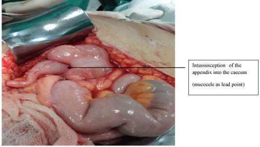
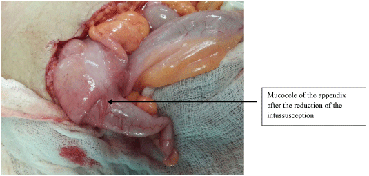
A 40 year female presented with pain in right lower side of abdomen since 5 days associated with nausea, vomiting and fever. Patient was hemodynamically stable. X-ray abdomen was normal with no signs suggestive of intestinal obstruction. Ultrasonography was suggestive of an inflamed appendix with an appendicular duplication cyst. Computed tomography scan of abdomen was suggestive of either an illeocolic intussusceptions or appendiceal intussusceptions with cystic lesion possibly a mucocele as a lead point.The patient was explored through midline incision. Intraoperatively, a mucoid lesion was seen in right iliac fossa identified to be within the inferior part of the caecum. Only the tip of the appendix was visualized and the rest (base) was found to be telescoping into the caecum (Figure 1). On milking the cystic lesion out of the caecum inferiorly the CT scan finding of a mucocoele of the appendix was confirmed (Figure 2). The base of the appendix and the caecum were normal in vascularise. No evidence of any peritoneal deposit of mucoid material was found. There was no evidence of abdominal lymphadenopathy or any other growth visible at any other site. The intraoperative findings were suggestive of a benign pathology. The frozen section of the specimen of appendisectomy after draining the mucocoele confirmed the benign nature of the disease. The mucoid contents were drained with a nick near the base of appendix under aseptic precautions avoiding intraperitoneal spillage of the mucoid content. A thorough peritoneal lavage was given before abdominal closure. Thus the surgery was restricted to a simple appendisectomy. Post operative recovery was uneventful at 3 months follow up.

Figure 1: Appendicular tip noted with mucocele at its base.

    Figure 2:  Appendiceal mucocoele after reduction of the intussuscepted out
of the caecum.
Discussion
Appendiceal mucocele has an estimated incidence of 0.2%-0.3%of all appendectomies performed and 8%-10% of all appendiceal tumors [1].
Mucocele on histopathological basis classified as (1) simple accumulation of mucus within the lumen of appendix due to outflow obstruction with normal epithelium i.e. retention cyst. (2) Hyperplastic epithelium with luminal dilatation. (3) Luminal dilatation upto 6cm with villous adenomatous changes which is benign in nature. (4) Maignant mucinous cystadenocarcinoma with gross dilatation associated with stromal invasion with peritoneal deposits [2,3].
Preoperative diagnosis may help in deciding the correct surgical approach for the patient. Ultrasonographic picture that should raise the suspicion of a mucocoele is a cystic, encapsulated lesion, firmly attached to the cecum, with liquid content and an internal variable echogenicity related to mucus density [4], CT-Scan abdomen findings of an appendiceal mucocele include a round, low-density, thin-walled, encapsulated mass, communicating with the cecum with cup and ball appearance [5,6]. Colonoscopy is a useful investigation and may show a ‘sign of the volcano’, i.e., an erythematous, soft mass with a central crater, from which mucus is discharged suggestive of an appendiceal mucocoele [7]. In this case ultrasonography showed a mass in right iliac fossa and CT-abdomen confirmed cystic lesion at the base of appendix, with appendiceal intussusception into the caecum with cystic lesion as the lead point of the intussusceptions.
Appendiceal mucocele is known to produce complications like bleeding, fistula, intussuseption, obstruction, perforation, volvulus and fatal complication like pseudomyxoma peritonei caused by its spontaneous rupture in peritoneal cavity [8-10].
Surgery is the best treatment for appendiceal intussusceptions [8]. Some cases are reported as colonoscopic reduction of intussusceptions caused by mucocele but carries high risk of peritoneal contamination, venous embolism and perforation [11]. Simple appendisectomy is the treatment of choice for uncomplicated mucocele without any evidence of perforation and peritoneal deposits [12], without enlarged mesenteric lymph nodes and essentially normal bowel.
In this case, the patient had an uncomplicated mucocele which was managed by appendisectomy after drainage of mucin content and confirmation of its benign nature on frozen section. The final histopath report of the appendisectomy specimen was suggestive of a chronic persistent appendicitis i.e. grade 1 [2,3].
It can thus be concluded that- Appendiceal mucocele presenting as an intussusceptions should be evaluated with urgency to identify signs of complication such as bleeding, obstruction and perforation which may warrant an aggressive surgical approach include a right hemicolectomy, cytoreductive surgery, early postoperative intraperitoneal chemotherapy, heated intraperitoneal chemotherapy. However early diagnosis through a high index of suspicion and appropriate investigations may limit the surgery to a less morbid, simple appendicectomy. The use of frozen section to reasonably exclude malignancy as the cause of appendiceal mucocoele and avoids more morbid procedures is also significant.
References
- Deans GT, Spence RA. Neoplastic lesions of the appendix. Br J Surg. 1995; 82: 299-306.
- Higa E, Rosai J, Pizzimbono CA, Wise L. Mucosal hyperplasia, mucinous cystadenoma, and mucinous cystadenocarcinoma of the appendix. A re-evaluation of appendiceal "mucocele". Cancer. 1973; 32: 1525-1541.
- Crawford J. Tumors of the appendix. In: Cotran R, Kumar V, Robbins S, editors. Pathologic basis of disease. Philadelphia Saunders. 1994; 824-825.
- Kim SH, Lim HK, Lee WJ, Lim JH, Byun JY. Mucocele of the appendix: ultrasonographic and CT findings. Abdom Imaging. 1998; 23: 292-296.
- Zissin R, Gayer G, Kots E, Apter S, Peri M, Shapiro-Feinberg M. Imaging of mucocoele of the appendix with emphasis on the CT findings: a report of 10 cases. Clin Radiol. 1999; 54: 826-832.
- Pickhardt PJ, Levy AD, Rohrmann CA Jr, Kende AI. Primary neoplasms of the appendix: radiologic spectrum of disease with pathologic correlation. Radiographics. 2003; 23: 645-662.
- Hamilton DL, Stormont JM. The volcano sign of appendiceal mucocele. Gastrointest Endosc. 1989; 35: 453-456.
- Ruiz-Tovar J, Teruel DG, Castineiras VM, Dehesa AS, Quindós PL, Molina EM. Mucocele of the appendix. World J Surg. 2007; 31: 542-548.
- Rudloff U, Malhotra S. Volvulus of an appendiceal mucocele: report of a case. Surg Today. 2007; 37: 514-517.
- Cois A, Pisanu A, Pilloni L, Uccheddu A. Intussusception of the appendix by mucinous cystadenoma. Report of a case with an unusual clinical presentation. Chir Ital. 2006; 58: 101-104.
- Park JK, Kwon TH, Kim HK, Park JB, Kim K, Suh JI. Adult intussusception caused by an appendiceal mucocele and reduced by colonoscopy. Clin Endosc. 2011; 44: 133-136.
- Dhage-Ivatury S, Sugarbaker PH. Update on the surgical approach to mucocele of the appendix. J Am Coll Surg. 2006; 202: 680-684.