Special Article - Surgical Case Reports
Austin J Surg. 2015;2(2): 1055.
Dual Gallbladders with Dual Cholecystitis
Szczech EC, Parikh SP* and Beniwal J
Department of Surgery, St. Joseph’s Regional Medical Center, USA
*Corresponding author: Parikh SP, Department of Surgery, St. Joseph’s Regional Medical Center, 703 Main Street, Paterson, NJ 0750, USA
Received: February 02, 2015; Accepted: March 20, 2015; Published: March 26, 2015
Abstract
Dual gallbladders, or gallbladder duplication are an uncommon entity encountered by surgeons. When problems arise, such as cholecystitis, these anomalies are uncovered during an operation. Often the problem is unknown until it is uncovered intra-operatively due to lack of pre-operative suspicion.
A 26 year old female presented with signs and symptoms of acute cholecystitis. Pain had been intermittent for the prior 3 months and biliary colic was originally diagnosed. Elevated liver function test warranted preoperative imaging which showed a possible septated gallbladder and non-filling of the gallbladder consistent with cholecystitis. A decision was made to procedure with a laparoscopic cholecystectomy. Upon operative intervention dual gallbladders were encountered, both with associated inflammation. The dual gallbladders each had their own cystic duct and artery and were removed in a minimally invasive fashion. The patient did well in the peri-operative period and was discharged home without complication. Pathology was consistent with cholecystitis in both gallbladders-one of acute nature, and one consistent with chronic cholecystitis.
If suspected pre-operatively, magnetic resonance cholangiopancreatography can be utilized to ascertain the presence of dual gallbladders; however, the significance of this is debated. If encountered in the setting of single gallbladder cholecystitis controversy exists as to whether the normal gallbladder should be removed. At present, the recommendation is to remove both gallbladders in order to avoid leaving behind the “symptomatic” gallbladder. This also allows the surgeon to avoid re-exploration in a previously operated field in case of persistent symptoms from recurrent cholecystitis. This case represents a rare challenge in the diagnosis and treatment of a congenital anomaly with the acquired affliction of dual cholecystitis.
Keywords: Gallbladder; Minimally invasive; Congenital abnormality
Introduction
Dual gallbladders, or gallbladder duplication are an uncommon entity encountered by surgeons. The approximate incidence of dual gallbladders is 1/4000; however, most are never seen due to their asymptomatic nature [1]. When problems arise, such as cholecystitis, these anomalies are uncovered. Here we present the case of a female with gallbladder duplication along with dual cholecystitis, an exotic condition rarely seen in literature.
Case Presentation
A 26 year old Hispanic female with no significant past medical or surgical history presented to the emergency department complaining of one day of sharp right upper quadrant abdominal pain associated with nausea and vomiting. She had prior episodes of pain for three months and in the past had cholelithiasis demonstrated on abdominal Ultrasonography (US). Her symptoms were intermittent, but increasing in frequency and severity which prompted her to seek urgent medical care.
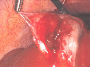
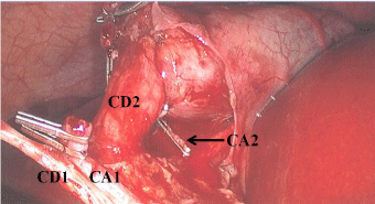
On examination she was mildy tender in the right upper quadrant with a negative Murphy’s sign. Her vital signs were normal. Her laboratory evaluation was unremarkable except for elevated Alkaline Phosphatase, AST and ALT to 123 IU/L, 213 U/L, and 211 U/L respectively. Total bilirubin was 1.1 mg/dL. In light of these laboratory findings suggestive of a biliary obstruction, pre-operative imaging was performed. Abdominal ultrasound demonstrated cholecystitis with the possibility of septated gallbladder or adjacent cyst. Magnetic Resonance Cholangiopancreatography (MRCP), based on the US findings, demonstrated no common bile duct obstruction or duplication of the biliary tree. During the operation, it was noted that the patient had two separate gallbladders, each with their own cystic duct (Figures 1, 2). The common bile duct and common hepatic duct were clearly identified; therefore an intra-operative cholangiogram was not performed. Pathology report returned as consistent with chronic cholecystitis. The patient had an unremarkable postoperative course and was discharged home maintaining all scheduled followup appointments.

Figure 1: Two separate gallbladders seen.

    Figure 2:  Dual Cystic Ducts (CD1/CD2) with dual clipped Cystic Arteries
(CA1/CA2).
Discussion
Pathology confirmed that the patient had two cystic ducts with acute cholecystitis in one gallbladder, and chronic cholecystitis in the other. Duplicate gallbladders were first reported in medicine from an autopsy report in 1674 [2]. Congenital duplication of the gallbladder is rarely seen in clinical practice [3]. Gallbladder duplication can be seen preoperatively on imaging studies such as ultrasonography or MRCP when warranted [2]. Having dual gallbladders does not predispose to cholecystitis or the associated symptoms of biliary colic. Gallbladders duplication can be subdivided into type 1 and type 2 duplications. Type 1 duplications involve gallbladders that share a cystic duct. Type 2 duplications involve dual gallbladders with individual non-communicating cystic ducts [4]. Each gallbladder may be predisposed to cholelithiasis if risk factors are present (i.e. obesity, cirrhosis, female gender, family history etc.). In our case, cholecystitis was suspected based on patient history, clinical examination, and preoperative imaging; however, gallbladder duplication was not suspected at the time of operation.
Intraoperatively when gallbladder duplication is seen both should be removed even if single gallbladder cholecystitis is seen. This would decrease the amount of inflammation, and make the “critical view” easier than if reoperation is necessary in the future [5]. Another method to reduce risk of missed intraoperatively ductal injury is a cholangiogram. A cholangiogram will differentiate both cystic ducts and make identification of the common bile duct easier; therefore, decrease major ductal injury rates.
At present, the recommendation is to remove both gallbladders in order to avoid leaving behind the “symptomatic” gallbladder. This also allows the surgeon to avoid re-exploration in a previously operated field in case of persistent symptoms from recurrent cholecystitis.
Conclusion
The case above represents a rarity in the medical field. When a disease process is seen in the setting of a rare anomaly caution must be taken to clearly identify anatomy. If not for fastidious inspection in our case, our patient would have required a second operation in the immediate post-operative period.
References
- Gross RE. Congenital anomalies of the gall-bladder. A review of 148 cases with a report of double gall-bladder. Arch Surg. 1936; 32: 131-159.
- Kim RD, Zendejas I, Velopulos C, Fujita S, Magliocca JF, Kayler LK, et al. Duplicate gallbladder arising from the left hepatic duct: report of a case. Surg Today. 2009; 39: 536-539.
- Cummiskey RD, Champagne LP. Duplicate gallbladder during laparoscopic cholecystectomy. Surg Laparosc Endosc. 1997; 7: 268-270.
- Singh B, Ramsaroop L, Allopi L, Moodley J, Satyapal KS. Duplicate gallbladder: an unusual case report. Surg Radiol Anat. 2006; 28: 654-657.
- Gigot J, Van Beers B, Goncette L, Etienne J, Collard A, Jadoul P, et al. Laparoscopic treatment of gallbladder duplication. A plea for removal of both gallbladders. Surg Endosc. 1997; 11: 479-482.